| [1] 蒋挺大.甲壳素[M].北京:化学工业出版社,2001: 15-19.
[2] Synowiecki J,Al-Khateeb NA.Production, properties, and some new applications of chitin and its derivatives .Crit Rev Food Sci Nutr.2003; 43: 145-171.
[3] Enríquez de Salamanca A,Diebold Y,Calonge M,et al. Chitosan nanoparticles as a potential drug delivery system for the ocular surface toxicity, uptake mechanism and in vivo tolerance.Invest Ophth Vis Sci.2006;47(4): 1416-1425.
[4] Zheng LY,Zhu JF.Study on antimicrobial activity of chitosan with different molecular weights.Carbohyd Polym.2003;54: 527-530.
[5] Yasunori M,Yoshiyuki K. Antitumor effects of various low-molecular-weight chitosans are due to increased natural killer activity of intestinal intraepithelial lymphocytes in sarcoma 180-bearing mice.J Nutr.2004; 134(4):945-950.
[6] Xie Y,Zhou NJ,Gong YF,et al.Th immune response induced by H pylori vaccine with chitosan as adjuvant and its relation to immune protection.World J Gastroenterol.2007;13(10): 1547-1553.
[7] Yuan XB,Yuan YB,Jiang W,et al.Preparation of rapamycin-loaded chitosan/PLA nanoparticles for immunosuppression in corneal transplantation.Int J Pharm. 2008;349(1-2): 241-248.
[8] Khatri K,Goyal AK,Gupta PN,et al.Plasmid DNA loaded chitosan nanoparticles for nasal mucosal immunization against hepatitis B.Int J Pharm.2008; 354(1-2): 235-241.
[9] Dailey RA,Chavez MR,Choi D.Use of a chitosan-based hemostatic dressing in dacryocystorhinostomy. Ophthal Plast Reconstr Surg.2009; 25(5): 350-353.
[10] 刘昊,张永刚,郭全义,等.新型脱细胞骨基质-壳聚糖骨组织工程支架的制备及性能评价[J].军医进修学院学报,2011,32(6): 616-619.
[11] 刘巍,朱新辉,张洁,等.Matrigel凝胶和壳聚糖/磷酸甘油凝胶两种支架体外构建组织工程软骨的比较[J].江苏医药,2011,37(6): 627-629.
[12] Madhumathi K,Shalumon KT,Rani VV,et al. Wet chemical synthesis of chitosan hydrogel-hydroxyapatite composite membranes for tissue engineering applications.Int J Biol Macromol.2009;45(1):12-15.
[13] Christenson EM,Anseth KS,van den Beucken JJ,et al. Nanobiomaterial applications in orthopedics.J Orthop Res.2007;25(1):11-22.
[14] Qin F,Ye Y,Yao X,et al.Effects of nano-selenium on the capability of learning memory and the activity of Se-protein of mice. Wei Sheng Yan Jiu. 2008; 37(4):502-504.
[15] Song S,Zhou F,Nordquist RE,et al.Glycated chitosan as a new non-toxic immunological stimulant.Immunopharmacol Immunotoxicol.2009;31(2):202-208.
[16] Zhang C,Qu G,Sun Y,et al.Biological evaluation of N-octyl-O-sulfate chitosan as a new nano-carrier of intravenous drugs. Eur J Pharm Sci.2008;33 (4-5):415-423.
[17] Nasti A,Zaki NM,de Leonardis P,et al.Chitosan/TPP and chitosan/TPP-hyaluronic acid nanoparticles: systematic optimisation of the preparative process and preliminary biological evaluation. Pharm Res. 2009; 26(8):1918-1930.
[18] Kashiwazaki H,Kishiya Y,Matsuda A,et al.Fabrication of porous chitosan/hydroxyapatite nanocomposites: Their mechanical and biological properties. Biomed Mater Eng. 2009;19 (2):133-140.
[19] Yoksan R,Chirachanchai S.Amphiphilic chitosan nanosphere: Studies on formation, toxicity, and guest molecule incorporation. Bioorg Med Chem. 2008; 16(5):2687-2696.
[20] Elmore S.Apoptosis: A Review of Programmed Cell Death. Toxicol Pathol. 2007;35: 4495-516.
[21] Mao HQ,Roy K,Troung-Le VL,et al.Chitosan-DNA nanoparticles as gene carriers:synthesis, characterization and transfection efficiency.J Controlled Release.2001; 70(3): 399-421.
[22] Qi LF,Xu ZR,Jiang X,et al.Cytotoxic activities of chitosan nano particles and copper- loaded nanoparticles.Bioorg Med Chem Lett.2005;15(5):1397-1399.
[23] 汪冰,丰伟悦,赵字亮,等.纳米材料生物效应及其毒理学研究进展[J].中国科学B辑,2005,35(1): 1-10.
[24] Wang B,Feng WY,Zhu MT,et al.Neurotoxicity of low-dose repeatedly intranasal instillation of nano- and submicron-sized ferric oxide particles in mice.J Nanopart Res.2009;11: 41-53.
[25] Inoue K,Takano H,Yanagisawa R,et al.Size effects of latex nanomaterials on lung inflammation in mice.Toxicol Appl Pharmacol.2009;234 (1):68-76.
[26] Inoue K,Takano H,Koike E,et al.Effects of pulmonary exposure to carbon nanotubes on lung and systemic inflammation with coagulatory disturbance induced by lipopolysaccharide in mice.Exp Biol Med.2008;233(12): 1583-1590.
[27] Wang B,Feng WY,Wang TC,et al.Acute toxicity of nano- and micro-scale zinc powder in healthy adult mice.Toxicol Lett. 2006;161(2):115-123.
[28] Wei SL,Yi X,Chuan CH,et al.Toxicity of nano- and micro-sized ZnO particles in human lung epithelial cells.J Nanopart Res. 2009;11: 25-39.
[29] Meirelles L,Melin L,Peltola T.Effect of hydroxyapatite and titania nanostructures on early in vivo bone response.Clin Implant Dent Relat Res. 2008; 10(4):245-54.
[30] 彭黎明,王曾里.细胞凋亡的基础与临床[M].北京:人民卫生出版社,2003:363.
[31] Awasthi KK,John PJ,Awasthi A,et al. Multi walled carbon nano tubes induced hepatotoxicity in Swiss albino mice. Micron. 2013;44: 359-364.
[32] 陈伙德,贾振斌,邱敏,等.纳米材料在医药领域中的应用与展望[J].广东化工,2008,35(10):93-95.
[33] Griffitt RJ,Hyndman K,Denslow ND,et al.Comparison of Molecular and Histological Changes in Zebrafish Gills Exposed to Metallic Nanoparticles. Toxicol Sci.2009;107(2): 404-415.
[34] Watari F,Abe S,Koyama C,et al.Behavior of in vitro, in vivo and internal motion of micro/nano particles of titanium, titanium oxides and others.J Ceram Soc Jpn.2008; 116(1349): 1-5.
[35] Nel A,Xia T,Madler L,et al.Toxic Potential of Materials at the Nanolevel. Science. 2006;(311):622-627.
[36] Khang D,Liu-Snyder P,Pareta R,et al.Reduced responses of macrophages on nanometer surface features of altered alumina crystalline phases.Acta Biomater. 2009; 5(5): 1425-1432.
[37] Waters KM,Masiello LM,Zangar RC,et al.Macrophage Responses to Silica Nanoparticles are Highly Conserved Across Particle Sizes.Toxicol Sci.2009; 107(2):553-569.
[38] Jin CH,Jin YH,Wang J,et al.Comparative study of the effect on oxidative damage in rats inhaled by nano-sized and micro-sized silicon dioxide. Wei Sheng Yan Jiu.2008;37(1): 16-8,36.
[39] Li R,Ning Z,Cui J,et al.Ultrafine particles from diesel engines induce vascular oxidative stress via JNK activation.Free Radic Biol Med.2009; 46(6):775-782.
[40] Barmo C,Ciacci C,Canonico B,et al.In vivo effects of n-TiO(2) on digestive gland and immune function of the marine bivalve Mytilus galloprovincialis. Aquat Toxicol.2013;132-133C:9-18.
[41] Min KY,Se WP.Exposing Zebrafish to Silver Nanoparticles during Caudal Fin Regeneration Disrupts Caudal Fin Growth and p53 Signaling.Mol Cell Toxicol.2008;4(4):311-317.
[42] Park MR,Gurunathan S,Choi YJ,et al.Chitosan nanoparticles cause pre- and postimplantation embryo complications in mice.Biol Reprod.2013;88(4):88.
[43] Yin N,Liu Q,Liu J,et al.Silver Nanoparticle Exposure Attenuates the Viability of Rat Cerebellum Granule Cells through Apoptosis Coupled to Oxidative Stress. Small. 2013; 9(9-10):1831-1841.
[44] Xu P,Xu J,Liu S,et al.Nano copper induced apoptosis in podocytes via increasing oxidative stress.J Hazard Mater. 2012;30 (241):279-286.
[45] 胡瑜兰,王淇,高建青,等.壳聚糖纳米粒的斑马鱼胚胎毒性及其机制研究[C].2010年中国药学大会暨第十届中国药师周论文集
[46] 黄琴琴,王永禄,李学明.纳米载体材料毒理学效应及其作用机制研究进展[J].中国药房,2011,22(21): 2004-2007.
[47] 纪宗斐,张丹瑛,沈锡,等.碳纳米管的毒性研究进展[J].复旦学报:医学版,2011; 38(6): 556-559.
[48] Klaus U,Catrin A,Lars-0liver K,et al.Cellular response to nanoparticles: target structures and mechanisms. Nanotoxicology. 2007;1(1): 52-71.
[49] 周国强,陈春英,李玉锋,等.纳米材料生物效应研究进展[J].生物化学与生物物理进展,2008,35(9): 998-1006.
[50] 韩雁,崔国权,董淑英,等.纳米氧化锌诱导血管内皮细胞凋亡及氧化应激[J].中国公共卫生,2012,28(4):94-96. |
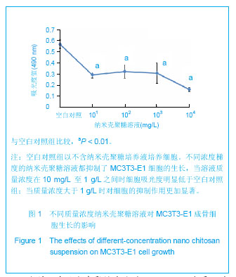
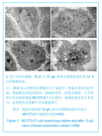
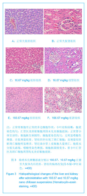
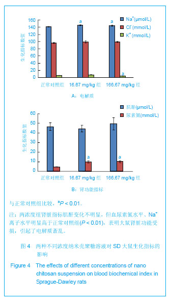
.jpg)