中国组织工程研究 ›› 2017, Vol. 21 ›› Issue (3): 418-422.doi: 10.3969/j.issn.2095-4344.2017.03.017

• 骨与关节图像与影像 bone and joint imaging • 上一篇    下一篇

磁共振T2 mapping和T1ρ成像研究群养成年恒河猴腰椎间盘的退变过程

陈江波1,潘希敏2,陈应明2,吴志强1,蒙仲猛1,陈立强1,庄文权1   

  1. 1中山大学附属第一医院介入放射科,广东省广州市 510080;2中山大学附属第一医院东院放射科,广东省广州市   510700
  • 修回日期:2016-11-29 出版日期:2017-01-28 发布日期:2017-03-14
  • 通讯作者: 庄文权,教授,硕士生导师,中山大学附属第一医院介入放射科,广东省广州市 510080
  • 作者简介:陈江波,男,1988年生,河南省开封市人,汉族,中山大学在读硕士,主要从事介入放射学研究。
  • 基金资助:

    国家自然科学基金资助项目(81272041);广东省科技计划基金资助项目(2013B021800116)

Magnetic resonance T2 mapping and T1ρ imaging of adult rhesus monkeys with lumbar intervertebral disc degeneration in free-range population  

Chen Jiang-bo1, Pan Xi-min2, Chen Ying-ming2, Wu Zhi-qiang1, Meng Zhong-meng1, Chen Li-qiang1, Zhuang Wen-quan1   

  1. 1Department of Interventional Radiology, the First Affiliated Hospital of Sun Yet-sen University, Guangzhou 510080, Guangdong Province, China; 2Department of Radiology, the First Affiliated Hospital East Campus of Sun Yet-sen University, Guangzhou 510700, Guangdong Province, China
  • Revised:2016-11-29 Online:2017-01-28 Published:2017-03-14
  • Contact: Zhuang Wen-quan, Professor, Master’s supervisor, Department of Interventional Radiology, the First Affiliated Hospital of Sun Yet-sen University, Guangzhou 510080, Guangdong Province, China
  • About author:Chen Jiang-bo, Studying for master’s degree, Department of Interventional Radiology, the First Affiliated Hospital of Sun Yet-sen University, Guangzhou 510080, Guangdong Province, China
  • Supported by:

    the National Natural Science Foundation of China, No. 81272041; the Guangdong Science and Technology Plan Project, No. 2013B021800116

摘要:

文章快速阅读:


文题释义:
磁共振T2 mapping和T1ρ成像技术:是检测腰椎间盘早期退变的新技术之一,T2-map值对椎间盘胶原蛋白的变化较敏感,而T1ρ值能敏感地反映蛋白聚糖的降解。
灵长类动物猴:被认为是模拟人类椎间盘退变的最理想实验动物,但有关群养种属的恒河猴腰椎间盘磁共振T2 mapping和T1ρ成像研究尚未见有报道。
 
摘要
背景:灵长类动物猴被认为是模拟人类椎间盘退变的最佳动物,但有关猴腰椎间盘退变规律的研究极少。
目的:通过磁共振T2mapping和T1ρ成像技术探讨群养种属成年恒河猴腰椎间盘退变过程及其退变特点。
方法:在1.5T MR扫描仪上采集符合研究条件的63只群养种属恒河猴腰椎间盘矢状位的T2WI、T2 mapping和T1ρ图像,利用后处理工作站测定腰椎间盘髓核组织感兴趣区的T2-map值和T1ρ值。

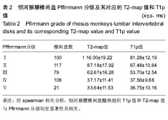
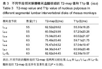
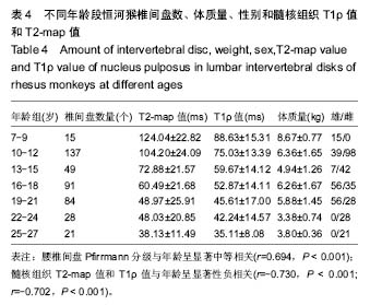
结果与结论:①研究获得425个质量良好的成年恒河猴腰椎间盘磁共振图像,不同人员对髓核组织T2-map值和T1ρ值的测量高度一致,两者Kappa系数达0.93以上;②恒河猴腰椎间盘髓核组织T1ρ值和T2-map值与Pfirrmann分级呈显著负相关(r=-0.842,P < 0.01;r=-0.896,P < 0.01)。PfirrmannⅠ-Ⅳ级之间恒河猴腰椎间盘髓核组织T1ρ值和T2-map值差异均有显著性意义(P < 0.001,P < 0.001)。PfirrmannⅡ-Ⅲ级(退变早期)和PfirrmannⅣ-Ⅴ级(退变晚期)的髓核组织T1ρ值与Pfirrmann分级呈显著负相关(r=-0.517, P < 0.01r=-0.499,P < 0.01);退变早期和退变晚期的髓核组织T2-map值与Pfirrmann分级也呈显著负相关(r=-0.617,P < 0.01;r=-0.652,P < 0.01);③L1-2和L2-3椎间盘髓核组织T2-map值明显低于L6-7和L7-S1椎间盘髓核组织T2-map值(P < 0.05);④不同年龄段恒河猴腰椎间盘髓核组织T2-map值和T1ρ值差异有显著性意义(r=-0.730,P < 0.001;r=-0.702,P < 0.001);⑤结果说明,磁共振T2 mapping和T1ρ成像技术可客观量化敏感地评估恒河猴腰椎间盘髓核组织的退变过程;成年恒河猴L1-2和L2-3椎间盘较L6-7和L7-S1椎间盘易发生退变且退变程度重;年龄是影响成年恒河猴腰椎间盘退变的重要因素之一。    

中国组织工程研究杂志出版内容重点:人工关节;骨植入物;脊柱骨折;内固定;数字化骨科;组织工程
ORCID: 0000-0002-7345-1940(陈江波)

关键词: 骨科植入物, 脊柱植入物, 骨与关节图像和影像, 磁共振成像, T2 mapping, T1ρ, 椎间盘退变, 恒河猴, 群养种属, 国家自然科学基金

Abstract:

BACKGROUND: Primates are considered to be the most appropriate animal model of lumbar intervertebraldisc degeneration, but the disc degenerated characteristics of monkeys were rarely reported.

OBJECTIVE: To verify the degenerated regularity and characteristics of lumbar intervertebral disks in rhesus monkeys with magnetic resonance T2 mapping and T1ρ imaging technology.
METHODS: The sagittal lumbar intervertebral disc magnetic resonance T2 weighted imaging,T2 weighted mapping imaging and T1ρ weighted imaging of 63 adult rhesus monkeys were acquired on 1.5T magnetic resonance equipment. The T2-map value and T1ρ value of lumbar intervertebral disc regions of interest were calculated on the post-processing workstation.
RESULTS AND CONCLUSION: (1) This study obtained 425 better magnetic resonance images of lumbar intervertebral disks in adult rhesus monkeys. T2-map value and T1ρ value of nucleus pulposus were most consistent by different persons, and the Kappa coefficient was more than 0.93. (2) The T2-map value and T1ρ value of nucleus pulposus were both negatively correlated significantly with Pfirrmann grades (r=-0.842, P < 0.01; r=-0.896, P < 0.01). The T1ρ value and T2-map value of nucleus pulposus were significantly statistically different between Pfirrmann grades I-IV (P < 0.001, P < 0.001). The T1ρ value of nucleus pulposus was negatively correlated significantly with Pfirrmann grade II-III (r=-0.517, P < 0.01) and Pfirrmann grade IV-V (r=-0.499, P < 0.01). The T2-map value of nucleus pulposus was also negatively correlated significantly with Pfirrmann grade II-III (r=-0.617, P < 0.01) and Pfirrmann grade IV-V (r=-0.652, P < 0.01). (3) The T2-map value of L1-2 and L2-3 segments nucleus pulposus were significantly lower than that in L6-7 and L7-S1 segments (P < 0.05). (4) There were significant differences in age among the T1ρ value and T2-map value of nucleus pulposus (r=-0.702, P < 0.001, r=-0.730, P < 0.001). (5) It is concluded that magnetic resonance T2 mapping and T1ρimaging technology can objectively and sensitively assess the degenerated process of nucleus pulposus in rhesus monkeys. The degeneration in upper lumbar segments (L1-2 and L2-3) was earlier and more severe than that in lower lumbar segments (L6-7 and L7-S1) in rhesus monkeys. Age is one of the most important factors in lumbar intervertebral disc degeneration of adult rhesus monkeys.

中国组织工程研究杂志出版内容重点:人工关节;骨植入物;脊柱骨折;内固定;数字化骨科;组织工程

Key words: Intervertebral Disk, Magnetic Resonance Imaging, Macaca mulatta, Tissue Engineering

中图分类号: