中国组织工程研究 ›› 2017, Vol. 21 ›› Issue (2): 187-191.doi: 10.3969/j.issn.2095-4344.2017.02.005

• 组织工程骨及软骨材料 tissue-engineered bone and cartilage materials • 上一篇    下一篇

聚甲基丙烯酸甲酯骨水泥对肺癌细胞毒性作用的体外实验

潘元星,米  川,施学东,王  冰,崔云鹏,林云飞
  

  1. 北京大学第一医院骨科,北京市   100083
  • 收稿日期:2016-10-21 出版日期:2017-01-18 发布日期:2017-02-27
  • 通讯作者: 米川,主任医师,副教授,北京大学第一医院骨科骨与软组织肿瘤病房,北京市 100083
  • 作者简介:潘元星,男,1985年生,广东省汕头市人,博士,主要从事骨肿瘤方面相关研究。

In vitro toxic effect of polymethylmethacrylate bone cement on lung cancer cells

Pan Yuan-xing, Mi Chuan, Shi Xue-dong, Wang Bing, Cui Yun-peng, Lin Yun-fei
  

  1. Department of Orthopaedics, Peking University First Hospital, Beijing 100083, China
  • Received:2016-10-21 Online:2017-01-18 Published:2017-02-27
  • Contact: Mi Chuan, Chief physician, Associate professor, Department of Orthopaedics, Peking University First Hospital, Beijing 02110003, China
  • About author:Pan Yuan-xing, M.D., Department of Orthopaedics, Peking University First Hospital, Beijing 02110003, China

摘要:

文章快速阅读:


文题释义:
聚甲基丙烯酸甲酯(PMMA)骨水泥
:是一种重要的可塑性高分子材料,具有较好的透明性、化学稳定性、较高的抗张强度和可塑性,与人体组织有适应性,已广泛应用于骨科手术中,是椎体成形术中填充材料的主要成分。
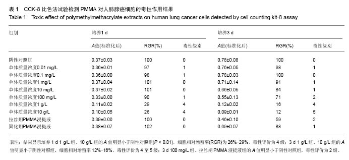
细胞毒性:是由细胞或化学物质导致的细胞杀伤事件,是不依赖于凋亡或坏死的细胞死亡。实验采用的量化细胞毒性评价方法是cck-8比色法,细胞增殖越多越快,则颜色越深;细胞增殖越慢,则颜色越浅。通过测定细胞的吸光度(A)值计算细胞相对增殖率(RGR)来评价细胞毒性。

背景:经皮椎体成形是治疗脊柱转移瘤的一种微创手术方法,但术中注射的聚甲基丙烯酸甲酯(polymethylmethacrylate,PMMA)骨水泥对肿瘤的治疗机制目前尚不明确。
目的:分析PMMA骨水泥及其单体对肿瘤细胞的细胞毒作用。
方法:将固化期、拉丝期PMMA骨水泥的浸提液及不同质量浓度单体的稀释液与人肺腺癌细胞spc-A1接触培养1,3 d后,采用细胞形态直接观察法和CCK-8比色法测定细胞吸光度(A)值,计算细胞相对增殖率(RGR),评价PMMA骨水泥及其单体对肿瘤细胞的毒性。
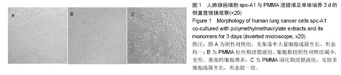
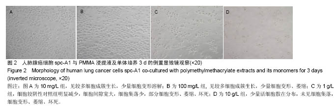
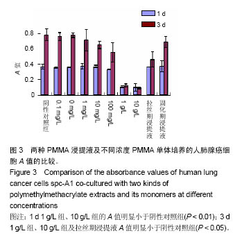
结果与结论:①1 d 1 g/L组、10 g/L组的A值明显小于阴性对照组(P < 0.01);3 d 1 g/L组、10 g/L组及拉丝期浸提液A值明显小于阴性对照组(P < 0.05);②结果显示培养1 d 1 g/L组、10 g/L组的A值明显小于阴性对照组(P < 0.01),细胞相对增殖率为26%-29%,毒性评价为4级;3 d 1 g/L组、10 g/L组的A值明显小于阴性对照组,细胞相对增殖率12%-16%,毒性评价为4至5级;3 d 100 mg/L组、拉丝期PMMA浸提液组的A值明显小于阴性对照组,毒性评价为2级。③结果表明,拉丝期PMMA骨水泥浸提液及PMMA单体质量浓度为100 mg/L-10 g/L时对肿瘤细胞有明显的毒性,说明经皮椎体成形过程中拉丝期PMMA骨水泥注入椎体后其释放的单体可能对肿瘤细胞存在细胞毒作用。

关键词: 生物材料, 骨生物材料, 经皮椎体成形术, 聚甲基丙烯酸甲酯, 骨水泥, 细胞毒, 肺癌细胞, 体外试验, 脊柱转移瘤

Abstract:

BACKGROUND: Percutaneous vertebroplasty is a minimally invasive treatment for spinal metastasis tumor, but the mechanism of the injected polymethylmethacrylate (PMMA) bone cement in the treatment of tumor is not fully understood.
OBJECTIVE: To explore the cytotoxicity of PMMA bone cement and its monomers on tumor cells.
METHODS: PMMA extracts in the wire drawing and curing periods and different mass concentrations of monomer dilutions were co-cultured with human lung cancer cells spc-A1. The cell morphology was observed using inverted microscope, the absorbance (A) values were detected by cell counting kit- 8 assay, the relative growth rate was calculated, and the toxicity of PMAA bone cement and its monomers was evaluated at 1 and 3 days of culture, respectively.
RESULTS AND CONCLUSION: At 1 day of culture, the absorbance values in the 1 and 10 g/L groups were significantly lower than that in the negative control group (P < 0.01). The absorbance values in the 1 g/L, 10 g/L, 100 mg/L and wire drawing extract groups were significantly lower than those in the negative control group at 3 days of culture (P < 0.05). The relative growth rate in the 1 and 10 g/L groups was 26%-29%, and the level of toxicity was grade 4 after 1-day culture; at 3 days, the relative growth rate was decreased to 12%-16%, and the level of toxicity was grades 4-5. After 3-day culture, the level of toxicity reached to grade 2 in the 100 mg/L and wire drawing extract groups. These results indicate that PMMA bone cement in wiredrawing period and its monomers exert toxic effects on tumor cells.

Key words: Polymethacrylic Acids, Vertebroplasty, Neoplasms, Tissue Engineering

中图分类号: