| [1] 徐焰,徐修礼,马越云,等.GHD矮小儿童血清IGF-1水平与血铅的相关性研究及其机制探讨[J].中华检验医学杂志, 2015,38(4):238-242.[2] 李萍,保丽丽,李政良,等.特发性矮小儿童生长激素治疗前后胰岛素样生长因子-1、胰岛素样生长因子结合蛋白-3的对比研究[J].临床荟萃,2013,28(8):918-919.[3] 陈宝定,贾俊静,刘丽,等.生长激素受体基因表达的研究进展[J].中国畜牧兽医,2010,37(12):127-130.[4] 单路娟,刘越坚,邱阳,等.肝细胞生长因子对神经干细胞分化的影响[J].立体定向和功能性神经外科杂志,2008, 12(6):351-354.[5] Bjornsson BT, Taranger GL, Hansen T, et al. The interrelation between photoperiod, growth-hormone, and sexual-maturation of adult atlantic salmon (Salmo salar). Gen Comp Endocrinol. 1994; 93(1):70-81.[6] Nugent AG, Leung KC, Sullivan D, et al. Modulation by progestogens of the effects of oestrogen on hepatic endocrine function inpostmenopausal women. Clin Endocrinol (Oxf). 2003;59(6): 690-698.[7] Melamed P, Eliahu N, Ofir M, et al. The effects of gonadal development and sex steroids on growth hormone secretion in the male tilapiahybrid (Oreochromis niloticus x o. aureus).Fish Physiol Biochem. 1995;14:267-277.[8] Reynolds BA, Weiss S. Generationof neuronsandastri cytesfromisolatedcells of the adult mammaliannervous system. Science. 1992;255(5052): 1707-1710.[9] 潘灏,章翔,刘卫平,等.不同浓度胰岛素对小鼠神经干细胞分化的影响[J]. 立体定向和功能性神经外科杂志,2005, 18(4): 204-206.[10] Vescovi AL, Parati EA, Gritti A, et al. Isolationandcloning of multipotential stemcells from the embryonic human CNS and establishment of transplantable humanneural stemcell linesby epigenetic stimulation. Exp Neurol.1999;156:71-83.[11] Nunez J. Primary Culture of Hippocampal Neurons from P0 Newborn Rats. J Vis Exp. 2008;29 (19): 3791-3895.[12] Gilyarov AV, Nestin in central nervous system cells. Neurosci Behav Physiol. 2008; 38(2):165-169.[13] 胡智兴,耿菊敏,梁道明,等.肝细胞生长因子促进人胚胎干细胞向神经前体细胞分化[J].中国病理生理杂志,2010, 26(4):730-736.[14] 潘灏,章翔,刘卫平,等.神经干细胞原代培养及胰岛素对其增殖与分化的作用[J].中华神经外科疾病研究杂志,2005, 4(4):316-319.[15] 上官芳芳,施建农.生长激素/IGF-1对认知功能的影响(综述)[J].中国心理卫生杂志,2007,21(8):568-570.[16] Martinez-Moreno CG, Giterman D, Henderson D, et al. Secretagogueinduction of GH release in QNR/D cells: Prevention ofcell death. Gen Comp Endocrinol. 2014; 203: 274-280.[17] Srimontri P, Hirota H, Kanno H, et al. Infusion of growth hormoneinto the hippocampus induces molecular and behavioral responses in mice. Exp Brain Res. 2014; 232(9): 2957-2966.[18] Alba- Betancourt C, Luna- Acosta JL, Ramirez- Martinez CE,et al. Neuro- protective effects of growth hormone (GH) after hypoxia-ischemia injury in embryonic chicken cerebellum.Gen Comp Endocrinol. 2013;183: 17-31.[19] 刘海涛,白宏英,曾志磊,等.重组人生长激素对脑缺血/再灌注损伤细胞凋亡及Nestin 表达的影响[J].中国实用神经疾病杂志, 2010, 13(1): 39-41.[20] 张孟玲,孙向荣,郭菲菲,等.生长激素释放肽对全脑缺血/再灌注损伤大鼠海马组织的保护作用及对谷氨酸/γ-氨基丁酸敏感神经元放电活动的影响[J].中华危重病急救医学. 2016, 28(5): 455-459. |
.jpg) 文题释义:
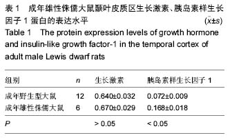
生长因子、胰岛素样生长因子1:除了垂体分泌生长因子生长激素外,大脑的其他部位如中脑、皮质、海马、下丘脑、纹状体、嗅球和小脑等脑区均检测到生长激素。实验中在垂体生长激素分泌缺乏的Lewis侏儒大鼠颞叶皮质中检测到了生长激素、胰岛素样生长因子1,进一步证实了大脑某些区域能独立于脑垂体的生长激素和外周血胰岛素样生长因子1而产生生长激素、胰岛素样生长因子1。
海马神经元干细胞:胎鼠的小脑半、中脑、海马、皮质、纹状体均可分离神经干细胞。成年大鼠中枢神经系统的广泛区域存在具有多项分化潜能的神经干细胞。实验中从成年SD大鼠海马分离神经元干细胞,在加入不同浓度生长激素共培养后发现生长激素能促进海马神经元干细胞向神经元细胞分化。
文题释义:
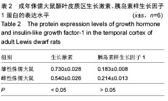
生长因子、胰岛素样生长因子1:除了垂体分泌生长因子生长激素外,大脑的其他部位如中脑、皮质、海马、下丘脑、纹状体、嗅球和小脑等脑区均检测到生长激素。实验中在垂体生长激素分泌缺乏的Lewis侏儒大鼠颞叶皮质中检测到了生长激素、胰岛素样生长因子1,进一步证实了大脑某些区域能独立于脑垂体的生长激素和外周血胰岛素样生长因子1而产生生长激素、胰岛素样生长因子1。
海马神经元干细胞:胎鼠的小脑半、中脑、海马、皮质、纹状体均可分离神经干细胞。成年大鼠中枢神经系统的广泛区域存在具有多项分化潜能的神经干细胞。实验中从成年SD大鼠海马分离神经元干细胞,在加入不同浓度生长激素共培养后发现生长激素能促进海马神经元干细胞向神经元细胞分化。
.jpg) 文题释义:
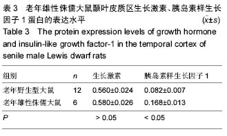
生长因子、胰岛素样生长因子1:除了垂体分泌生长因子生长激素外,大脑的其他部位如中脑、皮质、海马、下丘脑、纹状体、嗅球和小脑等脑区均检测到生长激素。实验中在垂体生长激素分泌缺乏的Lewis侏儒大鼠颞叶皮质中检测到了生长激素、胰岛素样生长因子1,进一步证实了大脑某些区域能独立于脑垂体的生长激素和外周血胰岛素样生长因子1而产生生长激素、胰岛素样生长因子1。
海马神经元干细胞:胎鼠的小脑半、中脑、海马、皮质、纹状体均可分离神经干细胞。成年大鼠中枢神经系统的广泛区域存在具有多项分化潜能的神经干细胞。实验中从成年SD大鼠海马分离神经元干细胞,在加入不同浓度生长激素共培养后发现生长激素能促进海马神经元干细胞向神经元细胞分化。
文题释义:
生长因子、胰岛素样生长因子1:除了垂体分泌生长因子生长激素外,大脑的其他部位如中脑、皮质、海马、下丘脑、纹状体、嗅球和小脑等脑区均检测到生长激素。实验中在垂体生长激素分泌缺乏的Lewis侏儒大鼠颞叶皮质中检测到了生长激素、胰岛素样生长因子1,进一步证实了大脑某些区域能独立于脑垂体的生长激素和外周血胰岛素样生长因子1而产生生长激素、胰岛素样生长因子1。
海马神经元干细胞:胎鼠的小脑半、中脑、海马、皮质、纹状体均可分离神经干细胞。成年大鼠中枢神经系统的广泛区域存在具有多项分化潜能的神经干细胞。实验中从成年SD大鼠海马分离神经元干细胞,在加入不同浓度生长激素共培养后发现生长激素能促进海马神经元干细胞向神经元细胞分化。