中国组织工程研究 ›› 2016, Vol. 20 ›› Issue (51): 7672-7677.doi: 10.3969/j.issn.2095-4344.2016.51.011
• 组织构建细胞学实验 cytology experiments in tissue construction • 上一篇 下一篇
一种新生SD大鼠皮质源性神经元的培养方法
王东艳1,杨金伟2,程敬茹3,马 微1,李兴统1,郭建辉2,李力燕1
- 1昆明医科大学神经科学研究所,云南省昆明市 650500;
2云南省第一人民医院普外二科,云南省昆明市 650032;3昆明理工大学医学院,云南省昆明市 650500
A culture method for cortical neurons derived from neonatal Sprague-Dawley rats
Wang Dong-yan1, Yang Jin-wei2, Cheng Jing-ru3, Ma Wei1, Li Xing-tong1, Guo Jian-hui2, Li Li-yan1
- 1Institute for Neuroscience, Kunming Medical University, Kunming 650500, Yunnan Province, China; 2Second Department of General Surgery, the First People’s Hospital of Yunnan Province, Kunming 650032, Yunnan Province, China; 3Medical Faculty of Kunming University of Science and Technology, Kunming 650500, Yunnan Province, China
摘要:
文章快速阅读:
.jpg) 文题释义:
大脑皮质:是大脑的表层,由灰质构成,其厚度为1-4 mm,其下方大部分由白质构成。大脑皮质的神经元都是多极神经元,按其细胞的形态分为锥体细胞、颗粒细胞和梭形细胞三大类。大脑皮质是调节躯体运动或者说控制躯体运动的最高级中枢。
神经元:又称神经细胞,是构成神经系统结构和功能的基本单位。具有长突起,由细胞体和细胞突起构成。细胞体位于脑、脊髓和神经节中,细胞突起可延伸至全身各器官和组织中。神经元的核大而圆,位于细胞中央,染色质少,核仁明显。常见的形态为星形、锥体形、梨形和圆球形状等。
文题释义:
大脑皮质:是大脑的表层,由灰质构成,其厚度为1-4 mm,其下方大部分由白质构成。大脑皮质的神经元都是多极神经元,按其细胞的形态分为锥体细胞、颗粒细胞和梭形细胞三大类。大脑皮质是调节躯体运动或者说控制躯体运动的最高级中枢。
神经元:又称神经细胞,是构成神经系统结构和功能的基本单位。具有长突起,由细胞体和细胞突起构成。细胞体位于脑、脊髓和神经节中,细胞突起可延伸至全身各器官和组织中。神经元的核大而圆,位于细胞中央,染色质少,核仁明显。常见的形态为星形、锥体形、梨形和圆球形状等。
文题释义:
大脑皮质:是大脑的表层,由灰质构成,其厚度为1-4 mm,其下方大部分由白质构成。大脑皮质的神经元都是多极神经元,按其细胞的形态分为锥体细胞、颗粒细胞和梭形细胞三大类。大脑皮质是调节躯体运动或者说控制躯体运动的最高级中枢。
神经元:又称神经细胞,是构成神经系统结构和功能的基本单位。具有长突起,由细胞体和细胞突起构成。细胞体位于脑、脊髓和神经节中,细胞突起可延伸至全身各器官和组织中。神经元的核大而圆,位于细胞中央,染色质少,核仁明显。常见的形态为星形、锥体形、梨形和圆球形状等。摘要
背景:神经元的体外原代培养在研究神经系统的发育、再生、信号转导机制、神经药理学以及基因表达方面具有极其重要的地位。
目的:建立一种操作更加简单且能够得到较高纯度新生SD大鼠皮质神经元原代培养的方法。
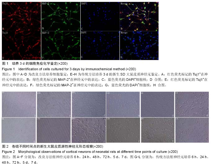
方法:取1 d新生SD大鼠皮质。传统方法实验组:取整个皮质;改良方法实验组:取SD大鼠表面2.0-3.0 mm处皮质。木瓜蛋白酶消化后离心,制备成单细胞悬液,以1×10⁵/孔的浓度接种至含神经元培养液的24孔培养板进行原代培养。培养3 d时采用免疫细胞化学染色方法,使用神经元特异性标志物Tuj1与MAP-2双标记法鉴定所培养的细胞;采用倒置相差显微镜观察6,24,48,72 h及5,7 d细胞数和突起长度并记录。
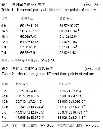
结果与结论:①所培养的细胞可表达神经元特异性标志物Tuj1与MAP-2,因此所培养细胞为神经元,可用于之后实验;②实验组培养至第3天时神经元的纯度已达到峰值92%,而普通实验组神经元的纯度为51%;③2组细胞培养至6 h均已贴壁且长处小突起,培养至第3天时,细胞已初步形成神经网络,培养至5 d时神经网络密集;④研究所用方法简便能够稳定的培养出纯度较高的神经元,可以用于SD大鼠皮质源性神经元的相关实验研究。
中国组织工程研究杂志出版内容重点:组织构建;骨细胞;软骨细胞;细胞培养;成纤维细胞;血管内皮细胞;骨质疏松;组织工程
ORCID: 0000-0002-8961-5807(王东艳)
中图分类号:
.jpg) 文题释义:
大脑皮质:是大脑的表层,由灰质构成,其厚度为1-4 mm,其下方大部分由白质构成。大脑皮质的神经元都是多极神经元,按其细胞的形态分为锥体细胞、颗粒细胞和梭形细胞三大类。大脑皮质是调节躯体运动或者说控制躯体运动的最高级中枢。
神经元:又称神经细胞,是构成神经系统结构和功能的基本单位。具有长突起,由细胞体和细胞突起构成。细胞体位于脑、脊髓和神经节中,细胞突起可延伸至全身各器官和组织中。神经元的核大而圆,位于细胞中央,染色质少,核仁明显。常见的形态为星形、锥体形、梨形和圆球形状等。
文题释义:
大脑皮质:是大脑的表层,由灰质构成,其厚度为1-4 mm,其下方大部分由白质构成。大脑皮质的神经元都是多极神经元,按其细胞的形态分为锥体细胞、颗粒细胞和梭形细胞三大类。大脑皮质是调节躯体运动或者说控制躯体运动的最高级中枢。
神经元:又称神经细胞,是构成神经系统结构和功能的基本单位。具有长突起,由细胞体和细胞突起构成。细胞体位于脑、脊髓和神经节中,细胞突起可延伸至全身各器官和组织中。神经元的核大而圆,位于细胞中央,染色质少,核仁明显。常见的形态为星形、锥体形、梨形和圆球形状等。