Design
Grouping and control study.
Time and setting
The experiment was performed in the Central Laboratory of Plastic Surgery Hospital from October 2009 to June 2011.
Materials
The amniotic membranes were harvested from the human placenta after cesarean section. Maternal donors had given informed consent and had been tested negative for infectious diseases such as syphilis, HIV, hepatitis B and C before delivery. The amniotic membranes were further disposed within 4 hours.
.jpg)
Methods
Preparation of amniotic membranes
Human placentas were obtained under sterile conditions from planned, uneventful cesarean sections. Immediately after surgery, the placenta was cleared of blood clots with sterile PBS, and the amniotic membrane was removed from the chorion by blunt dissection under the laminar flow hood. Pieces of the membrane were cut on as scale of
1 cm×1 cm and sutured with the epithelial side up onto sterile nitrocellulose membranes (Raucocel, Lohmann-Rauscher, Neuwied, Germany). Then, the nitrocellulose membranes with the amniotic membranes were taken for storage experiments[13].
Cryopreserving and thawing procedures
To study the effect of cryopreservation, two different processes were applied. Deep-frozen cryopreservation: the nitrocellulose membranes with the amniotic membranes were deposited into cryo-tube containing glycerol/DMEM (1:1) at 4 ℃ for 1 hour and then at
-80 ℃[14]. Vitrification: the nitrocellulose membranes with the amniotic membranes were deposited into cryo-tube containing vitreous solutions (80% DMEM and 10% dimethyl sulfoxide and 10% propylee glycol at 4 ℃ for 1 hour, then plunged into liquid nitrogen (-196 ℃). After 3 and 6 months of cryopreservation, the cryo-tubes were taken out and floated in a water bath at 37 ℃ until their content became liquid. The amniotic membrane was immersed in the DMEM containing 10% fetal bovine serum at room temperature for 30 minutes, and then processed for tests of ultrastructure and viability.
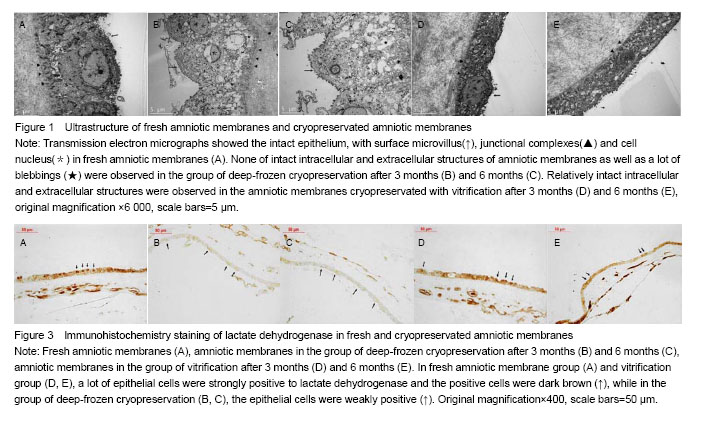
Assessment of amniotic membrane ultrastructure using transmission electron microscopy
The amniotic membranes were collected after thawed and pre-fixed in 2.5% glutaraldehyde at 4 ℃ overnight. The amniotic membranes were washed three times with deionized water and post-fixed with 1% osmium tetroxide at 4 ℃ for 1 hour, and then serially dehydrated with increasing gradient of acetone. The amniotic membranes were permeated and embedded with epoxy resin. Finally, the amniotic membranes were made into half-thin slice up for transmission electron microscopy viewing. Amniotic membrane ultrastructure was observed and collected.
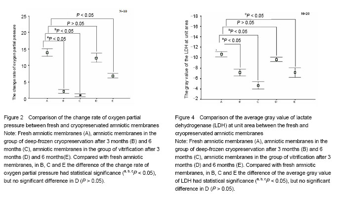
Determination of oxygen partial pressure of amniotic membranes
Both of the fresh and the thawed amniotic membranes were immersed into DMEM containing 10% fetal bovine serum at 37 ℃ for 1 minute, and then cleaned out of the water on the surface. Opening the microcomputer analysis system for biological oxygen consumption, the epithelial side of the amniotic membrane was kept close to the sensor. Oxygen partial pressure was examined at 5 and 65 seconds, the change rate of oxygen consumption was calculated by ΔPO2%=(PO2Sec5-PO2Sec65)/PO2Sec5×100%.
Histology and immunohistochemistry
After fixed and dehydrated, all amniotic membranes were subsequently embedded in paraffin and cut into 4 μm sections. After heat fixation, deparaffinization antigen unmasking and blocking, the slides were incubated with the primary antibody of the corresponding antigen. Therefore, the mouse anti-human lactate dehydrogenase antibody was used at a dilution of 1:100. The subsequent incubation was carried out at 4 ℃ overnight. Afterward, the slides were rinsed with PBS several times and incubated with a corresponding biotinylated secondary antibody for 30 minutes at room temperature. After the washing procedure, sections were incubated in streptavidin conjugated for 30 minutes at room temperature. The slides were rinsed again with PBS and not counterstained. As a semiquantitative score for lactate dehydrogenase, gray value of the lactate dehydrogenase at unit area was used. Photographs of stained sections were taken and processed using Leica microscope and Leica QWin image analysis system. The average gray value of the lactate dehydrogenase at unit area was measured from 20 positive epithelial cells of each sample chosen randomly.
Main outcome measures
Ultrastructure change, oxygen partial pressure and lactate dehydrogenase viability of amniotic membrane.
Statistical analysis
All data are presented as mean±SD for n=10. Statistical analyses were carried out by Paired t test. The data analyses were performed with SPSS 13.0 software. The level of statistical significance was taken to be P < 0.05.