中国组织工程研究 ›› 2015, Vol. 19 ›› Issue (15): 2371-2375.doi: 10.3969/j.issn.2095-4344.2015.15.015

• 组织构建基础实验 basic experiments in tissue construction • 上一篇    下一篇

三维重建颈内动脉形态:虹吸部解剖测量与管壁钙化分布

周乐夫1,陈丽君2,段少银2   

  1. 1深圳市龙岗区中医院,广东省深圳市  518172
    2厦门大学附属中山医院,福建省厦门市  361004
  • 修回日期:2015-03-06 出版日期:2015-04-09 发布日期:2015-04-09
  • 通讯作者: 段少银,博士, 教授,主任医师,厦门大学附属中山医院,福建省厦门市 361004
  • 作者简介:周乐夫,男,1963年生,湖南省沅江市人,汉族,2004年解放军第一军医大学毕业,硕士,副主任医师,主要从事CT和MR研究。
  • 基金资助:

    国家自然科学基金项目(81071214)

Three-dimensional reconstruction of the carotid artery: anatomical measurement and calcification distribution of the carotid siphon

Zhou Le-fu1, Chen Li-jun2, Duan Shao-yin2   

  1. 1Shenzhen Longgang Hospital of TCM, Shenzhen 518172, Guangdong Province, China
    2Zhongshan Hospital of Xiamen University, Xiamen 361004, Fujian Province, China
  • Revised:2015-03-06 Online:2015-04-09 Published:2015-04-09
  • Contact: Duan Shao-yin, M.D., Professor, Chief physician, Zhongshan Hospital of Xiamen University, Xiamen 361004, Fujian Province, China
  • About author:Zhou Le-fu, Master, Associate chief physician, Shenzhen Longgang Hospital of TCM, Shenzhen 518172, Guangdong Province, China
  • Supported by:

    the National Natural Science Foundation of China, No. 81071214

摘要:

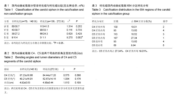
背景:颈内动脉虹吸部结构复杂,由于颅底骨结构的遮挡,二维影像学观察存在一定的困难。数字减影血管造影(DSA)有利于显示颈内动脉的行程,但缺乏解剖标志。CT三维成像有利于显示颈内动脉管壁、壁内外结构与测量,为相关研究提供了有效的新手段。
目的:明确颈内动脉虹吸部相关形态及钙化分布特点,为相关医学临床与基础研究提供客观依据。
方法:选择50岁以上行头颈CT血管造影检查无明显病变或变异者206例作为研究对象,分为无钙化组70例、钙化组136例。在图像工作站上重建颈内动脉虹吸部,观察其分型(根据虹吸部的解剖形态分为“U”型,“V”型,“C”型,“S” 型)与钙化分布,测量C4、C5弯曲的折角及管腔内径。
结果与结论:颈内动脉虹吸部无钙化组70例,年龄为(59.17±10.27)岁,虹吸部“U”型占35.7%、“V”型30.7%、“C”型占27.2%、“S”型占6.4%,其中两侧分型相同者占33.3%(25/70例);管腔内径为(4.92±0.63) mm,虹吸部C4、C5折角分别为(87.23±16.66)°、(49.21±16.01)°;钙化组136例,年龄为(67.39±9.32)岁,虹吸部“U”型占41.5%、“V”型33.1%、“C”型占24.3%、“S”型占1.1%,其中两侧分型相同者占43.4%(59/136例);管腔内径为(4.90±0.44) mm,虹吸部C4、C5折角分别为(84.44±17.20)°、(52.57±14.16)°。统计学分析两组间年龄、S型分型有显著性差异(P < 0.05),虹吸部管腔内径、C4、C5段折角无显著性差异(P > 0.05)。无钙化组及钙化组C4折角内、外弯区钙化分别占13.60%、19.01%;C5折角占27.34%、16.52%;C6段占9.94%、13.60%。结果表明,颈内虹吸部形态以“U”、“V”、“C”型多见,两侧分型不同超过50%。管壁钙化以C5折角内弯区最多、C4折角外弯区其次,钙化随年龄增大而发生率增多,其中钙化虹吸部S型发生率较低,钙化与管腔、折角大小无明显关系。


中国组织工程研究
杂志出版内容重点:组织构建;骨细胞;软骨细胞;细胞培养;成纤维细胞;血管内皮细胞;骨质疏松组织工程


全文链接:

关键词: 组织构建, 组织工程, 颈动脉, 三维CT, 影像解剖, 钙化, 国家自然科学基金

Abstract:

BACKGROUND: The carotid siphon has a complex structure, which is difficult for two-dimensional imaging observation due to shelter from the basicranial bone. Digital subtraction angiography is conducive to display the course of carotid siphon, but there is a lack of anatomical landmarks. Three-dimensional CT imaging is beneficial to show the structure of carotid siphon and have its measurement, which provides a new and effective means in relevant studies.
OBJECTIVE: To clarify the morphology and calcification distribution of the carotid siphon, thereby providing an objective basis for relevant clinical and basic research.
METHODS: A total of 206 patients, over 50 years old, with no lesions or variation under head and neck CT angiography were selected and divided into non-calcification group (n=70) and calcification group (n=136). The carotid siphon was reconstructed on the imaging workstation to observe the classification (“U”, “V”, “C” and “S” types) and calcification distribution, the bending angles of C4 and C5 segments as well as lumen diameter were measured.
RESULTS AND CONCLUSION: The 70 patients in the non-calcification group were at a mean age of (59.17±10.27) years, and the “U” type accounted for 35.7%, “V” type for 30.7%, “C” type for 27.2%, and “S” type for 6.4%; Among them, 33.3% patients (25/70) had the same type at both sides; the lumen diameter was (4.92±0.63) mm, and the blending angles of C4 and C5 were (87.23±16.66)° and (49.21±16.01)°. In the calcification group, the mean age was (67.39±9.32) years, and there were 41.5% of “U” type, 33.1% of “V” type, 24.3% of “C” type, 1.1% of 24.3%, and among the 136 patients, 43.4% (59/136) showed the same type at both sides; the lumen diameter was (4.90±0.44) mm, and the blending angles of C4 and C5 segments were (84.44±17.20)° and (52.57±14.16)°. There were significant differences in age and “S” type between the two groups (P < 0.05), but no statistical difference in the lumen diameter and blending angles of C4 and C5 (P > 0.05). In the calcification group, the calcification percentage of inwardly and outwardly curved regions was 13.60% and 19.01% for C4, 27.34% and 16.52% for C5, 9.94% and 13.60% for C6. The morphology of the carotid siphon is mostly seen as “U”, “V”, “C” types, and over 50% types are different at both sides. Calcification of the lumen wall is seen most in the inwardly curved region of C5 segment, followed by the outwardly curved region of C4 segment. The incidence of calcification is increased with age, which is lower in the S-type carotid siphon. The lumen diameter and blending angle of the carotid siphon show no correlation with calcifications.


中国组织工程研究
杂志出版内容重点:组织构建;骨细胞;软骨细胞;细胞培养;成纤维细胞;血管内皮细胞;骨质疏松组织工程


全文链接:

Key words: Carotid Arteries, Imaging, Three-Dimensional, Dissection

中图分类号: