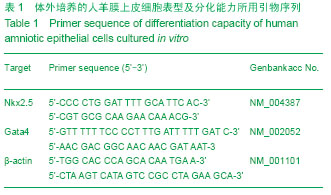
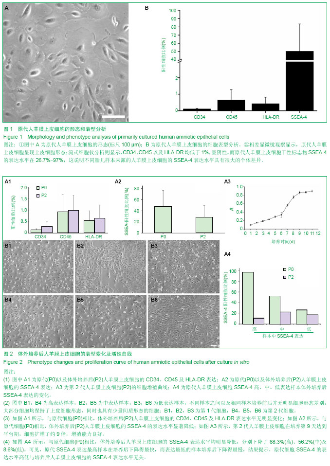
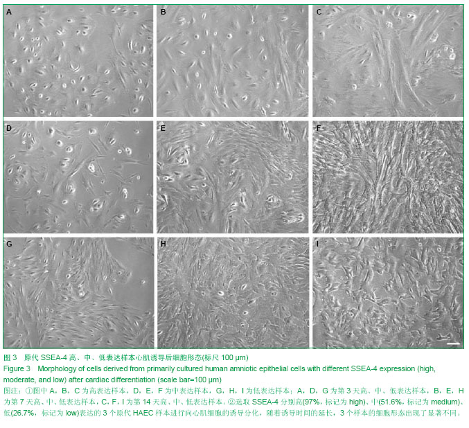
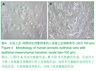
| [1] 罗晓婷,李舒梅,文道源等.新鲜制备及中长期保存的羊膜细胞外基质结构特征[J].中国临床康复.2006,10(1):79-81.[2] 高振,罗晓婷,邓炜等.羊膜与羊膜细胞外基质在组织工程中的应用[J].中国组织工程研究与临床康复,2007,11(32):6450-6453.[3] Heeger PS. Amnion and Chorion Cells as Therapeutic Agents for Transplantation and Tissue Regeneration:A Field in Its Infancy. Transplantation.2004;78(10):1411-1412.[4] Akle CA, Adinolfi M,Welsh KI, et al.Immunogenicity of human amniotic epithelial cells after transplantation into volunteers. Lancet.1981;2(8254):1003-1005. [5] Adinolfi M, Akle CA, Mccoll I, et al.Expression of HLA antigens,beta 2-microglobulinand enzymes by human amniotic epithelial cells. Nature.1982;295 (5847):325-327.[6] Sakuragawa N, Tohyama J, Yamamoto H. Immunostaining of human amniotic epithelial cells: possible use as a transgene carrier in gene therapy for inborn errors of metabolism. Cell Transplant.1995;4(3):343-346.[7] Hancheran S, Michalska A, Peh G, et al. Stem cells derived from fetal membranes display multi-lineage differentiation potential. BiolReprod. 2007;77(3):577-588.[8] Ilancheran S, Moodley Y, Manuelpillai U. Human fetal membranes: a source of stem cells for tissue regeneration and repaire. Placenta. 2009;30(1):2-10.[9] Akle CA, Adinolfi M, Welsh KI, et al. Immunogenicity of human amniotic epithelial cells after transplantation into volunteers. Lancet. 1981;2(8254):1003-1005.[10] Sakuragawa N, Tohyama J, Yamamoto H. Immunostaining of human amniotic epithelial cells: possible use as a transgene carrier in gene therapy for inborn errors of metabolism. Cell Transplant. 1995;4(3):343-346.[11] Miki T, Lehmann T, Cai H, et al.Stem cell characteristics of amniotic epithelial cells. Stem Cells. 2005;23(10):1549-1559.[12] Hou Y, Huang Q, Liu T, et al. Human amnion epithelial cells can be induced to differentiate into functional insulin-producing cells. ActaBiochimBiophys Sin. 2008; 40(9): 830-839.[13] Ursula M, Jorge T, Dinushka L, et al. Transplantation of human amnion epithelial cells reduces hepatic fibrosis in immunocompetent CCl4-treated mice. Cell Transplant. 2010; 19(9):1157-1168.[14] Wei JP, Zhang TS, Kawa S, et al. Human amnion-isolated cells normalize blood glucose in streptozotocin-induced diabetic mice.Cell Transplant. 2003;12(5):545-552.[15] Toda A, Okabe M, Yoshida T, et al. The potential of amniotic membrane/ amnio- derived cells for regeneration of various tissues. Pharmacol Sci.2007;105(3):215-228.[16] Cheng H, Qiu L, Ma J, et al. Replicative senescence of human bone marrow and umbilical cord derived mesenchymal stem cells and their differentiation to adipocytes and osteoblasts. MolBiol Rep. 2011;38(8): 5161-5168.[17] Majore I, Moretti P, Stahl F, et al. Growth and differentiation properties of mesenchymalstromal cell populations derived from whole human umbilical cord. Stem Cell Rev. 2011; 7(1): 17-31.[18] PratamaG,VaghjianiV,Tee JY, et al. Changes in culture expanded human amniotic epithelial cells: Implications for potential therapeutic applications. PloS one. 2011;6(11): e26136.[19] 张路,方宁,陈代雄,等.人羊膜上皮细胞有向心肌样细胞分化的特性[J].中国组织工程研究与临床康复,2008,12(3):401-405.[20] 王建,彭琳,卢光琇.人羊膜上皮细胞干细胞特性及向胰岛素分泌细胞分化的研究[J].南方医科大学学报,2011,31(9):1484-1487.[21] 朱梅,陈东,孟晓婷,等.羊膜上皮细胞移植治疗帕金森病大鼠的实验研究[J].中国老年学杂志,2006,2(26):227-230.[22] Kakishita K, Elwan MA, Nalkao N, et al. Human amniotic epithelial cells produce dopamine and survive after implantation into the striatum of a rat model of parkinson's disease: A potential source of donor for transplantation therapy. ExpNeurol. 2000;165(1):27-34.[23] Enosawa S, Sakuragawa N, Suzuki S. Possible use of amniotic cells for regenerative medicine. Nippon Rinsho. 2003;61(3):396-400.[24] KakishitaK,NakaoN,Sakuragawa N, et al. Implantation of human amniotic epithelial cells prevents the degeneration of nigral dopamine neurons in rars with 6-hydroxydopamine lesions. Brain Res. 2003;980(1):48-56.[25] Lv S, Liu Y, Lian J, et al. Proliferation, Stemness and Cardiac Differentiation Properties of Umbilical Cord Wharton's Jelly-derived Mesenchymal Stem Cells during the Continuous Passages. Hard tissue biol. 2013;22(2):261-266.[26] Brivanlou AH,Gage FH,Jaenisch R,et a1.Stem cell.Setting standards for human embryonic stem cells.Science. 2003; 300(5621):913-916.[27] Chambem I,Colby D,Robertson M,et al.Functional Expression Cloning of Nanog,aPluripotency Sustaining Factor in Embryonic Stem Cells.Cell. 2003;13(5):643-655.[28] Izumi M,Pazin BJ,Minervini CF, et al. Quantitative comparison of stem cell marker-positive cells in fetal and term human amnion. Reprod Immunol.2009;81(1):39-43.[29] Diaz-Prado S, Muinos-Lopez E, Hermida-Gomez T, et al. Multilineage differentiation potential of cells isolated from the human amniotic membrane. Cell Biochem.2010;111(4): 846-857.[30] Simat SF,Chua KH,AbdulRaham H,et a1.The stemness gene expression of cultured human amniotic epithelial cells in serial passages.Med J Malaysia.2008;63 Suppl A:53-54.[31] Tamagawa T,Ishiwata I,Saito S.Establishment and characterization of a pluripotent stem cell line derived from human amniotic membranes and initiation of germ layers in vltro.Hum Cell.2004;17(3):125-130.[32] Sakuragawa N,Kakinumm K, Kikuchi A,et a1.Human amnion mesenchyme cells express phenotypes ofneuroglial progenitor cells. Neurosci Res.2004;78(2):208-214. |