中国组织工程研究 ›› 2019, Vol. 23 ›› Issue (2): 245-250.doi: 10.3969/j.issn.2095-4344.1512

• 膜生物材料 membrane biomaterials • 上一篇    下一篇

面向皮肤支架3D生物打印喷头的CFD模拟与实验

陈冬冬1,张 琦1,张鹏飞2,周骥平1,姜亚妮3   

  1. 1扬州大学机械工程学院,江苏省扬州市 225127;2江苏牧羊控股有限公司,江苏省扬州市 255120;3扬州大学动物科学与技术学院,江苏省扬州市 225009
  • 收稿日期:2018-07-11 出版日期:2019-01-18 发布日期:2019-01-18
  • 通讯作者: 张琦,博士,硕士生导师,副教授,扬州大学机械工程学院农业机械及其自动化系,江苏省扬州市 225127
  • 作者简介:陈冬冬,男,1992年生,江苏省东台市人,汉族,扬州大学在读硕士,主要从事3D生物打印的研究。
  • 基金资助:

    国家自然科学基金(81770018);扬州市-扬州大学科技合作资金项目(SCX2017020015,项目负责人:周骥平);江苏省研究生教育教学改革研究与实践课题(JGLX16-109);扬州大学创新创业教育改革项目(yzucx2016-3C,项目负责人:张琦)

3D bioprinted nozzle for skin scaffolds: CFD simulation and experiments

Chen Dongdong1, Zhang Qi1, Zhang Pengfei2, Zhou Jiping1, Jiang Yani3   

  1. 1College of Mechanical Engineering, Yangzhou University, Yangzhou 225127, Jiangsu Province, China; 2Su Shepherd Holding Co. (Jiangsu Province) Yangzhou 255120, Jiangsu Province, China; 3College of Animal Science and Technology, Yangzhou University, Yangzhou 225009, Jiangsu Province, China
  • Received:2018-07-11 Online:2019-01-18 Published:2019-01-18
  • Contact: Zhang Qi, PhD, Master’s supervisor, Associate professor, College of Mechanical Engineering, Yangzhou University, Yangzhou 225127, Jiangsu Province, China
  • About author:Chen Dongdong, Master candidate, College of Mechanical Engineering, Yangzhou University, Yangzhou 225127, Jiangsu Province, China
  • Supported by:

    the National Natural Science Foundation of China, No. 81770018; Yangzhou Municipality-Yangzhou University Science and Technology Cooperation Project, No. SCX2017020015 (to ZJP); Research and Practice of Postgraduate Education Teaching Reform in Jiangsu Province, No. JGLX16-109; Yangzhou University Innovation and Entrepreneurship Education Reform Project, No. yzucx2016-3C (to ZQ)

摘要:

文章快速阅读:


文题释义:
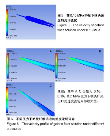
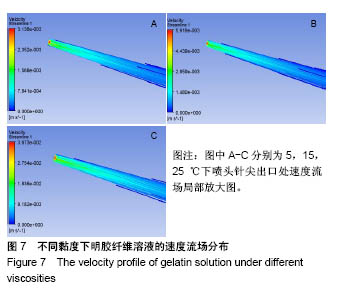
流场模拟:通过限元软件Fluent模拟3D生物打印机在不同压力、温度和不同喷头挤出直径下,实际的喷头流速变化情况。
气动挤压:通过气泵提供动力的来源,可设置不同的挤出压力大小,可以挤出明胶纤维溶液。
3D生物打印技术:是将3D打印技术应用到组织工程中,可实现具有复杂生理结构的一种制造方法,是3D打印技术的一个分支,通过增材制造原理将复杂的器官结构离散成一系列的二维层状结构,这种构建方法显著降低了复杂器官的构建难度,特别在一些异质性组织的构建方面,3D生物打印技术突破了传统制造技术的局限。
 
 
背景:随着组织工程的发展,3D生物打印技术可通过打印生物质材料来构建皮肤支架,但工艺参数不明确,打印过程中经常出现堆积和不连续情况。
目的:通过3D生物打印机和有限元软件Fluent进行数值模拟,以获得合理的挤出压强、黏度、喷头行走速度、喷头直径、温度参数,解决明胶纤维打印过程中的堆积和不连续问题。
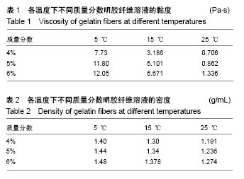
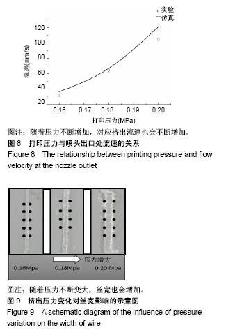
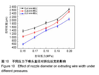
方法:以5%明胶纤维素溶液为打印材料,采用3D生物打印机和限元软件Fluent进行数值模拟,设置打印温度分别为5,15,25 ℃,打印压力分别为0.16,0.18,0.2 MPa,喷头直径分别为0.21,0.26,0.41 mm,检测喷头针尖处的流速与流量;以合适的参数3D打印明胶纤维皮肤生物支架,扫描电镜观察支架结构。

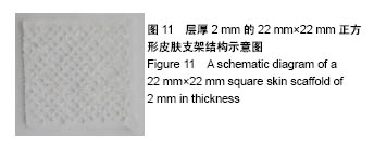
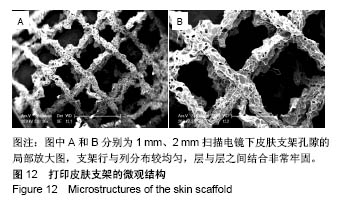
结果与结论:3D生物打印明胶纤维皮肤生物支架过程中,设置打印温度在15 ℃、喷头行走速度为30 mm/s、压力为0.18 MPa、喷头直径为0.21 mm时,可保证喷头挤出的明胶纤维材料不会堆积或者断丝,连续性挤出明胶纤维,并且在上述打印条件下打印的丝宽1 400 μm左右;将此条件下打印的生物支架进行扫描电镜观察,可见支架的行与列分布较均匀,层与层之间的结合部位黏结牢固,不易变形,支架孔隙率约57%。

ORCID: 0000-0001-8332-9693(陈冬冬) 

关键词: 明胶纤维, CFD模拟, 3D生物打印, 组织工程, 皮肤支架, 实验验证, 生物材料

Abstract:

BACKGROUND: With the development of tissue engineering, 3D bioprinting technology is used to prepare skin scaffolds. The process parameters are however unclear, and stacking and discontinuities often occur during the printing process.

OBJECTIVE: To conduct the numerical simulation by 3D bio-printer and finite element software Fluent so as to obtain reasonable extrusion pressure, viscosity, nozzle walking speed, nozzle diameter and temperature parameters to solve the problem of accumulation and discontinuity in the printing process of gelatin fibers.
METHODS: Using 5% gelatin cellulose solution as the printing material, the numerical simulation was carried out by using 3D bio-printer and software Fluent. The printing temperature was set at 5, 15, 25 oC, and the printing pressure was 0.16, 0.18, 0.2 MPa, respectively. The flow rate and flow at the tip of the nozzle were measured at 0.21, 0.26, and 0.41 mm. The gelatin fiber skin bio-scaffold was 3D-printed with appropriate parameters, and the scaffold structure was observed by scanning electron microscopy.
RESULTS AND CONCLUSION: When the printing temperature was set at 15 oC, the nozzle walking speed was 30 mm/s, the pressure was 0.18 MPa, and the nozzle diameter was 0.21 mm, the gelatin fiber material could be continuously extruded but not stacked or broken. Under the above printing conditions, the width of printed filament was about 1 400 μm. For the biological scaffold printed under this condition, the row and column distribution of the scaffold was more uniform shown by the scanning electron microscope, and the bonding site between the layers was firmly bonded and not easily deformed. The porosity of the scaffold was about 57%. 

Key words: Gelatin, Tissue Engineering

中图分类号: