中国组织工程研究 ›› 2019, Vol. 23 ›› Issue (20): 3182-3187.doi: 10.3969/j.issn.2095-4344.1197

• 骨与关节图像与影像 bone and joint imaging • 上一篇    下一篇

肌骨超声评价类风湿性指关节炎的病理特征及炎性病变

胡小丽,谷 颖,蔡 燕,谢 谨,刘 婵   

  1. 贵州医科大学附属医院超声中心,贵州省贵阳市 550004
  • 出版日期:2019-07-18 发布日期:2019-07-18
  • 通讯作者: 谷颖,硕士,主任医师,贵州医科大学附属医院,贵州省贵阳市 550004
  • 作者简介:胡小丽,女,1975年生,2013年贵阳医学院毕业,硕士,副主任医师,主要从事肌骨超声研究。
  • 基金资助:

    贵州省科技计划支持项目(黔科合 LH字[2017]7207号)

Musculoskeletal ultrasound evaluates the pathological features and inflammatory lesions of rheumatoid finger arthritis  

Hu Xiaoli, Gu Ying, Cai Yan, Xie Jin, Liu Chan   

  1. Ultrasound Center, Affiliated Hospital of Guizhou Medical University, Guiyang 550004, Guizhou Province, China
  • Online:2019-07-18 Published:2019-07-18
  • Contact: Gu Ying, Master, Chief physician, Ultrasound Center, Affiliated Hospital of Guizhou Medical University, Guiyang 550004, Guizhou Province, China
  • About author:Hu Xiaoli, Master, Associate chief physician, Ultrasound Center, Affiliated Hospital of Guizhou Medical University, Guiyang 550004, Guizhou Province, China
  • Supported by:

    the Science and Technology Supporting Project of Guizhou Province, No. LH[2017]7207

摘要:

文章快速阅读:


文题释义:
类风湿性指关节炎:作为一种动态变化的病理现象,选择合适的影像学检测方法密切关注其病理特征,了解关节炎性病变程度,对准确评估患者关节损伤病情、评价疾病进展、制定治疗重点方案具有重要的意义。
肌骨超声:是近年来在影像学技术发展进程不断推进背景下应运而生的新兴超声检查技术,具有费用低廉、无创、无辐射、操作简单、禁忌证少、短期内可反复检查及实现实时动态观察等显著优点。
 
摘要
背景:类风湿性关节炎作为一种动态变化的病理现象,选择合适的影像学检测方法对评价疾病进展、制定治疗重点方案具有重要的意义,而肌骨超声作为新兴超声检查技术,在疾病的诊疗中具有良好的应用价值。
目的:探讨肌骨超声评价类风湿性指关节炎病理特征及炎性病变的临床意义。
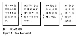
方法:以60例类风湿性指关节炎患者为研究对象,分别予以患者肌骨超声和MRI检查,共检查手指关节450个。统计2种方法对滑膜增生、关节积液、骨侵蚀3种典型病理特征的检出率;检测患者血清指标(超敏C-反应蛋白、红细胞沉降率和类风湿因子),对比分析不同关节滑膜厚度分级和滑膜血流信号分级患者的超敏C-反应蛋白、红细胞沉降率、类风湿因子值。另外对比分析活动期和非活动期患者的滑膜内动脉阻力指数。

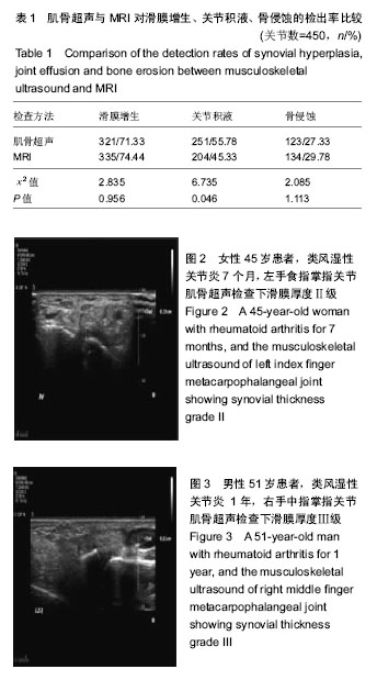
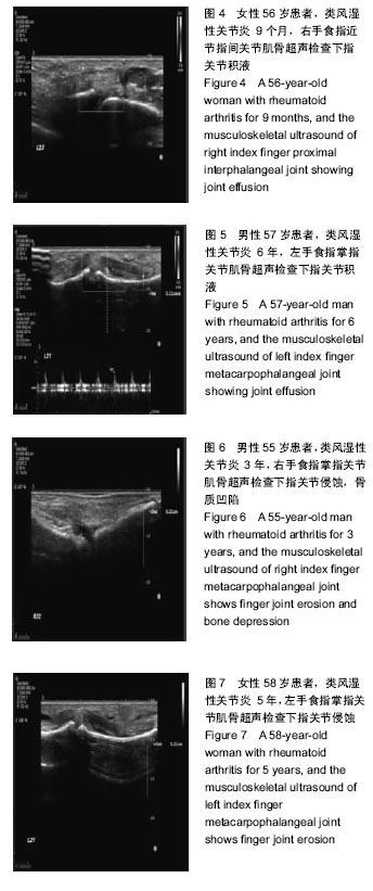
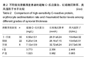
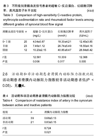
结果与结论:①肌骨超声对滑膜增生、关节积液、骨侵蚀的检出率依次为71.33%,55.78%及27.33%,MRI依次为74.44%,45.33%及29.78%,2种方法对滑膜增生、骨侵蚀的检出率比较差异无显著性意(P > 0.05),但肌骨超声对关节积液的检出率要高于MRI(P < 0.05);②Spearman相关性分析显示,滑膜厚度与超敏C-反应蛋白、红细胞沉降率、类风湿因子水平均无相关性(r=0.463,0.813,0.459,P > 0.05),滑膜血流信号与超敏C-反应蛋白、红细胞沉降率、类风湿因子水平均呈正相关(r=0.623,0.498,0.591,P < 0.05);③活动期患者滑膜内动脉阻力指数值较非活动期患者低(P < 0.05);④综上,肌骨超声在类风湿性指关节炎病理特征的评价上与MRI具有相当的效益,但其具有更高的应用价值;且能够通过滑膜厚度、血流信号、滑膜内动脉阻力指数的综合检测反映患者炎性病变情况,对评估病情、制定治疗方案具有重要的意义。

中国组织工程研究杂志出版内容重点:人工关节;骨植入物;脊柱骨折;内固定;数字化骨科;组织工程
ORCID: 0000-0003-1253-773X(胡小丽)

关键词: 肌骨超声, 类风湿性指关节炎, 病理特征, 炎性病变, 滑膜增生, 骨膜厚度, 关节积液, 骨侵蚀

Abstract:

BACKGROUND: Pathological change of rheumatoid arthritis is a dynamic process, and selecting appropriate radiology is of great significance for assessing disease progress and making treatment scheme. Musculoskeletal ultrasound as an emerging technology has good application value in the diagnosis and treatment of disease.

OBJECTIVE: To study the clinical significance of musculoskeletal ultrasound in the evaluation of pathological features and inflammatory lesions of rheumatoid arthritis.
METHODS: Sixty patients with rheumatoid arthritis were selected, underwent musculoskeletal ultrasound and MRI, and 450 fingers were examined. The detection rates of three typical pathological features of synovial hyperplasia, joint effusion and bone erosion were recorded. The serum indexes (high-sensitivity C-reactive protein, erythrocyte sedimentation rate and rheumatoid factor) were detected. The hypersensitivity C-reaction protein, erythrocyte sedimentation rate and rheumatoid factor of the patients with different grades of synovial thickness and synovial blood flow signal were compared. The resistance index of artery in the synovium was compared between active and inactive patients.
RESULTS AND CONCLUSION: (1) The detection rates of synovial hyperplasia, joint effusion and bone erosion by musculoskeletal ultrasound were 71.33%, 55.78% and 27.33%, respectively. The detection rates by MRI were 74.44%, 45.33% and 29.78% respectively. There was no significant difference in the detection rate of synovial hyperplasia and bone erosion between two methods (P > 0.05), but the detection rate of joint effusion by musculoskeletal ultrasound was higher than that by MRI (P < 0.05). (2) Spearman correlation analysis showed that synovial thickness was not correlated with high-sensitivity C-reactive protein, erythrocyte sedimentation rate and rheumatoid factor (r=0.463, 0.813, 0.459, P > 0.05), and synovial blood flow signal was positively correlated with high-sensitivity C-reactive protein, erythrocyte sedimentation rate and rheumatoid factor (r=0.623, 0.498, 0.591, P < 0.05). (3) The resistance index of artery in the synovium of active patients was lower than that of inactive patients (P < 0.05). (4) To conclude, musculoskeletal ultrasound has considerable benefit for the evaluation of pathological features of rheumatoid arthritis with MRI, but it has higher application value. Musculoskeletal ultrasound can reflect the inflammatory condition through detecting the thickness of the synovial membrane, blood flow signal and the resistance index of artery in the synovium, and it is important to evaluate the disease condition and make the treatment plan.

Key words: musculoskeletal ultrasound, rheumatoid finger arthritis, pathological features, inflammatory lesions, synovial hyperplasia, periosteal thickness, joint effusion, bone erosion

中图分类号: