中国组织工程研究 ›› 2026, Vol. 30 ›› Issue (26): 6880-6891.doi: 10.12307/2026.401
• 材料生物相容性 material biocompatibility • 上一篇 下一篇
聚乳酸/胶原蛋白静电纺双层引导组织再生膜的生物相容性评价
宋沐泽1,刘楚怡2,3,唐庆娟1,代元坤2,3,宋文山2,李八方2,王园园2
- 中国海洋大学,1食品科学与工程学院,3医药学院,山东省青岛市 266003;2青岛海洋生物医药研究院,山东省青岛市 266000
-
接受日期:2025-12-17出版日期:2026-09-18发布日期:2026-03-12 -
通讯作者:王园园,高级工程师,青岛海洋生物医药研究院,山东省青岛市 266000 -
作者简介:宋沐泽,男,2000年生,山东省荣成市人,汉族,硕士,主要从事生物材料与组织工程研究。
Biocompatibility evaluation of polylactic acid/collagen electrospinning bilayer guided tissue regeneration membrane
Song Muze1, Liu Chuyi2, 3, Tang Qingjuan1, Dai Yuankun2, 3, Song Wenshan2, Li Bafang2, Wang Yuanyuan2
- 1School of Food Science and Engineering, 3School of Medicine and Pharmacy, Ocean University of China, Qingdao 266003, Shandong Province, China; 2Marine Biomedical Research Institute of Qingdao, Qingdao 266000, Shandong Province, China
-
Accepted:2025-12-17Online:2026-09-18Published:2026-03-12 -
Contact:Wang Yuanyuan, Senior engineer, Marine Biomedical Research Institute of Qingdao, Qingdao 266000, Shandong Province, China -
About author:Song Muze, MS, School of Food Science and Engineering, Ocean University of China, Qingdao 266003, Shandong Province, China
摘要:

文题释义:
静电纺丝:是一种利用聚合物溶液或熔体在强电场作用下形成喷射流进行纺丝加工的工艺。静电纺丝的基本原理是将聚合物溶液或熔体置于高压静电场中,使其带电并产生形变,在喷头末端处形成泰勒锥液滴,当液滴表面的电荷斥力超过表面张力时,在液滴表面会高速喷射出聚合物微小液滴,简称“射流”,这些射流经过电场力的高速拉伸、溶剂挥发和固化,最终沉积在接收板上,形成聚合物纤维。
引导组织再生:是一种被广泛应用于骨缺损临床治疗的技术,一般将屏障膜置于牙龈软组织与骨缺损之间阻止非成骨性细胞向骨缺损区生长,确保骨缺损区的成骨过程不受干扰。
背景:海洋胶原蛋白可以促进成牙周膜成纤维细胞增殖分化、促进血管内皮细胞增殖,但是单纯胶原膜的机械强度低、降解速度快,一般需要复合其他材料。聚乳酸是被美国食品药品监督管理局批准可植入体内的降解性医用材料,与胶原蛋白复合可以改善单纯胶原蛋白机械强度不足的问题。
目的:制备聚乳酸/胶原蛋白静电纺双层引导组织再生膜,探讨该引导组织再生膜的生物相容性。
方法:以7%聚乳酸溶液作为致密层纺丝液,14%聚乳酸-胶原蛋白溶液为疏松层纺丝液,采用静电纺丝技术制备聚乳酸/胶原蛋白双层引导组织再生膜,表征膜的微观形貌、孔径和孔隙率。分别采用戊二醛蒸汽、戊二醛溶液和碳化二亚胺/羟基琥珀酰亚胺3种交联方法对膜进行交联,通过拉伸实验筛选力学性能提高效果最佳的膜进行后续实验。通过表征水接触角评估聚乳酸/胶原蛋白双层引导组织再生膜的亲疏水性能。通过细胞毒性实验、热源实验、溶血实验、急性全身毒性实验、亚慢性全身毒性实验、致敏实验、皮内刺激实验,评价聚乳酸/胶原蛋白双层引导组织再生膜的生物相容性。
结果与结论:聚乳酸/胶原蛋白静电纺双层引导组织再生膜的致密层纤维直径为(0.45±0.11) μm,疏松层纤维直径为(0.85±0.19) μm,致密层孔径为(2.43±1.31) μm、孔隙率为(29.86±2.89)%,疏松层孔径为(11.71±4.41) μm、孔隙率为(48.54±1.33)%。综合拉伸实验的拉伸强度、弹性模量与断裂伸长率结果,最终选择戊二醛蒸汽交联法进行交联,该方法的交联度为(17.42±1.67)%。聚乳酸/胶原蛋白双层引导组织再生膜的疏松层表现出亲水性,致密层表现为疏水性。聚乳酸/胶原蛋白静电纺双层引导组织再生膜无细胞毒性、不溶血、无致热性、无潜在毒性、无刺激性和无致敏性,具有良好的生物相容性。
https://orcid.org/0009-0000-4683-6717 (宋沐泽)
中国组织工程研究杂志出版内容重点:生物材料;骨生物材料;口腔生物材料;纳米材料;缓释材料;材料相容性;组织工程
中图分类号:
引用本文
宋沐泽, 刘楚怡, 唐庆娟, 代元坤, 宋文山, 李八方, 王园园. 聚乳酸/胶原蛋白静电纺双层引导组织再生膜的生物相容性评价[J]. 中国组织工程研究, 2026, 30(26): 6880-6891.
Song Muze, Liu Chuyi, , Tang Qingjuan, Dai Yuankun, , Song Wenshan, Li Bafang, Wang Yuanyuan. Biocompatibility evaluation of polylactic acid/collagen electrospinning bilayer guided tissue regeneration membrane[J]. Chinese Journal of Tissue Engineering Research, 2026, 30(26): 6880-6891.
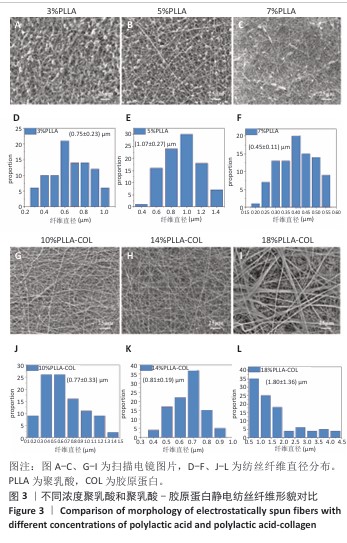
使用聚乳酸-胶原蛋白溶液进行纺丝时均无液滴出现,10%聚乳酸-胶原蛋白组纤维存在弯曲牵伸不足的现象,并且纤维直径不均一,平均直径为(0.77±0.33) μm;14%聚乳酸-胶原蛋白组纤维不存在弯曲,纤维直径分布呈现正态分布,说明纤维尺寸均一性较好,平均直径为(0.81±0.19) μm;18%聚乳酸-胶原蛋白组静电纺丝时会出现泰勒锥不稳定、纺丝液堵塞针头的情况,调整电压和推注速度均无法解决,纺丝纤维可以看到出现纤维黏附情况,纤维直径分布不规律且不均一。
综合以上结果,7%聚乳酸溶液和14%聚乳酸-胶原蛋白溶液制备的纺丝纤维直径粗细有别,分别为(0.45±0.11) μm和(0.81±0.19) μm,大体可见两者孔隙大小不一,可以达到相对致密和相对疏松的要求,并且纤维直径符合正态分布,因此选择7%聚乳酸溶液和14%聚乳酸-胶原蛋白溶液分别作为致密层纺丝液和疏松层纺丝液,用作后续双层膜的制备。
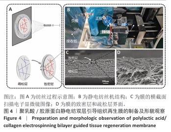
2.2 静电纺丝双层口腔修复膜的制备 以7%聚乳酸溶液作为致密层纺丝液、14%聚乳酸-胶原蛋白溶液疏松层纺丝液进行静电纺丝,纺丝过程、纺丝设备和截面观察如图4所示。图4A展示了双层膜的制备过程,先进行疏松层静电纺丝,然后在致密层基础上进行致密层静电纺丝。截面扫描电子显微镜结果如图4C,D所示,可以看出致密层紧贴疏松层,双层结合紧密,疏松层占主体,成功完成了双层膜的制备。聚乳酸/胶原蛋白静电纺双层引导组织再生膜的致密层孔径为(2.43±1.31) μm、孔隙率为(29.86±2.89)%,疏松层孔径为(11.71±4.41) μm、孔隙率为(48.54±1.33)%。
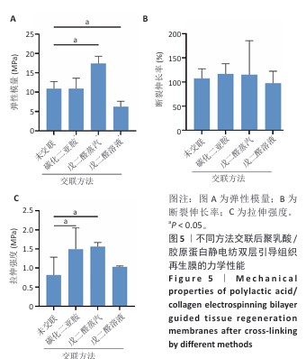
2.3 静电纺丝双层口腔修复膜的力学性能检测结果 弹性模量是衡量材料产生弹性变形难易程度的指标,拉伸强度是评价材料力学强度性能的特征值,断裂伸长率是纤维受外力作用至拉断时拉伸后伸长长度与拉伸前长度的比值。交联前后聚乳酸/胶原蛋白静电纺双层引导组织再生膜的力学性能参数结果,如图5所示。戊二醛蒸汽交联后膜的弹性模量为(17.38±1.88) MPa,拉伸强度为(1.57±0.11) MPa,相比未交联膜分别提高了59.60%和92.60%(P < 0.05)。戊二醛溶液交联后膜的弹性模量为(6.21±1.45) MPa,相比未交联膜降低了43.04%(P < 0.05),拉伸强度为(1.03±0.03) MPa,相比未交联膜提高了26.64%。碳化二亚胺/羟基琥珀酰亚胺交联后膜的弹性模量为(10.86±2.73) MPa,与未交联膜相比无显著差异(P > 0.05),拉伸强度为(1.49±0.55) MPa,
相比未交联膜提高了83.61%(P < 0.05)。3种交联方法后膜的断裂伸长率与未交联膜相比无显著差异(P > 0.05)。引导组织再生膜需要具备良好的机械性能,不易破碎且抗拉伸,才能更好地适应组织再生复杂的内环境。戊二醛蒸汽交联后膜的弹性模量和拉伸强度显著提高,综合以上结果表明,戊二醛蒸汽交联方法对膜的力学性能提高效果最佳,因此后续实验采用戊二醛蒸汽交联法进行交联,该方法交联度为(17.42±1.67)%。
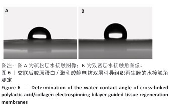
2.4 静电纺丝双层口腔修复膜的亲疏水性能检测结果 戊二醛蒸汽交联后聚乳酸/胶原蛋白静电纺双层引导组织再生膜的致密层和疏松层接触角,如图6所示,致密层水接触角为(118.74±0.59)°,疏松层水接触角为(87.00±1.04)°,疏松层表现出亲水性,致密层表现为疏水性。
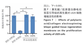
2.5 细胞毒性实验结果 聚乳酸/胶原蛋白静电纺双层引导组织再生膜浸提液的细胞毒性实验结果,如图7所示。阳性对照组细胞相对增殖率仅为(15.37±1.56)%,50%,100%样品浸提液组细胞相对增殖率分别为(104.94±12.94)%和(95.44±10.33)%,50%样品浸提液组细胞相对增殖率高于100%样品浸提液组,并且4组细胞相对增殖率都超过70%,证明聚乳酸/胶原蛋白静电纺双层引导组织再生膜无细胞毒性。
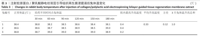
2.6 热源实验结果 聚乳酸/胶原蛋白静电纺双层引导组织再生膜热源实验结果,如表3所示。3只兔体温最高升温分别为0.4,0.4和0.2 ℃,均不高于0.6 ℃,升温总和为1.0 ℃,不超过1.3 ℃,说明聚乳酸/胶原蛋白静电纺双层引导组织再生膜无致热作用。
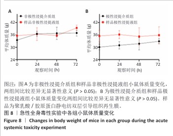
2.8 急性全身毒性实验结果 样品极性浸提液组和样品非极性浸提液组注射浸提液后即刻小鼠轻微运动迟缓;注射后2,48,72 h,4组小鼠均未出现死亡,小鼠运动正常,未出现虚脱震颤、呼吸困难、反应迟缓、腹泻等症状,4组小鼠临床观察无显著差异,均未见毒性反应。各组小鼠体质量变化如图8所示,实验结束后小鼠体质量未下降,样品极性浸提液组与极性浸提介质组小鼠体质量相比均无显著差异(P > 0.05),样品非极性浸提液组与非极性浸提介质组小鼠体质量相比均无显著差异(P > 0.05)。
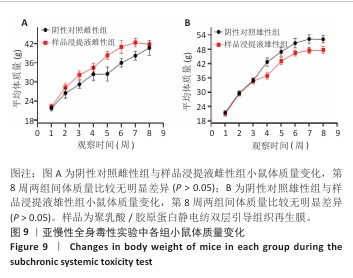
2.9 亚慢性全身毒性实验结果 实验周期内4组小鼠均未出现死亡,小鼠运动正常,未出现虚脱震颤、呼吸困难、反应降低、腹泻等症状,临床观察未见毒性反应。各组小鼠体质量变化如图9所示,样品浸提液雄性组与阴性对照雄性组小鼠体质量变化趋势一致,第8周实验结束后两组小鼠体质量相比无显著差异(P > 0.05);样品浸提液雌性组与阴性对照雌性组小鼠体质量变化趋势一致,第8周实验结束后两组小鼠体质量相比无显著差异(P > 0.05)。
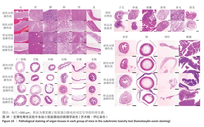
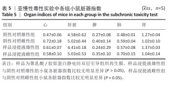
各组小鼠脏器组织病理学染色结果如图10所示,样品浸提液雄性组和样品浸提液雌性组小鼠脏器均未见异常。各组小鼠脏器指数结果如表5所示,样品浸提液雄性组与阴性对照雄性组小鼠各脏器指数比较无明显差异(P > 0.05),样品浸提液雌性组与阴性对照雌性组小鼠各脏器指数比较无明显差异(P > 0.05)。
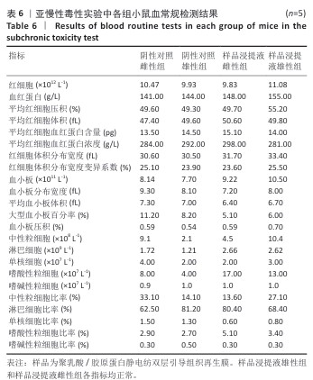
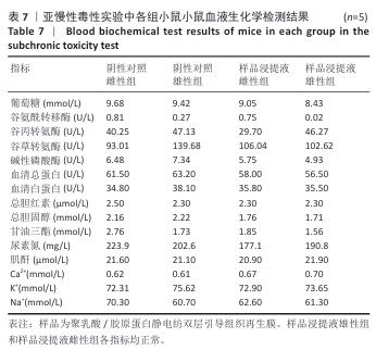
各组小鼠眼球取血后测定血常规与血液生化学,血常规结果如表6所示,样品浸提液雄性组和样品浸提液雌性组小鼠各指标均正常,未见异常指标;血液生化学结果如表7所示,样品浸提液雄性组和样品浸提液雌性组各指标均正常。
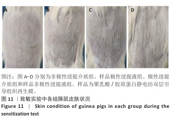
2.10 致敏实验结果 实验周期内所有豚鼠敷贴牢固、未脱落,实验结束拆除敷贴后豚鼠腹部皮肤状况如图11所示,皮肤均未出现红斑、焦痂、水肿反应,对照分级标准,分级平均为0级。聚乳酸/胶原蛋白静电纺双层引导组织再生膜极性和非极性浸提液诱导豚鼠皮肤后并未出现任何过敏反应,与对照组(极性浸提介质组、非极性浸提介质组)豚鼠皮肤状况相同。
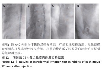
2.11 皮内刺激实验结果 聚乳酸/胶原蛋白静电纺双层引导组织再生膜样品皮内刺激实验结果,如图12所示,注射24,48,72 h后,样品极性浸提液组和样品非极性浸提液组均未出现水肿和红斑,未见任何刺激反应,皮肤状况与极性浸提介质组、非极性浸提介质组一致,并且皮内反应计分均为0分。
| [1] XIA D, YANG F, ZHENG Y. Research status of biodegradable metals designed for oral and maxillofacial applications: A review. Bioact Mater. 2021;276(11):4186-4208. [2] ALQAHTANI AM. Guided Tissue and Bone Regeneration Membranes: A Review of Biomaterials and Techniques for Periodontal Treatments. Polymers (Basel). 2023;15(16):3355. [3] GALLI M, YAO Y, GIANNOBILE WV, et al. Current and future trends in periodontal tissue engineering and bone regeneration. Plast Aesthet Res. 2021;8:3. [4] CHEN H, XU J, DUN Z, et al. Emulsion electrospun epigallocatechin gallate-loaded silk fibroin/polycaprolactone nanofibrous membranes for enhancing guided bone regeneration. Biomed Mater. 2024;19(5). doi: 10.1088/1748-605X/ad6dc8. [5] KARAHALILOGLU Z, ERCAN B, TAYLOR EN, et al. Antibacterial Nanostructured Polyhydroxybutyrate Membranes for Guided Bone Regeneration. J Biomed Nanotechnol. 2015;11(12):2253-2263. [6] KARFELDSULZER LS, GHAYOR C, SIEGENTHALER B, et al. Comparative study of NMP-preloaded and dip-loaded membranes for guided bone regeneration of rabbit cranial defects. J Tissue Eng Regen Med. 2017;11(2):425-433. [7] LINDNER C, ALKILDANI S, STOJANOVIC S, et al. In Vivo Biocompatibility Analysis of a Novel Barrier Membrane Based on Bovine Dermis-Derived Collagen for Guided Bone Regeneration (GBR). Membranes. 2022;12(4):378. [8] 刘蓉,潘涛华,朱丽雷.侧向转位瓣联合GTR治疗牙龈瘤伴根分叉病变患牙1例[J].临床口腔医学杂志,2023,12(39):753-754. [9] 杨浩然,刘青,徐力群,等.应用引导组织再生术及植骨术治疗种植体周围炎1例[J].山东大学学报,2024,62(4):101-107. [10] SHAOQIONG L, CHAUSANG L, KUN L, et al. Swee Hin Teoh,Marine collagen scaffolds in tissue engineering. Curr Opin Biotechnol. 2022;74: 92-103. [11] TIAN Z, SIWEI C, XINXIN D, et al. Fabrication and Characterization of Collagen/PVA Dual-Layer Membranes for Periodontal Bone Regeneration. Front Bioeng Biotechnol. 2021;9:630977. [12] ANDONEGI M, IRASTORZA A, IZETA A, et al. A Green Approach towards Native Collagen Scaffolds: Environmental and Physicochemical Assessment. Polymers. 2020;12(7):1597. [13] AKHTER M, AHMAD I, ALSHAHRAN M, et al. Drug Delivery Challenges and Current Progress in Nanocarrier-Based Ocular Therapeutic System. Gels. 2022;8(2):82. [14] SHAKOOR S, KIBBLE E, El-JAWHARI JJ. Bioengineering Approaches for Delivering Growth Factors: A Focus on Bone and Cartilage Regeneration.Bioengineering. 2022;9(5):223. [15] ZHANG KR, GAO HL, PAN XF, et al. Multifunctional Bilayer Nanocomposite Guided Bone Regeneration Membrane. Matter. 2019;1(3):770-781. [16] YOSHIMOTO I, SASAKI JI, TSUBOI R, et al. Development of layered PLGA membranes for periodontal tissue regeneration. Dent Mater. 2018;34(3):538-550. [17] ABE GL, SASAKI JI, KATATA C, et al. Fabrication of novel poly(lactic acid/caprolactone) bilayer membrane for GBR application. Dent Mater. 2020;36(5):626-634. [18] FENELON M, GALVEZ P, KALBERMATTEN D, et al. Emerging Strategies for the Biofabrication of Multilayer Composite Amniotic Membranes for Biomedical Applications. Int J Mol Sci. 2023;24(19):14424. [19] LIU C, ZHANG X, ZHAO L, et al. Multilayer amnion-PCL nanofibrous membrane loaded with celecoxib exerts a therapeutic effect against tendon adhesion by improving the inflammatory microenvironment. Heliyon. 2023;9(12):e23214. [20] MA M, LIU Q, YE C, et al. Preparation of P3HB4HB/(Gelatin + PVA) Composite Scaffolds by Coaxial Electrospinning and Its Biocompatibility Evaluation. Biomed Res Int. 2017;2017:9251806. [21] YE K, KUANG H, YOU Z, et al. Electrospun Nanofibers for Tissue Engineering with Drug Loading and Release. Pharmaceutics. 2019;11(4):182. [22] DZOBO K, TURNLEY T, WISHART A, et al. Fibroblast-Derived Extracellular Matrix Induces Chondrogenic Differentiation in Human Adipose-Derived Mesenchymal Stromal/Stem Cells in Vitro. Int J Mol Sci. 2016;17(8):1259. [23] WANG Y, LIU Y, ZHANG X, et al. Engineering Electrospun Nanofibers for the Treatment of Oral Diseases. Front Chem. 2021;9:797523. [24] 李可,李未扬,唐毓婧,等.引导组织再生膜的研究进展[J].石油化工,2024,53(8):1163-1169. [25] ALI IH, KHALIL IA, EL-SHERBINY IM. Design, development, in-vitro and in-vivo evaluation of polylactic acid-based multifunctional nanofibrous patches for efficient healing of diabetic wounds. Sci Rep. 2023;13(1):3215. [26] SROUJI S, BENDAVID D, LOTAN R, et al. Slow-release human recombinant bone morphogenetic protein-2 embedded within electrospun scaffolds for regeneration of bone defect: in vitro and in vivo evaluation. Tissue Eng Part A. 2011;17(3-4):269-277. [27] HUNTER K, MA T. In vitro evaluation of hydroxyapatite-chitosan-gelatin composite membrane in guided tissue regeneration. J Biomed Mater Res A. 2013;101(4):1016-1025. [28] HIGUCHI J, FORTUNATO G, WOZNIAK B, et al. Polymer Membranes Sonocoated and Electrosprayed with Nano-Hydroxyapatite for Periodontal Tissues Regeneration. Nanomaterials. 2019;9(11):1625. [29] THOMAS NG, SANIL GP, GOPIMOHAN R, et al. Biocompatibility and cytotoxic evaluation of drug-loaded biodegradable guided tissue regeneration membranes. J Indian Soc Periodontol. 2012;16(4):498-503. [30] XUE J, HE M, LIU H, et al. Drug loaded homogeneous electrospun PCL/gelatin hybrid nanofiber structures for anti-infective tissue regeneration membranes. Biomaterials. 2014;35(34):9395-9405. [31] ZHOU T, CHEN S, DING X, et al. Fabrication and Characterization of Collagen/PVA Dual-Layer Membranes for Periodontal Bone Regeneration. Front Bioeng Biotechnol. 2021;9:630977. [32] CARDENAS-AGUAZACO W, CAMACHO B, GOMEZ-PACHON EY, et al. Electrospun Scaffolds of Polylactic Acid, Collagen, and Amorphous Calcium Phosphate for Bone Repair. Pharmaceutics. 2023;15(11):2529. [33] HALL BARRIENTOS IJ, PALADINO E, SZABO P, et al. Electrospun collagen-based nanofibres: A sustainable material for improved antibiotic utilisation in tissue engineering applications. Int J Pharm. 2017;531(1): 67-79. [34] ZHANG S, CHEN L, JIANG Y, et al. Bi-layer collagen/microporous electrospun nanofiber scaffold improves the osteochondral regeneration. Acta Biomater. 2013;9(7):7236-7247. [35] HUANG WY, HIBINO T, SUYE SI, et al. Electrospun collagen core/poly-l-lactic acid shell nanofibers for prolonged release of hydrophilic drug. RSC Adv. 2021;11(10):5703-5711. [36] XU Y, CUI W, ZHANG Y, et al. Hierarchical Micro/Nanofibrous Bioscaffolds for Structural Tissue Regeneration. Adv Healthc Mater. 2017;6(13):10. [37] CHEN H, ZHANG H, SHEN Y, et al. Instant in-situ Tissue Repair by Biodegradable PLA/Gelatin Nanofibrous Membrane Using a 3D Printed Handheld Electrospinning Device. Front Bioeng Biotechnol. 2021;9:684105. [38] NING Y, SHEN W, AO F. Application of blocking and immobilization of electrospun fiber in the biomedical field. RSC Adv. 2020;10(61): 37246-37265. [39] KONG B, LIU R, GUO J, et al. Tailoring micro/nano-fibers for biomedical applications. Bioact Mater. 2023;19:328-347. [40] GREINER A, WENDORFF JH. Electrospinning: a fascinating method for the preparation of ultrathin fibers. Angew. Chem. 2007;46(30):5670-5703. [41] APABLAZA JA, LEZCANO MF, LOPEZ MARQUEZ A, et al. Main Morphological Characteristics of Tubular Polymeric Scaffolds to Promote Peripheral Nerve Regeneration-A Scoping Review. Polymers. 2021;13(15):2563. [42] SUN D, CAO R, WU H, et al. Harsh Environmental-Tolerant and High-Performance Triboelectric Nanogenerator Based on Nanofiber/Microsphere Hybrid Membranes. Materials (Basel, Switzerland). 2023; 16(2):562. [43] VANHEUSDEN C, VANMINSEL J, REDDY N, et al. Fabrication of poly(3-hydroxybutyrate-co-3-hydroxyhexanoate) Fibers Using Centrifugal Fiber Spinning: Structure, Properties and Application Potential. Polymers. 2023;15(5):1181. [44] ZHENG W, ZHANG W, JIANG X. Precise control of cell adhesion by combination of surface chemistry and soft lithography. Adv Healthcare Mater. 2013;2(1):95-108. [45] DALBY M J, GADEGAARD N, TARE R, et al. The control of human mesenchymal cell differentiation using nanoscale symmetry and disorder. Nat Mater. 2007;6(12):997-1003. [46] KARAGEORGIOU V, KAPLAN D. Porosity of 3D biomaterial scaffolds and osteogenesis. Biomaterials. 2005;26(27):5474-5491. [47] DOS SANTOS VI, MERLINI C, ARAGONES Á, et al. In vitro evaluation of bilayer membranes of PLGA/hydroxyapatite/β-tricalcium phosphate for guided bone regeneration. Mater Sci Eng C Mater Biol Appl. 2020; 112:110849. [48] CHANG, HI, WANG Y. Cell Responses to Surface and Architecture of Tissue Engineering Scaffolds. Regen Med Tissue Eng-Cells Biomater. 2010;8(29).doi: 10.5772/21983. [49] BAZGIR M, ZHANG W, ZHANG X, et al. Degradation and Characterisation of Electrospun Polycaprolactone (PCL) and Poly(lactic-co-glycolic acid) (PLGA) Scaffolds for Vascular Tissue Engineering. Materials. 2021; 14(17):4773. [50] RUHUNAGE C, DHAWAN V, NAWARATHEN CP, et al. Evaluation of Polymer-Coated Carbon Nanotube Flexible Microelectrodes for Biomedical Applications. Bioengineering. 2023;10(6):647. [51] LEE DY, SONG WH, LIM YS, et al. Fish Collagen Peptides Enhance Thymopoietic Gene Expression, Cell Proliferation, Thymocyte Adherence, and Cytoprotection in Thymic Epithelial Cells via Activation of the Nuclear Facto-κB Pathway, Leading to Thymus Regeneration after Cyclophosphamide-Induced Injury. Mar Drugs. 2023;21(10):531. [52] SREENA R, NATHANAEL AJ. Biodegradable Biopolymeric Nanoparticles for Biomedical Applications-Challenges and Future Outlook. Materials(Basel). 2023;16(6):2364. [53] CASTANEDA-RODRIGUES S, GONZALEZ-TORRES M, RIBAS-APARICIO RM, et al. Recent advances in modified poly (lactic acid) as tissue engineering materials. J Biol Eng. 2023;17(1):21. [54] QIAO Y, YU L, YANG P, et al. Spatiotemporal Immunomodulation and Biphasic Osteo-Vascular Aligned Electrospun Membrane for Diabetic Periosteum Regeneration. Adv Sci (Weinh). 2023;10(36):e2302874. [55] KAUR D, SHARMA RR, MARWAHA N. Defining an appropriate leucoreduction strategy by serial assessment of cytokine levels in platelet concentrates prepared by different methods. Asian J Transfus Sci. 2015; 9(1):31-35. [56] KRAMER M, PLUM PS, VELAZQUAZ O, et al. Cell type-specific transcriptomics of esophageal adenocarcinoma as a scalable alternative for single cell transcriptomics. Mol Oncol. 2020;14(6):1170-1184. [57] CHECKOURI E, REIGNIER F, ROBERTSilva C, et al. Evaluation of Polyphenol Content and Antioxidant Capacity of Aqueous Extracts from Eight Medicinal Plants from Reunion Island: Protection against Oxidative Stress in Red Blood Cells and Preadipocytes. Antioxidants. 2020;9(10):959. [58] FADILAH NIM, AHMAT N, HAO LQ, et al. Biological Safety Assessments of High-Purified Ovine Collagen Type I Biomatrix for Future Therapeutic Product: International Organisation for Standardisation (ISO) and Good Laboratory Practice (GLP) Settings. Polymers. 2023;15(11):2436. [59] WANI AL, ARA A, USMANI JA. Lead toxicity: a review. Interdiscip. Toxicol. 2015;8(2): 55-64. [60] XIONG W, WANG P, HU J, et al. A new sensitizer DVDMS combined with multiple focused ultrasound treatments: an effective antitumor strategy. Sci Rep. 2021;11(1):17288. [61] KIM TH, HEO SY, OH GW, et al. Biocompatibility and sub-chronic toxicity studies of phlorotannin/polycaprolactone coated trachea tube for advancing medical device applications. Sci Rep. 2024;14(1):3945. [62] MACHONA O, MUTANGA M, CHIDZWONDO F, et al. Sub-chronic toxicity determination of powdered Tenebrio molitor larvae as a novel food source. Toxicol Rep. 2024;12:111-116. [63] SINGH C, TIWRI KN, KUMAR P, et al. Toxicity profiling and antioxidant activity of ethyl acetate extract of leaves of Premna integrifolia L. for its application as protective agent against xenobiotics. Toxicol Rep. 2021;8:196-205. [64] HAN X, SUN Y, HUANGFU B, et al. Ultra-high-pressure passivation of soybean agglutinin and safety evaluations. Food Chem X. 2023;18: 100726. [65] HUZUM B, PUHA B, NECOARA RM, et al. Biocompatibility assessment of biomaterials used in orthopedic devices: An overview (Review). Exp Ther Med. 2021;22(5):1315. |
| [1] | 杨琼琼, 刘 玮. 氧化锆与钛种植体的性能及临床效果对比[J]. 中国组织工程研究, 2026, 30(8): 2063-2071. |
| [2] | 孙 蕾, 张 琦, 张 宇. 绿原酸蛋白微球/聚己内酯静电纺丝膜的促成骨效应[J]. 中国组织工程研究, 2026, 30(8): 1877-1884. |
| [3] | 杨 淇, 向 蹊, 王 涵, 邹 圳, 张伦慈, 米热阿德力·阿布力米提, 廖 悦, 李新志. 天然口服水凝胶在药物递送系统中的开发与应用[J]. 中国组织工程研究, 2026, 30(26): 6930-6936. |
| [4] | 杨 光, 印治涛, 许 燕. 3D打印异烟肼脂质体光热支架及性能评价[J]. 中国组织工程研究, 2026, 30(26): 6701-6709. |
| [5] | 赵张红, 金东升, 阮世强, 黄文良, 万 喻, 田仁元, 邓 江. 淫羊藿苷缓释微球三维支架的体外促成骨与抗炎性能[J]. 中国组织工程研究, 2026, 30(26): 6710-6718. |
| [6] | 皮志龙, 李嘉源, 谭志超, 陆小梅, 张志强, 叶翔凌. 3D打印新补骨脂异黄酮涂层支架调节成骨/破骨细胞活性促进骨再生[J]. 中国组织工程研究, 2026, 30(26): 6736-6743. |
| [7] | 周云圻, 刘 旭, 肖东琴, 李兴平, 匙 峰, 张 波, 蒲 超, 罗栩伟, 张成栋. 兼具抗菌与促成骨功能水凝胶的制备与表征[J]. 中国组织工程研究, 2026, 30(26): 6768-6778. |
| [8] | 张 静, 何丽萍, 温 玉, 付 航. 负载成纤维细胞外泌体的水凝胶促进内皮细胞功能恢复和糖尿病创面修复[J]. 中国组织工程研究, 2026, 30(26): 6798-6806. |
| [9] | 陈 刚, 葛彩军, 陈建澎, 王元斌, 王前亮. 载铁抑素1水凝胶治疗腰椎间盘突出症的机制[J]. 中国组织工程研究, 2026, 30(26): 6807-6813. |
| [10] | 赵文博, 缪 鑫, 王 洋, 刘 浩, 李胜发, 陶崎峰. 锶/比拉瑞塞共载生物活性玻璃调控骨微环境治疗骨质疏松症[J]. 中国组织工程研究, 2026, 30(26): 6814-6825. |
| [11] | 吕天阳, 李 宁, 黄 硕, 刘昌奎, 郭亚媛, 胡开进. 奥当卡替载药微球-凝胶复合缓释载体的制备及生物相容性[J]. 中国组织工程研究, 2026, 30(26): 6840-6848. |
| [12] | 李宇津, 倪关森, 茅伟青, 汤嘉宇, 李学庆. 国产3D打印微创钨合金针形电极的生物相容性和临床前实验[J]. 中国组织工程研究, 2026, 30(26): 6859-6867. |
| [13] | 周丽静, 王 双, 向谨姣, 王会超, 柴雪姣 . 体外环境下C-Root BP材料根尖封闭性及抗力强度[J]. 中国组织工程研究, 2026, 30(26): 6868-6872. |
| [14] | 余金烨, 蒋 南, 赵一浔, 黄梦静, 杨 洁, 孙 瑞, 冯所兰, 蒋 卉, 杨 军. 用于细胞三维培养的即用型海藻酸钠@纸材料[J]. 中国组织工程研究, 2026, 30(26): 6873-6879. |
| [15] | 陈 颖, 孙盱衡, 刘 青, 肖 聪, 蒋虹婧, 林展翼. 促进组织工程血管移植物早期阶段形成的无血清培养基[J]. 中国组织工程研究, 2026, 30(20): 5093-5102. |
骨缺损形成后,周围纤维组织会占据骨再生的空间,导致骨组织的再生效果并不理想。因此,有学者提出利用生物膜将牙周组织与牙槽骨进行隔离从而阻止软组织长入牙槽骨缺损,以此完成牙槽骨的缺损修复[3],这是最早的引导组织再生假设。引导组织再生膜的理想特质为:具有一定的力学性能和机械强度,在体内复杂环境中持续作用需要保持膜的结构特征,防止膜的断裂塌陷;发挥屏障作用,能在骨修复时隔绝纤维组织长入骨缺损;具有一定的降解性能,降解时间需要与骨再生与组织修复的时间相匹配;具有良好的生物相容性,要求植入体内不能诱发机体任何不良反应。越来越多的研究表明,引导组织再生膜在骨再生过程中发挥重要的作用[4-7],因此具备良好生物相容性的引导组织再生膜备受学者关注。
胶原蛋白是细胞外基质的主要成分之一,具备良好的生物相容性,并且降解产物功能活性高,已被用于制备多种生物材料并完成了产品转化。吉特瑞可吸收胶原膜[8]、Bio-Guide胶原膜等产品在临床试验中显示出了良好的组织相容性[9]。市售产品主要使用哺乳动物胶原为原料,可能会存在人畜共患病的传播风险和宗教信仰的使用限制[10],海洋胶原蛋白则不存在以上问题,而且可以促进成牙周膜成纤维细胞增殖分化、促进血管内皮细胞增殖[11]。但是单纯胶原膜的机械强度低、降解速度快,一般需要复合其他材料[12]。聚乳酸是一种高分子聚合物,是被美国食品药品监督管理局批准可植入体内的降解性医用材料[13],与胶原蛋白复合可以改善单纯胶原蛋白机械强度不足的问题[14]。
生物材料在组织工程中不断发展,引导组织再生膜的结构设计逐渐从单一均质结构向多相结构发展,多相支架由其结构(孔隙率、孔隙组织等)和化学成分的差异定义。传统单一结构材料无法达到功能区分,在牙周伤口愈合过程中,引导组织再生膜不仅要求起到屏障作用,抑制上皮组织和软组织组织向缺陷区域内生长,还需要可以促进牙周组织的再生,这是单一结构不能达到的,因此,新型引导组织再生膜往往被设计成多相结构。例如,ZHANG等[15]制备了由致密层和多孔层组成的双层功能膜用于引导骨再生;YOSHIMOTO等[16]通过改变冷冻干燥温度制备了聚乳酸-羟基乙酸双层膜,能够控制每层的厚度,发现聚乳酸-羟基乙酸双层膜可促进骨再生。相关的体内研究还表明,与单层膜相比,双层膜具有更好的机械性能[17]。新型材料制备技术也是引导组织再生材料的研究方向之一,静电纺丝技术可以得到微纳米级纤维膜,具有高比表面积、高孔隙率的特点,得到的纤维网状结构类似于细胞外基质,目前越来越多的研究采用静电纺丝技术构建组织工程支架[18-21]。课题组制备的聚乳酸/胶原蛋白静电纺双层引导组织再生膜,预期用于口腔骨缺损修复,以GB/T 16886《医疗器械生物学评价》和YY/T 0127《口腔材料生物学评价》系列标准为依据对该膜进行生物相容性评价,为骨组织工程选择适宜的材料提供理论参考。
1.2 时间及地点 实验于2023年12月至2024年8月在青岛海洋生物医药研究院完成。
1.3 材料
1.3.1 实验动物 健康KM小鼠40只,4周龄,雄性30只、雌性10只,体质量17-23 g,用于急性全身毒性实验和亚慢性全身毒性实验;健康雌性新西兰兔3只,3月龄,体质量2.0-2.5 kg,用于溶血实验、热源实验和皮内刺激实验;健康白化雌性未孕豚鼠20只,4周龄,体质量300-350 g,用于致敏实验。小鼠购自济南朋悦动物繁育有限公司,许可证号:SCXK(鲁)2022-0006;新西兰兔和豚鼠购自山东艾莱克生物科技有限公司,许可证号:SCXK(鲁)2019- 0006。
动物实验已通过青岛海洋生物医药研究院动物伦理委员会批准,批准文号:E-MBYYDB-2024-4-132。
1.3.2 主要试剂及仪器 去端肽鱼皮胶原蛋白由青岛海洋生物医药研究院提供;左旋聚乳酸(DG-LH200,济南岱罡生物工程有限公司,中国);六氟异丙醇(920-66-1,源叶,中国);碳化二亚胺(A298745,阿拉丁,中国);羟基琥珀酰亚胺(N164060,阿拉丁,中国);戊二醛(G359127,阿拉丁,中国);三硝基苯磺酸(P2297-10ML,Sigma-Aldrich公司,美国);弗氏完全佐剂剂(S30541,上海源叶,中国);生理氯化钠溶液(四川科伦药业股份有限公司,中国);棉籽油(S25957,上海源叶,中国);MEM基础培养基(PM150411,武汉普诺赛,中国);胎牛血清(164210-50,武汉普诺赛,中国);青链霉素混合液(P1400,索莱宝,中国);苯酚(108-95-2,吉至生化科技有限公司,中国);MTT(M8180,索莱宝,中国);柠檬酸钠采血管(浏阳市三力医用科技发展有限公司,中国);离心机(H1-16KR可成,中国);酶标仪(VersaMax and SpectraMax 340PC,Molecular Devices公司,美国);CO2培养箱(CCL-170B-8,艺思高科技有限公司,新加坡);双人净化工作台(SW-CJ-2D,浙江苏净净化设备有限公司,中国);扫描电子显微镜(S4800,Hitachi公司,日本);电子万能试验机(WDW-1,济南友创试验机有限公司,中国);静电纺丝机
(ET-2535X,北京永康乐业科技发展有限公司,中国)。
1.4 实验方法
1.4.1 静电纺丝液浓度对纤维的影响 研究不同浓度聚乳酸纺丝液和聚乳酸-胶原蛋白共混静电纺丝液的纺丝情况及纤维孔隙与直径情况,以期找到可以使纺丝层达到相对致密和相对疏松的静电纺丝液浓度。以六氟异丙醇为溶剂,分别配制3%聚乳酸、5%聚乳酸、7%聚乳酸、10%聚乳酸-胶原蛋白、14%聚乳酸-胶原蛋白、18%聚乳酸-胶原蛋白静电纺丝液进行静电纺丝。将得到的纤维膜裁剪成合适大小,喷金后用导电胶粘在样品台上,扫描电子显微镜下观察纤维微观形貌并拍照,利用Image J软件分析纤维直径。每张图随机选取100根纤维,利用Origin软件分析并得出直径分布直方图。
1.4.2 双层口腔修复膜的制备及形貌观察 利用静电纺丝机进行静电纺丝,以粘贴锡箔纸的接收板作为接收器进行纤维收集,连接设备负极,用2.5 mL注射器吸取疏松层静电纺丝液(14%聚乳酸-胶原蛋白溶液),用23号不锈钢平头针头,连通设备正极,具体静电纺丝参数为正电压12 kV,负电压2 kV,针头与接收器接收距离15 cm,推注速度1.5 mm/min;致密层更换致密层静电纺丝液(7%聚乳酸溶液),在疏松层接收板上直接静电纺丝,具体静电纺丝参数为正电压10 kV,负电压2 kV,接收距离10 cm,推注速度1 mm/min。静电纺丝受空气湿度影响较大,需要控制纺丝环境的空气相对湿度在20%-30%。静电纺丝结束之后将接收板上的锡箔纸取下,将膜从锡箔纸上取下进行裁剪。裁剪后的膜浸泡在液氮中,在液氮中用刀片完成纵切,对截面进行真空喷金,扫描电子显微镜下观察。
孔径和孔隙率测定:根据致密层和疏松层扫描电子显微镜图像,使用Image J软件分析孔,将扫描电子显微镜图像转为8位灰度图像,调整对比度使得纤维和孔隙区域更清晰,将图像转为黑白二值图像,区分黑色纤维部分和白色孔隙部分,调整阈值为40%,每张图随机选取100处进行孔径测量,计算孔隙区域面积、孔隙率。
1.4.3 化学交联 分别采用戊二醛蒸汽、戊二醛溶液和碳化二亚胺/羟基琥珀酰亚胺3种交联方法对膜进行交联,通过拉伸实验筛选力学性能提高效果最佳的膜进行后续实验。
戊二醛蒸汽交联法:在干燥器底部倒入25%戊二醛溶液,将膜用隔板与戊二醛溶液隔开,利用挥发的戊二醛蒸汽室温交联24 h。
戊二醛溶液法:将膜置于0.5%戊二醛溶液中,室温摇床振荡交联24 h。
碳化二亚胺/羟基琥珀酰亚胺法:将50 mmol/L 2-N-吗啡啉乙磺酸溶液(pH=5.5)与纯水按体积比2∶3配置成缓冲液,取该缓冲液100 mL,加入膜在室温下浸泡30 min,然后依次向缓冲液中加入200 mg碳化二亚胺和50 mg羟基琥珀酰亚胺,室温摇床振荡交联24 h。
交联后清洗:戊二醛蒸汽和戊二醛溶液法交联后,先用0.02 mol/L甘氨酸溶液冲洗3次,再用0.05 mol/L 磷酸氢二钠溶液浸泡清洗1 h,最后用纯水浸泡清洗1 h;碳化二亚胺/羟基琥珀酰亚胺交联后不用甘氨酸溶液清洗,其余步骤同上。清洗后真空干燥48 h,真空包装机包装后高能电子辐射15 kGy灭菌处理。
力学性能测试:将不交联膜样品和交联后的3组膜样品裁剪成10 mm×20 mm条状,需将样品提前在PBS中浸泡1 h以模拟膜在体内湿态环境,测定时先将样品表面水分吸干,用螺旋测微器测定样品厚度,然后将样品固定在电子万能试验机上进行拉伸实验,设置拉伸速度为20 mm/min,每组测定5个样品。
交联度测定:精确称取2.0-3.0 mg未交联的和戊二醛蒸汽交联的膜样品,质量分别记为m1和m2,浸入1 mL 4%NaHCO3溶液中,随后加入1 mL 0.5% 2,4,6-三硝基苯磺酸溶液,于40 ℃下反应2 h;加入3 mL HCl溶液(6 mol/L)于60 ℃反应90 min,加入5 mL超纯水定容至10 mL,在345 nm波长处测定吸光度值,交联样品吸光度值记为A1,未交联样品吸光度值记为A2,计算交联度。
交联度(%)=(1-A1/m1)/(A2/m2)
1.4.4 亲疏水性能测定 将不交联膜样品与戊二醛蒸汽交联的膜样品裁剪成1 cm×1 cm大小,固定在样品台上并确保样品表面水平,使用微量注射器将3 μL超纯水滴在膜表面,8 s后捕获液滴图像,对样品的致密面和疏松面分别进行测定。
1.4.5 实验液准备 双层引导组织再生膜经15 kGy高能电子辐射灭菌后,按照3 cm2/mL的比例浸入浸提介质中,以MEM完全培养基(90%MEM基础培养基,体积分数10%胎牛血清,1%青链霉素混合液)作为浸提介质,置于37 ℃振荡培养箱(100 r/min)中浸提(24±2) h,得到的100%样品浸提液,用MEM完全培养基稀释1倍后得到50%样品浸提液,用于细胞毒性实验。
双层引导组织再生膜经15 kGy高能电子辐射灭菌后,按照3 cm2/mL的比例浸入浸提介质中,以极性介质无菌生理盐水和非极性介质棉籽油作为浸提介质,置于37 ℃振荡培养箱(100 r/min)中浸提(72±2) h,得到样品极性浸提液和样品非极性浸提液,极性浸提液用于溶血实验、热源实验、急性/亚慢性全身毒性实验、致敏实验、皮内刺激实验,浸提液现用现配。
1.4.6 细胞毒性实验
实验分组与处理:将L929小鼠成纤维细胞(CL-0137,武汉普诺赛,中国)按1×10⁴/孔的密度接种在96孔板上,置于培养箱培养24 h后,100%样品浸提液组加入100 μL 100%样品浸提液,阴性对照组加入100 μL MEM完全培养基,阳性对照组加入100 μL含0.64%苯酚的MEM完全培养, 50%样品浸提液组加入100 μL 50%样品浸提液,每组设置6个平行。继续培养24 h后弃去孔内液体,每孔加入50 μL MTT培养2 h;弃去液体加入100 μL DMSO,于37 ℃振荡培养箱中振荡30 min,用酶标仪检测570 nm波长处的吸光度值,计算细胞相对增殖率。
细胞相对增殖率=样品吸光度值/阴性对照组吸光度值×100%
结果判定:50%样品浸提液细胞相对增殖率应至少与100%样品浸提液相同或较高,否则应重复实验,100%样品浸提液细胞相对增殖率高于70%,认为材料不具有潜在细胞毒性。
1.4.7 溶血实验
实验分组与处理:取1只健康新西兰兔耳缘静脉血3 mL,加入含有柠檬酸钠抗凝剂的真空管中,取2 mL新鲜抗凝血加入2.5 mL生理盐水得到稀释兔血。取离心管,按照组别分别加入2.5 mL的生理盐水(阴性对照组)、蒸馏水(阳性对照组)和极性浸提液(样品组),每组设置3个平行,置于37℃水浴锅中孵育30 min,每管加入0.05 mL稀释兔血继续孵育60 min,800 ×g离心5 min,吸取上清液于96孔板中,测量各组在 545 nm波长下的吸光度值,计算溶血率。
溶血率(%)=(A1-A2)/(A3-A2)×100%
其中,A1为样品组吸光度值,A2为阴性对照组吸光度值,A3为阳性对照组吸光度值。
结果判定:样品组溶血率小于5%认为血液相容性合格。
1.4.8 热源实验
实验分组与处理:实验开始前在同一环境中饲养7 d,确保动物适应环境,并且不出现任何异常反应。实验当日每隔30 min测量体温1次,共测8次,8次体温均在38.0-39. 6 ℃范围内,并且最高与最低体温相差不超过0.4 ℃的新西兰兔,可进行热源实验。对筛选出的3只新西兰兔提前12 h禁食,保持实验室温度在25℃,实验全过程避免温度和噪声干扰,异氟烷吸入性麻醉后开始实验。实验开始前30 min,将样品极性浸提液水浴加热至38 ℃,并每10 min测定一次3只兔子肛温,保证兔最高与最低肛温不超过0.5 ℃。经兔耳缘静脉注射样品极性浸提液10 mL/kg,注射结束后每30 min测量一次肛温,共测量6次,以最高体温减去最低体温作为升高温度,计算3只兔升高温度总和。
结果判定:每只兔子肛温升高温度低于0.6 ℃,并且3只兔子升高温度总和低于1.3 ℃,认为样品的热源检查符合规定。
1.4.9 急性全身毒性实验
实验分组与处理:将20只雄性KM小鼠随机分为极性浸提介质组、非极性浸提介质组、样品极性浸提液组和样品非极性浸提液组,每组5只。异氟烷吸入性麻醉后开始实验,极性浸提介质组和样品极性浸提液组采用尾静脉注射法,以不超过0.1 mL/s的速度注射实验液,注射剂量为50 mL/kg;非极性浸提介质组和样品非极性浸提液组采用腹腔注射法注射实验液,注射剂量为50 mL/kg。实验开始前称小鼠体质量。注射完成后观察小鼠即时状态,注射后第24,48,72小时称小鼠体质量,并观察动物状态。记录小鼠的呼吸(有无呼吸急促或困难)、肌肉运动(有无嗜睡减轻或加重,反射是否消失)、眼症状(有无出现流泪,眼球突出)、胃肠(有无出现腹泻呕吐)、皮肤(有无水肿和红斑)等情况。
结果判定:在实验观察期间内,若样品组小鼠生物学反应不大于阴性对照组认为样品不会引起急性全身毒性;若样品组出现2只或2只以上小鼠死亡,或2只或2只以上出现抽搐或俯卧、3只或3只以上出现体质量下降超过10%,认为样品会引起急性全身毒性。
1.4.10 亚慢性全身毒性实验
实验分组与处理:取20只健康KM小鼠,雌雄各半,随机分为阴性对照雄性组、阴性对照雌性组、样品浸提液雄性组和样品浸提液雌性组,每组5只。实验开始前称小鼠体质量。异氟烷吸入性麻醉后开始实验,阴性对照雄性组、阴性对照雌性组肌内注射极性浸提介质,样品浸提液雄性组和样品浸提液雌性组肌内注射样品极性浸提液,每天注射1次,持续8周。每周记录小鼠体质量并观察动物状态。记录小鼠的呼吸(有无呼吸急促或困难)、肌肉运动(有无嗜睡减轻或加重,反射是否消失)、眼症状(有无出现流泪,眼球突出)、胃肠(有无出现腹泻呕吐)、皮肤(有无水肿和红斑)等情况。实验结束后,每组随机取1只小鼠,异氟烷吸入性麻醉后处死,取脏器进行苏木精-伊红染色;其余小鼠异氟烷吸入性麻醉后处死,眼球取血1 mL进行血常规和血液生化学测定,称量主要脏器质量,计算脏器指数。
脏器指数=脏器质量/小鼠体质量×100%
结果判定:通过动物体质量、血液学、临床生化、脏器系数、组织病理学检测等指标,综合分析样品的亚慢性全身毒性。
1.4.11 致敏实验
实验分组与处理:将20只雌性未孕白化豚鼠随机为极性浸提介质组、非极性浸提介质组、样品极性浸提液组和样品非极性浸提液组,每组5只。配制实验液A:弗氏完全佐剂与浸提介质(无菌生理盐水或棉籽油)1∶1体积比混合成稳定的乳化剂;实验液B:样品浸提液或浸提介质;实验液C:样品浸提液以体积比1∶1与体积比为1∶1的弗氏完全佐剂和溶剂配制成稳定的乳化剂。
实验步骤:异氟烷吸入性麻醉后开始实验。①皮内诱导Ⅰ阶段:实验前24 h用电动剃刀和脱毛膏去除豚鼠背部和肩胛骨部位的毛发,注射点位如图1所示,每只动物在去毛的肩胛骨内侧成对皮内注射0.1 mL相应实验液,极性浸提介质组、非极性浸提介质组注射位点C注射相应的实验液A。②皮内诱导Ⅱ阶段:诱导Ⅰ阶段后(7±1) d,去除豚鼠背部和肩胛骨部位的毛发,激发前(24±2) h将10%十二烷基硫酸钠在豚鼠实验部位按摩导入。24 h后贴敷含浸提液的敷贴,极性浸提介质组、非极性浸提介质组使用浸提介质的敷贴,用封闭式包扎带固定敷贴片,(48±2) h后除去包扎带和敷贴。③激发阶段:诱导(14±1) d后,激发前1 d除去上腹部毛发,将浸透浸提液和溶剂的敷贴贴敷在上腹部并用封闭式包扎带固定,24 h后除去封闭式包扎带。(24±2) h后观察豚鼠皮肤情况,按照反应分级表1进行分级。

结果判定:根据Magnusson和Kligman分级,若对照组(极性浸提介质组、非极性浸提介质组)豚鼠分级等级小于1,而实验组等级≥1说明样品致敏;若对照组(极性浸提介质组、非极性浸提介质组)豚鼠分级等级≥1,而实验组有任何豚鼠分级等级超过对照组豚鼠最高等级过敏反应,则认为样品存在致敏性。
1.4.12 皮内刺激实验
实验分组与处理:取3只新西兰兔,耳缘静脉注射2%戊巴比妥钠按照30 mg/kg麻醉后剃去背部脊柱两侧毛发,充分暴露皮肤,按图2所示位点皮内注射相应实验液,每种实验液注射5个位点,每个位点注射0.2 mL。
结果判定:注射完成后24,48,72 h观察注射位点,对每个位点的刺激反应进行评分,评分规则见表2。
1.5 主要观察指标 聚乳酸/胶原蛋白静电纺双层引导组织再生膜的生物相容性。

1.6 统计学分析 将实验数据在Excel表格中进行整理统计,采用SPSS 22.0统计软件进行数据分析,数据结果用x±s形式表示,多组间比较采用单因素方差分析,两两组间比较采用t检验,
P < 0.05为差异有显著性意义。该文统计学方法已经中国海洋大学生物统计学专家审核。
引导组织再生膜可以分为可被人体吸收膜与不可被人体吸收膜,其中不可吸收膜因需二次手术取出,会对机体造成二次损伤,已经逐渐被可吸收膜代替。可吸收膜根据原料不同又可分为胶原、透明质酸等天然高分子膜和聚乳酸、聚己内酯等合成高分子膜。因为原料不同,这种单一组分的可吸收膜优缺点也很明显,天然高分子膜组织生物相容性良好,但易降解且降解速度不可控,机械性能差;合成高分子膜机械性能良好但不具备生物活性。目前,中国引导组织再生膜市场份额逐年增加,但超过70%的国内市场被瑞士Geistlich公司的Bio-Gide®膜占据[24]。国产产品以海奥®口腔修复膜、膜瑞®可吸收生物膜和吉特瑞®医用胶原修复膜等胶原膜代表和杰圣博®聚乳酸可吸收生物膜代表的合成高分子膜,共同占据不到30%的国内市场。尽管胶原膜占据市场的主导地位,但在临床使用中胶原膜存在机械性能较差容易塌陷、降解过快无法匹配骨再生速率,而合成高分子膜力学性能佳但是生物相容性不如胶原膜,因此,为取得更好的治疗效果,开发具备优良性能的引导组织再生膜十分必要。
目前的研究一方面集中在针对膜的功能性进行设计,原料设计中添加生长因子[25-26]、磷酸钙颗粒等促成骨物质[27-28],负载四环素[29]、甲硝唑等药物起到抗菌作用[30];另一方面针对膜的结构进行设计,例如进行功能分级设计多层膜,ZHOU等[31]制备了胶原蛋白与聚乙烯醇双层功能膜用于牙周骨再生。在聚乳酸/胶原蛋白静电纺丝引导组织再生膜的研究中,CARDENAS-AGUAZACO等[32]通过负载羟基磷灰石和无定型磷酸钙来促进成骨分化;HALL BARRIENTOS等[33]证明了Ⅰ型胶原蛋白/聚乳酸静电纺丝可以很好地负载抗生素药物从而促进组织修复;ZHANG等[34]制备了胶原蛋白/聚乳酸双层支架并在支架上培养间充质干细胞以促进髌骨的再生,这些研究都设计出了各具特色的引导组织再生膜。在对膜生物相容性的研究中,HUANG 等[35]使用同轴静电纺丝技术制备了胶原蛋白/聚乳酸核壳结构纳米纤维,具备很好的药物缓释效果,但研究缺少对材料的生物相容性研究;XU等[36]对制备的胶原蛋白自组装聚乳酸静电纺丝膜进行了CCK-8细胞毒性实验;CHEN等[37]对聚乳酸/明胶静电纺丝材料在细胞毒性实验基础上进行活死细胞染色检测细胞活性,生物相容性评价还应包括在生物体内评价,现有研究对生物相容性评价并不全面。
此次研究利用静电纺丝技术设计制备了包含致密层与疏松层的双层引导组织再生膜,首先对致密层与疏松层进行了纤维设计,静电纺丝纤维膜的微观结构将影响膜上细胞的生长附着情况[38],在制备双层口腔修复膜之前,使用六氟异丙醇溶解制备聚乳酸和聚乳酸-胶原蛋白静电纺丝液进行纺丝。静电纺丝需要调节各项参数(如电压、接收距离、纺丝液浓度等)来保证纺丝过程的稳定性,静电纺丝过程不稳定就会导致纤维上存在类似串珠的不规则形貌,造成纤维直径不均、表面粗糙,影响纤维的机械性能和功能[39-40],而直径均匀的纤维通常具有更一致的机械性能(如拉伸强度、弹性模量)[41],适合需要高可靠性的应用,如组织工程支架等。因此,最终的双层引导组织再生膜两种纺丝液浓度及纺丝溶质的挑选原则为:纤维上串珠最少,纤维直径均一为最佳,与SUN等[42]在静电纺丝制备聚偏二氟乙烯膜和VANHEUSDEN等[43]在静电纺丝制备聚3-羟基丁酸-co-3-羟基己酸聚合物膜时的挑选原则一致。通过扫描电子显微镜表征表面纤维情况,最终选择7%聚乳酸和14%聚乳酸-胶原蛋白分别作为致密层纺丝液和疏松层纺丝液用作后续双层膜的制备。细胞在材料上的黏附、增殖和迁移高度依赖材料表面的物理化学特性[44],静电纺丝膜的孔径和孔隙率可以直接影响成纤维细胞和成骨细胞的行为。当静电纺丝膜的孔隙率较低(孔径< 5 μm)时,致密的高纤维密度网络形成物理屏障,成纤维细胞的直径通常为10-20 μm,较小的孔隙会限制细胞伪足的伸展和迁移能力,与DALBY等[45]提出的接触引导理论吻合。胶原蛋白/聚乳酸静电纺双层引导组织再生膜的致密层孔径为(2.43±1.31) μm,孔隙率为(29.86±2.89)%,将有效起到阻隔成纤维细胞的作用。有研究表明,较高的孔隙率将有利于成骨细胞向膜内迁移,KARAGEORGIOU等[46]通过对比不同孔隙率对成骨细胞的影响,得到高孔隙率有利于成骨细胞的渗透;DOS SANTOS等[47]静电纺丝聚乳酸-羟基乙酸得到孔隙率为38.2%的膜,其上培养的成骨细胞黏附良好并向膜内开始迁移。聚乳酸/胶原蛋白静电纺双层引导组织再生膜的疏松层孔径为(11.71±4.41) μm,孔隙率为(48.54±1.33)%,孔隙率较高将允许成骨细胞的长入。引导组织再生膜需要具备良好的机械性能,不易破碎且抗拉伸,才能更好地适应组织再生的内环境。胶原蛋白基生物材料通常需要交联以提高机械强度,因此对膜进行3种方法的交联,以挑选机械强度改善最佳的方法。戊二醛蒸汽交联时蒸汽多集中在材料表面,内部纤维交联程度不高,形成由表及里的梯度交联结构,交联后膜的弹性模量为(17.38±1.88) MPa,拉伸强度为(1.57±0.11) MPa,相比未交联分别提高了59.60%和92.60%,效果最佳。而戊二醛溶液交联后膜的弹性模量有所降低,可能是因为溶液浸透整个材料后对纤维交联过度导致材料脆化;碳化二亚胺/羟基琥珀酰亚胺交联对膜机械强度无显著提高,因此选择戊二醛蒸汽法进行交联。此外,生物医用材料的亲疏水性能是其重要性能之一,影响着细胞在材料表面的黏附、增殖和分化等行为,可以一定程度反映材料在细胞环境中的表现[48]。聚乳酸/胶原蛋白静电纺双层引导组织再生膜的致密层表现疏水性,疏松层表现为亲水性,两者差异是由于疏松层含有胶原蛋白,胶原蛋白的极性基团会暴露在材料表面增加材料的亲水性,因此疏松层具有一定的亲水性,而致密层则只含有高分子质量的疏水聚合物,与BAZGIR等[49]静电纺丝制备用于血管组织工程的聚己内酯和聚乳酸-羟基乙酸共聚物支架呈现疏水性结果一致。
生物相容性是生物材料在骨科临床应用的重要指标,因此,生物相容性评价是生物医用材料的重要组成部分,也是生物医用材料预期用于临床的首要要求[50]。近年来,胶原蛋白由于良好的生物降解性、无毒性、低免疫原性已在医疗、食品和化妆品等各种领域中广泛应用,它的研究已在全球范围内进行[51]。课题组以胶原蛋白复合聚乳酸进行引导组织再生膜的制备,已验证具备良好的机械性能,但它对机体的相容性尚不清楚,而生物材料的生物相容性验证至关重要,因此开展此次研究进行深入研究。
此次研究通过细胞毒性实验、热源实验、溶血实验、急性全身毒性实验、亚慢性全身毒性实验、致敏实验和皮内刺激实验对聚乳酸/胶原蛋白静电纺双层引导组织再生膜进行生物相容性评价。在细胞毒性实验中,50%和100%双层引导组织再生膜浸提液组细胞相对增殖率分别为(104.94±12.94)%和(95.44±10.33)%,引导组织再生膜以胶原蛋白和聚乳酸为原料,两者都是组织工程中常用原料[52],具有良好生物相容性,因此双层引导组织再生膜具有良好的细胞亲和性,与CASTANEDA-RODRIGUES等[53]和QIAO等[54]研究结果一致。材料中含有一定量的致热源时会作用于机体体温调节中枢,引起体温升高[55],通过热源实验对聚乳酸/胶原蛋白静电纺双层引导组织再生膜浸提液引起兔体温升高程度进行评价,评价材料是否具有致热性,结果显示兔耳注射浸聚乳酸/胶原蛋白静电纺双层引导组织再生膜提液后兔体温升高值为(0.30±0.12) ℃,3只兔体温升高总和小于1.3 ℃,表明符合生物材料的热源检测要求。膜经过手术植入会直接与血液接触,可能造成红细胞破裂导致血红蛋白释放,从而影响伤口恢复。溶血率表示红细胞裂解程度[56],反映了材料的血液相容性好坏,通过溶血实验评价聚乳酸/胶原蛋白静电纺双层引导组织再生膜浸提液对红细胞的破坏程度[57],结果显示膜浸提液接触稀释兔血后溶血率为(1.88±0.39)%,符合GB/T 16886.4规定生物材料溶血率不应超过5%的规定。除此之外,生物材料临床前还应在动物体内进行多个实验终点的毒性反应[58]。毒性实验中小鼠临床反应和死亡率是评价毒性反应的直接指标,实验期间小鼠死亡常被认为是肝脏、肾脏和神经系统等多个系统衰竭而导致的[59];除此之外,实验前后体质量的定量观察也是毒性检测的标准之一[60]。急性全身毒性实验结果显示聚乳酸/胶原蛋白静电纺双层引导组织再生膜浸提液组小鼠体体质量未见减轻,变化趋势与对照组一致,与KIM等[61]评价聚己内酯涂层的评价结果一致,说明材料不会引起机体全身毒性。为期8周的亚慢性全身毒性结果显示,聚乳酸/胶原蛋白静电纺双层引导组织再生膜浸提液组小鼠体质量也未见减轻;机体产生毒性反应后脏器会有不同程度衰竭,其中肝脏在外源性物质代谢中起着至关重要的作用,如果有任何毒性迹象通常会显得异常[62],结果显示聚乳酸/胶原蛋白静电纺双层引导组织再生膜浸提液组小鼠肝脏等其他脏器病理学染色均未见异常;血液生化学指标中尿素和肌酐可以反映肾功能[63],碱性磷酸酶、血清蛋白、谷草转氨酶、谷丙转氨酶和总胆红素可以反映肝脏功能[64],聚乳酸/胶原蛋白静电纺双层引导组织再生膜浸提液组小鼠各项指标均在正常范围内,说明长期接触聚乳酸/胶原蛋白静电纺双层引导组织再生膜浸提液不会影响小鼠的肝肾功能和血糖血脂水平,以上结果表明聚乳酸/胶原蛋白静电纺双层引导组织再生膜无潜在慢性毒性。刺激性实验是用于评估生物材料对身体暴露区域造成刺激的可能性[65],采用兔皮内刺激实验用于预测机体接触材料是否引起刺激反应,结果表明聚乳酸/胶原蛋白静电纺双层引导组织再生膜极性和非极性浸提液接触均未引起刺激反应,结果符合GB/T 16886.10要求,说明聚乳酸/胶原蛋白静电纺双层引导组织再生膜被认为无刺激性。豚鼠皮肤致敏实验用于预测胶原蛋白/聚乳酸静电纺双层引导组织再生膜极性和非极性浸提液诱导皮肤是否出现过敏反应,结果表明诱导阶段结束后豚鼠皮肤表现正常,无过敏症状,表明聚乳酸/胶原蛋白静电纺双层引导组织再生膜无致敏性。
根据上述结果可以得出结论,课题组制备的聚乳酸/胶原蛋白静电纺双层引导组织再生膜无细胞毒性、不溶血、无致热性、无潜在毒性、无刺激性和无致敏性,具有良好的生物相容性,这不仅为引导组织再生膜的生物相容性提供了完整评价方法,也为后续功效研究提供了基础。在此次实验基础上,课题组将进一步在细胞功能、降解性能、原位植入功效性等方面展开后续研究,设计出更适合骨组织工程的电纺聚乳酸/胶原蛋白静电纺双层引导组织再生膜。
①创新性双层结构设计与材料选择:研究采用静电纺丝技术构建双层膜结构,其中致密层(聚乳酸)与疏松层(聚乳酸/胶原蛋白)分别承担屏障功能与细胞引导作用。致密层纤维直径(0.45±0.11)μm和孔隙率(29.86%)可有效阻隔成纤维细胞迁移,而疏松层较大的孔径(11.71±4.41)μm和高孔隙率(48.54%)为成骨细胞渗透和血管化提供了理想微环境。同时,选用海洋胶原蛋白替代传统哺乳动物胶原,避免了人畜共患病风险,并利用其优异的生物活性促进细胞增殖与分化。②高效交联工艺与力学性能优化:通过戊二醛蒸汽交联法显著提升了膜的机械性能,弹性模量(17.38±1.88)MPa和拉伸强度(1.57±0.11)MPa较未交联膜分别提高59.60%和92.60%。该方法形成的梯度交联结构在增强材料稳定性的同时,避免了溶液交联导致的脆化问题,为临床应用提供了可靠的力学支撑。③系统性生物相容性评价体系:研究依据国际标准(GB/T 16886、YY/T 0127),开展了包括细胞毒性、溶血、热原、急性/亚慢性全身毒性、致敏及皮内刺激试验在内的全方位生物安全性评价。结果显示:细胞相对增殖率超过95%,溶血率仅1.88%,且无致热性(体温升高总和<1.3℃)。在动物实验中,小鼠未出现毒性反应,组织病理学与血液生化指标均正常,证实材料具备卓越的生物相容性。
| 阅读次数 | ||||||
|
全文 |
|
|||||
|
摘要 |
|
|||||