[1] RADDATZ MA, PERSHAD Y, PARKER AC, et al. Clonal Hematopoiesis of Indeterminate Potential and Cardiovascular Health. Cardiol Clin. 2025;43(1):13-23.
[2] GUSEV E, SARAPULTSEV A. Atherosclerosis and Inflammation: Insights from the Theory of General Pathological Processes. Int J Mol Sci. 2023;24(9):7910.
[3] RUIZ-LEON AM, LAPUENTE M, ESTRUCH R, et al. Clinical Advances in Immunonutrition and Atherosclerosis: A Review. Front Immunol. 2019;10:837.
[4] LIBBY P. The changing landscape of atherosclerosis. Nature. 2021; 592(7855):524-533.
[5] MACH F, BAIGENT C, CATAPANO AL, et al. 2019 ESC/EAS Guidelines for the management of dyslipidaemias: lipid modification to reduce cardiovascular risk. Eur Heart J. 2020;41(1):111-188.
[6] HERNANDO-REDONDO J, NIÑO OC, FITÓ M. Atherogenic low-density lipoprotein and cardiovascular risk. Curr Opin Lipidol. 2025;36(1):8-13.
[7] ITABE H, OBAMA T. The Oxidized Lipoproteins In Vivo: Its Diversity and Behavior in the Human Circulation. Int J Mol Sci. 2023;24(6):5747.
[8] LI Z, GUO J, BI L. Role of the NLRP3 inflammasome in autoimmune diseases. Biomed Pharmacother. 2020;130:110542.
[9] KELLEY N, JELTEMA D, DUAN Y, et al. The NLRP3 Inflammasome: An Overview of Mechanisms of Activation and Regulation. Int J Mol Sci. 2019;20(13):3328.
[10] FU JN, WU H. Structural Mechanisms of NLRP3 Inflammasome Assembly and Activation. Annu Rev Immunol. 2023;41:301-316.
[11] HA MT, PHAN TN, KIM JA, et al. Trichosanhemiketal A and B: Two 13,14-seco-13,14-epoxyporiferastanes from the root of Trichosanthes kirilowii Maxim. Bioorg Chem. 2019;83:105-110.
[12] HU XQ, SONG H, LI N, et al. Identification and analysis of miRNAs differentially expressed in male and female Trichosanthes kirilowii maxim. BMC Genomics. 2023;24(1):81.
[13] ZHANG Y, WANG K, HUANG Q, et al. Molecular cloning and characterization of an alpha-amylase inhibitor (TkAAI) gene from Trichosanthes kirilowii Maxim. Biotechnol Lett. 2022;44(10):1127-1138.
[14] HOU Z, ZHU L, MENG R, et al. Hypolipidemic and antioxidant activities of Trichosanthes kirilowii maxim seed oil and flavonoids in mice fed with a high-fat diet. J Food Biochem. 2020;44(8):e13272.
[15] 鲍友利,曹寅,吴鸿飞. “瓜蒌-薤白”药对诱导自噬抑制NLRP3炎症小体激活减轻RAW264.7巨噬细胞炎症反应[J].中国中药杂志, 2023,48(10):2820-2828.
[16] MU N, LI J, ZENG L, et al. Plant-Derived Exosome-Like Nanovesicles: Current Progress and Prospects. Int J Nanomedicine. 2023;18:4987-5009.
[17] CUI LS, PERINI G, PALMIERI V, et al. Plant-Derived Extracellular Vesicles as a Novel Frontier in Cancer Therapeutics. Nanomaterials. 2024; 14(16):1331.
[18] BUZAS EI. The roles of extracellular vesicles in the immune system. Nat Rev Immunol. 2023;23(4):236-250.
[19] ZHANG B, SIM WK, SHEN TL, et al. Engineered EVs with pathogen proteins: promising vaccine alternatives to LNP-mRNA vaccines. J Biomed Sci. 2024;31(1):9.
[20] WU J, MA Y, CHEN Y. Extracellular vesicles and COPD: foe or friend? J Nanobiotechnology. 2023;21(1):147.
[21] COLY PM, BOULANGER CM. Role of extracellular vesicles in atherosclerosis: An update. J Leukoc Biol. 2022;111(1):51-62.
[22] SHARMA S, MAHANTY M, RAHAMAN SG, et al. Avocado-derived extracellular vesicles loaded with ginkgetin and berberine prevent inflammation and macrophage foam cell formation. J Cell Mol Med. 2024;28(7):e18177.
[23] ZHANG S, XIA J, ZHU Y, et al. Establishing Salvia miltiorrhiza-Derived Exosome-like Nanoparticles and Elucidating Their Role in Angiogenesis. Molecules. 2024;29(7):1599.
[24] NIU W, XIAO Q, WANG X, et al. A Biomimetic Drug Delivery System by Integrating Grapefruit Extracellular Vesicles and Doxorubicin-Loaded Heparin-Based Nanoparticles for Glioma Therapy. Nano Lett. 2021;21(3):1484-1492.
[25] KIM J, LI S, ZHANG S, et al. Plant-derived exosome-like nanoparticles and their therapeutic activities. Asian J Pharm Sci. 2022;17(1):53-69.
[26] MADRIGAL-MATUTE J, FERNANDEZ-GARCIA CE, BLANCO-COLIO LM, et al. Thioredoxin-1/peroxiredoxin-1 as sensors of oxidative stress mediated by NADPH oxidase activity in atherosclerosis. Free Radic Biol Med. 2015;86:352-361.
[27] EL HADRI K, MAHMOOD DF, COUCHIE D, et al. Thioredoxin-1 promotes anti-inflammatory macrophages of the M2 phenotype and antagonizes atherosclerosis. Arterioscler Thromb Vasc Biol. 2012;32(6):1445-1452.
[28] KHARE HA, BINDERUP T, HAG AMF, et al. Longitudinal imaging of murine atherosclerosis with 2-deoxy-2-[18F]fluoro-D-glucose and [18F]-sodium fluoride in genetically modified Apolipoprotein E knock-out and wild type mice. Sci Rep. 2023;13(1):22983.
[29] WANG LH, GU ZW, LI J, et al. Isorhynchophylline inhibits inflammatory responses in endothelial cells and macrophages through the NF-κB/NLRP3 signaling pathway. BMC Complement Med Ther. 2023;23(1):80.
[30] GUPTA D, ZICKLER AM, EL ANDALOUSSI S. Dosing extracellular vesicles. Adv Drug Deliv Rev. 2021;178:113961.
[31] FENG WJ, TENG YT, ZHONG QP, et al. Biomimetic Grapefruit-Derived Extracellular Vesicles for Safe and Targeted Delivery of Sodium Thiosulfate against Vascular Calcification. Acs Nano. 2023;17(24):24773-24789.
[32] LI M, HOU XF, ZHANG J, et al. Applications of HPLC/MS in the analysis of traditional Chinese medicines. J Pharm Anal. 2011;1(2):81-91.
[33] 吴希泽,康健,李越,等.基于“浊气归心,淫精于脉”理论运用HPLC-Q-TOF-MS/MS和网络药理学探讨抵挡汤防治动脉粥样硬化和高脂血症的作用机制[J].中国中药杂志,2023,48(5):1352-1369.
[34] LIAN MQ, CHNG WH, LIANG J, et al. Plant-derived extracellular vesicles: Recent advancements and current challenges on their use for biomedical applications. J Extracell Vesicles. 2022;11(12):e12283.
[35] NEMATI M, SINGH B, MIR RA, et al. Plant-derived extracellular vesicles: a novel nanomedicine approach with advantages and challenges. Cell Commun Signal. 2022;20(1):69.
[36] CHEN Y, CUI FC, WU XY , et al. The expression and clinical significance of serum exosomal-long non-coding RNA DLEU1 in patients with cervical cancer. Ann Med. 2025;57(1):2442537.
[37] ALDALI F, DENG CC, NIE MB, et al. Advances in therapies using mesenchymal stem cells and their exosomes for treatment of peripheral nerve injury: state of the art and future perspectives. Neural Regen Res. 2025;20(11):3151-3171.
[38] ZHAO Y, ZHANG YD, LIU X, et al. Comparative proteomic analysis of plasma exosomes reveals the functional contribution of N-acetyl-alpha-glucosaminidase to Parkinson’s disease. Neural Regen Res. 2025; 20(10):2998-3012.
[39] YIN WJ, MA HY, QU Y, et al. Exosomes: the next-generation therapeutic platform for ischemic stroke. Neural Regen Res. 2025;20(5):1221-1235.
[40] WU GQ, SU TY, ZHOU P, et al. Engineering M2 macrophage-derived exosomes modulate activated T cell cuproptosis to promote immune tolerance in rheumatoid arthritis. Biomaterials. 2025:315:122943.
[41] JIANG MY, ZHANG K, MENG JF, et al. Engineered exosomes in service of tumor immunotherapy: From optimizing tumor-derived exosomes to delivering CRISPR/Cas9 system. Int J Cancer. 2025;156(5):898-913.
[42] RAIMONDO S, URZÌ O, MERAVIGLIA S, et al. Anti-inflammatory properties of lemon-derived extracellular vesicles are achieved through the inhibition of ERK/NF-kappaB signalling pathways. J Cell Mol Med. 2022;26(15):4195-4209.
[43] LI ZF, WANG HZ, YIN HR, et al. Arrowtail RNA for ligand display on ginger exosome-like nanovesicles to systemic deliver siRNA for cancer suppression. Sci Rep. 2018;8(1):14644.
[44] QIAO ZZ, ZHANG K, LIU J, et al. Biomimetic electrodynamic nanoparticles comprising ginger-derived extracellular vesicles for synergistic anti-infective therapy. Nat Commun. 2022;13(1):7164.
[45] KANG SJ, LEE JH, RHEE WJ. Engineered plant-derived extracellular vesicles for targeted regulation and treatment of colitis-associated inflammation. Theranostics. 2024;14(14):5643-5661.
[46] WATABE N, SUBSOMWONG P, YAMANE K, et al. Exosome-like nanoparticles from Arbutus unedo L. mitigate LPS-induced inflammation via JAK-STAT inactivation. Food Funct. 2024;15(22):11280-11290.
[47] BIAN YP, LI WZ, JIANG XQ, et al. Garlic-derived exosomes carrying miR-396e shapes macrophage metabolic reprograming to mitigate the inflammatory response in obese adipose tissue. J Nutr Biochem. 2023;113:109249.
[48] AL-HAWARY SIS, JASIM SA, ROMERO-PARRA RM, et al. NLRP3 inflammasome pathway in atherosclerosis: Focusing on the therapeutic potential of non-coding RNAs. Pathol Res Pract. 2023;246:154490.
[49] XUE Z, ZHANG Z, LIU H, et al. lincRNA-Cox2 regulates NLRP3 inflammasome and autophagy mediated neuroinflammation. Cell Death Differ. 2019;26(1):130-145.
[50] HAO H, CAO L, JIANG C, et al. Farnesoid X Receptor Regulation of the NLRP3 Inflammasome Underlies Cholestasis-Associated Sepsis. Cell Metab. 2017;25(4):856-867. e5.
[51] CHEN Y, QIN X, AN Q, et al. Mesenchymal Stromal Cells Directly Promote Inflammation by Canonical NLRP3 and Non-canonical Caspase-11 Inflammasomes. EBioMedicine. 2018;32:31-42.
[52] SHARMA BR, KANNEGANTI TD. NLRP3 inflammasome in cancer and metabolic diseases. Nat Immunol. 2021;22(5):550-559.
[53] TANASE DM, VALASCIUC E, GOSAV EM, et al. Portrayal of NLRP3 Inflammasome in Atherosclerosis: Current Knowledge and Therapeutic Targets. Int J Mol Sci. 2023;24(9):8162.
[54] WU P, WU W, ZHANG S, et al. Therapeutic potential and pharmacological significance of extracellular vesicles derived from traditional medicinal plants. Front Pharmacol. 2023;14:1272241.
[55] LIU X, LOU K, ZHANG Y, et al. Unlocking the Medicinal Potential of Plant-Derived Extracellular Vesicles: current Progress and Future Perspectives. Int J Nanomedicine. 2024;19:4877-4892.
[56] KIM M, PARK JH. Isolation of Aloe saponaria-Derived Extracellular Vesicles and Investigation of Their Potential for Chronic Wound Healing. Pharmaceutics. 2022;14(9):1905.
[57] 丁杨,胡容. NLRP3炎症小体激活及调节机制的研究进展[J].药学进展,2018,42(4):294-302.
[58] LUCAFÒ M, GRANATA S, BONTEN EJ, et al. Hypomethylation of NLRP3 gene promoter discriminates glucocorticoid-resistant from glucocorticoid-sensitive idiopathic nephrotic syndrome patients. Clin Transl Sci. 2021;14(3):964-975.
[59] JIAO Y, YAN Z, YANG A. Mitochondria in innate immunity signaling and its therapeutic implications in autoimmune diseases. Front Immunol. 2023;14:1160035.
[60] WU Y, NI T, ZHANG M, et al. Treatment with β-Adrenoceptor Agonist Isoproterenol Reduces Non-parenchymal Cell Responses in LPS/D-GalN-Induced Liver Injury. Inflammation. 2024;47(2):733-752.
[61] LIAO X, CHANG E, TANG X, et al. Cardiac macrophages regulate isoproterenol-induced Takotsubo-like cardiomyopathy. JCI Insight. 2022;7(3):e156236.
[62] RONG J, HAN C, HUANG Y, et al. Inhibition of xanthine oxidase alleviated pancreatic necrosis via HIF-1α-regulated LDHA and NLRP3 signaling pathway in acute pancreatitis. Acta Pharm Sin B. 2024;14(8): 3591-3604.
[63] MA J, CHEN L, ZHU X, et al. Mesenchymal stem cell-derived exosomal miR-21a-5p promotes M2 macrophage polarization and reduces macrophage infiltration to attenuate atherosclerosis. Acta Biochim Biophys Sin (Shanghai). 2021;53(9):1227-1236.
[64] AL-AHMADI W, WEBBERLEY TS, JOSEPH A, et al. Pro-atherogenic actions of signal transducer and activator of transcription 1 serine 727 phosphorylation in LDL receptor deficient mice via modulation of plaque inflammation. FASEB J. 2021;35(10):e21892.
[65] JAIPERSAD AS, LIP GY, SILVERMAN S, et al. The role of monocytes in angiogenesis and atherosclerosis. J Am Coll Cardiol. 2014;63(1):1-11.
[66] XIAO X, XU M, YU H, et al. Mesenchymal stem cell-derived small extracellular vesicles mitigate oxidative stress-induced senescence in endothelial cells via regulation of miR-146a/Src. Signal Transduct Target Ther. 2021;6(1):354. |

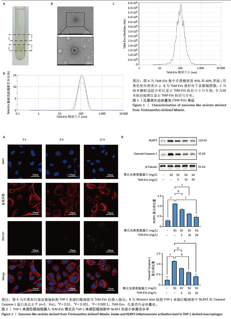
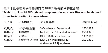
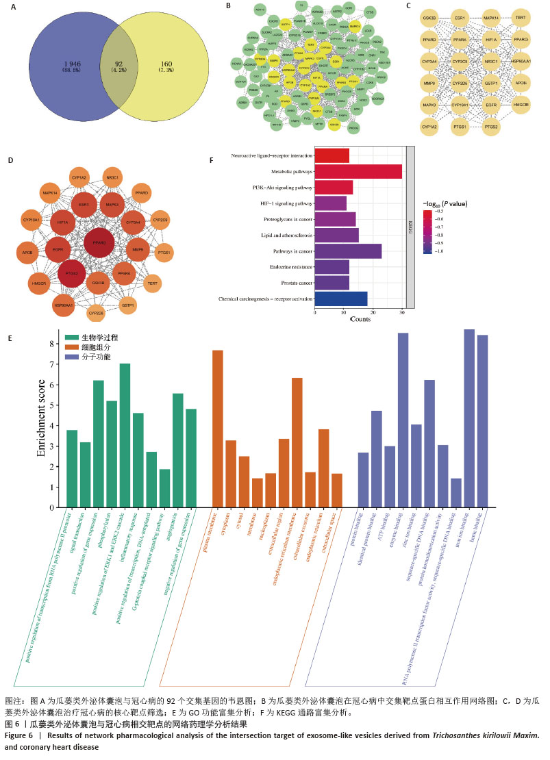
 为进一步探讨瓜蒌类外泌体囊泡治疗冠心病的潜在分子机制,该研究采用HPLC-MS筛选出了瓜蒌类外泌体囊泡中潜在的4种与NLRP3相关的化合物。通过网络药理学分析,筛选出92个与冠心病相关的靶点,并预测了23个关键靶点。这些关键靶点中,NR3C1、MAPK14、PPARG、PTGS2、HIF1A与NLRP3炎症小体的活化有着密切关系。研究发现,NR3C1是一种糖皮质激素受体,糖皮质激素通过NR3C1进入细胞后,能够抑制核因子κB通路以及NLRP3基因的表达,减少NLRP3炎症小体的活化[58]。PPARG是一种核受体,PPARG能通过抑制核因子κB和丝裂原活化蛋白激酶信号通路,有效减少NLRP3炎症小体的活化[59]。MAPK14是一种细胞信号传导激酶,在感染、氧化应激、细胞损伤等应激状态下,MAPK14可以激活转录因子(如ATF2、CHOP等),进而增强NLRP3炎症小体的形成和功能[60]。另有研究发现,PTGS2是一种参与前列腺素合成的关键酶,可以通过生成前列腺素E2等递质,激活NLRP3炎症小体,促进炎症反应[61]。HIF1A是一种缺氧诱导因子,在缺氧条件下,HIF1A能够诱导NLRP3上调,并增强NLRP3活性,这可能与细胞在低氧环境中的应激反应有关[62]。综上所述,瓜蒌类外泌体囊泡所含的化合物可能具有促进NR3C1、PPARG和抑制MAPK14、PTGS2、HIF1A表达的作用,通过多靶点的综合调控,抑制NLRP3炎症小体的活化,达到减轻血管炎症、减缓动脉粥样硬化进程的目的。筛选出的92个靶点,经GO和KEGG分析,预测其主要富集于以下几类通路:血管生成、正向调控ERK1和ERK2连锁反应、炎症反应、内质网功能、脂质代谢及动脉粥样硬化、低氧诱导因子1信号通路等。研究表明,ERK1/2信号通路的激活是动脉粥样硬化发展过程中的关键环节,而间充质干细胞衍生外泌体能够通过抑制该通路的激活有效减缓动脉粥样硬化的进展[63-64]。血管生成通常发生在缺氧环境中,白细胞与血管壁的相互作用通过一系列分子介导促进了炎症细胞向斑块微环境的迁移[65]。此外,已有研究表明,来自间充质干细胞的外泌体能够通过miR-146a/Src信号通路刺激血管生成,进而减缓内皮细胞衰老,最终延缓动脉粥样硬化的进程[66]。因此,这些研究揭示了瓜蒌类外泌体囊泡在治疗冠心病中可能的多重作用机制,为后续的分子机制研究提供了理论基础,并提示在后续研究中需要进一步探索瓜蒌类外泌体囊泡的下游分子与NLRP3之间的关系。