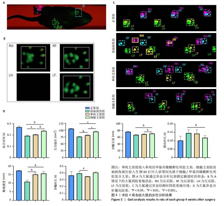
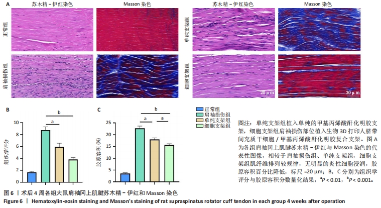
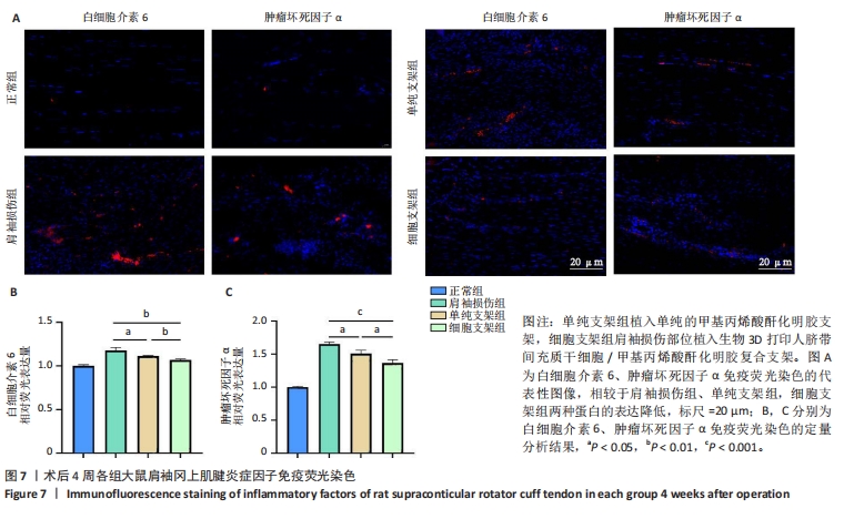
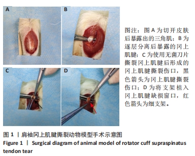
[1] LUI P, ZHANG P, CHAN K, et al. Biology and augmentation of tendon-bone insertion repair. J Orthop Surg Res. 2010;5:59.
[2] HERNIGOU J, VERTONGEN P, RASSCHAERT J, et al. Role of Scaffolds, Subchondral, Intra-Articular Injections of Fresh Autologous Bone Marrow Concentrate Regenerative Cells in Treating Human Knee Cartilage Lesions: Different Approaches and Different Results. Int J Mol Sci. 2021;22(8):3844.
[3] LIU Q, HATTA T, QI J, et al. Novel engineered tendon-fibrocartilage-bone composite with cyclic tension for rotator cuff repair. J Tissue Eng Regen Med. 2018;12(7):1690-1701.
[4] ZHU Q, MA ZJ, LI HY, et al. Enhancement of rotator cuff tendon-bone healing using combined aligned electrospun fibrous membranes and kartogenin. Rsc Adv. 2019;9(27):15582-15592.
[5] AICALE R, BISACCIA RD, OLIVIERO A, et al. Current pharmacological approaches to the treatment of tendinopathy. Expert Opin Pharmaco. 2020;21(12):1467-1477.
[6] GOVINDARAJU DT, CHEN CH, SHALUMON KT, et al. Bioactive Nanostructured Scaffold-Based Approach for Tendon and Ligament Tissue Engineering. Nanomaterials. 2023;13(12):35.
[7] CHEN J, ZHANG E, ZHANG W, et al. Fos Promotes Early Stage Teno-Lineage Differentiation of Tendon Stem/Progenitor Cells in Tendon. Stem Cells Transl Med. 2017;6(11):2009-2019.
[8] GE Z, LI W, ZHAO R, et al. Programmable DNA Hydrogel Provides Suitable Microenvironment for Enhancing TSPCS Therapy in Healing of Tendinopathy. Small. 2023;19(32):e2207231.
[9] MURCHISON ND, PRICE BA, CONNER DA, et al. Regulation of tendon differentiation by scleraxis distinguishes force-transmitting tendons from muscle-anchoring tendons. Development. 2007;134(14):2697-2708.
[10] TSUCHIYA A, TAKEUCHI S, WATANABE T, et al. Mesenchymal stem cell therapies for liver cirrhosis: MSCs as “conducting cells” for improvement of liver fibrosis and regeneration. Inflamm Regen. 2019;39(1): 6.
[11] WANG J, QIN W, ZHONG Y, et al. Injectable collagen hydrogel combines human umbilical cord mesenchymal stem cells to promote endometrial regeneration in rats with thin endometrium. Int J Biol Macromol. 2024;254(Pt 1):127591.
[12] CAO TT, CHEN H, PANG M, et al. Dose optimization of intrathecal administration of human umbilical cord mesenchymal stem cells for the treatment of subacute incomplete spinal cord injury. Neural Regen Res. 2022;17(8):1785-1794.
[13] WANG S, YAO Z, CHEN L, et al. Preclinical assessment of IL-1β primed human umbilical cord mesenchymal stem cells for tendon functional repair through TGF-β/IL-10 signaling. Heliyon. 2023;9(11):e21411.
[14] YUAN Z, YU D, WANG Y, et al. Early delivery of human umbilical cord mesenchymal stem cells improves healing in a rat model of Achilles tendinopathy. Regen Med. 2024;19(2):93-102.
[15] YEA JH, BAE TS, KIM BJ, et al. Regeneration of the rotator cuff tendon-to-bone interface using umbilical cord-derived mesenchymal stem cells and gradient extracellular matrix scaffolds from adipose tissue in a rat model. Acta Biomater. 2020;114:104-116.
[16] YAO Z, LI J, XIONG H, et al. MicroRNA engineered umbilical cord stem cell-derived exosomes direct tendon regeneration by mTOR signaling. J Nanobiotechnology. 2021;19(1):169.
[17] ZHANG J, XU W, LI C, et al. Tissue Engineering Microtissue: Construction, Optimization, and Application. Tissue Eng Part B Rev. 2022;28(2):393-404.
[18] ALMEIDA GHD, IGLESIA RP, ARAUJO MS, et al. Uterine Tissue Engineering: Where We Stand and the Challenges Ahead. Tissue Eng Part B Rev. 2022;28(4):861-890.
[19] ZHOU T, YUAN ZN, WENG JY, et al. Challenges and advances in clinical applications of mesenchymal stromal cells. J Hematol Oncol. 2021; 14(1):24.
[20] YUAN Z, YUAN X, ZHAO Y, et al. Injectable GelMA Cryogel Microspheres for Modularized Cell Delivery and Potential Vascularized Bone Regeneration. Small. 2021;17(11):e2006596.
[21] YAN XR, LI J, NA XM, et al. Mesenchymal Stem Cells Proliferation on Konjac Glucomannan Microcarriers: Effect of Rigidity. Chin J Polym Sci. 2022;40(9):1080-1089.
[22] LOUBIÈRE C, SION C, DE ISLA N, et al. Impact of the type of microcarrier and agitation modes on the expansion performances of mesenchymal stem cells derived from umbilical cord. Biotechnol Progr. 2019;35(6): 17.
[23] YAN X, ZHANG K, YANG Y, et al. Dispersible and Dissolvable Porous Microcarrier Tablets Enable Efficient Large-Scale Human Mesenchymal Stem Cell Expansion. Tissue Eng Part C Methods. 2020;26(5):263-275.
[24] GROSS G, HOFFMANN A. Therapeutic strategies for tendon healing based on novel biomaterials, factors and cells. Pathobiology. 2013; 80(4):203-210.
[25] FENG Y, ZHU S, MEI D, et al. Application of 3D Printing Technology in Bone Tissue Engineering: A Review. Curr Drug Deliv. 2021;18(7):847-861.
[26] JIANG H, LI X, CHEN T, et al. Bioprinted vascular tissue: Assessing functions from cellular, tissue to organ levels. Mater Today Bio. 2023; 23:100846.
[27] MATAI I, KAUR G, SEYEDSALEHI A, et al. Progress in 3D bioprinting technology for tissue/organ regenerative engineering. Biomaterials. 2020;226:119536.
[28] BANDYOPADHYAY A, MITRA I, BOSE S. 3D Printing for Bone Regeneration. Curr Osteoporos Rep. 2020;18(5): 505-514.
[29] WANG Z, WANG L, LI T, et al. 3D bioprinting in cardiac tissue engineering. Theranostics. 2021;11(16):7948-7969.
[30] ZHANG H, CHEN Y, FAN C, et al. Cell-subpopulation alteration and FGF7 activation regulate the function of tendon stem/progenitor cells in 3D microenvironment revealed by single-cell analysis. Biomaterials. 2022;280:121238.
[31] JIAO X, ZHANG Y, LI W, et al. HIF-1α inhibition attenuates severity of Achilles tendinopathy by blocking NF-κB and MAPK pathways. Int Immunopharmacol. 2022;106:108543.
[32] JIANG L, LIU T, LYU K, et al. Inflammation-related signaling pathways in tendinopathy. Open Life Sci. 2023;18(1):20220729.
[33] HUA T, YANG M, SONG H, et al. Huc-MSCs-derived exosomes attenuate inflammatory pain by regulating microglia pyroptosis and autophagy via the miR-146a-5p/TRAF6 axis. J Nanobiotechnology. 2022;20(1):324.
[34] SUN X, HAO H, HAN Q, et al. Human umbilical cord-derived mesenchymal stem cells ameliorate insulin resistance by suppressing NLRP3 inflammasome-mediated inflammation in type 2 diabetes rats. Stem Cell Res Ther. 2017;8(1):241.
[35] WU LL, PAN XM, CHEN HH, et al. Repairing and Analgesic Effects of Umbilical Cord Mesenchymal Stem Cell Transplantation in Mice with Spinal Cord Injury. BioMed Res Int. 2020;2020:10.
[36] WU J, CHU CC. Block copolymer of poly(ester amide) and polyesters: synthesis, characterization, and in vitro cellular response. Acta Biomater. 2012;8(12):4314-4323.
[37] MD FADILAH NI, MOHD ABDUL KADER JAILANI MS, BADRUL HISHAM MAI, et al. Cell secretomes for wound healing and tissue regeneration: Next generation acellular based tissue engineered products. J Tissue Eng. 2022;13:20417314221114273.
[38] FOO JB, LOOI QH, CHONG PP, et al. Comparing the Therapeutic Potential of Stem Cells and their Secretory Products in Regenerative Medicine. Stem Cells Int. 2021;2021:2616807.
[39] ASGHAR W, EL ASSAL R, SHAFIEE H, et al. Engineering cancer microenvironments for in vitro 3-D tumor models. Mater Today. 2015;18(10):539-553.
[40] JAYARAMAN P, LIM R, NG J, et al. Acceleration of Translational Mesenchymal Stromal Cell Therapy Through Consistent Quality GMP Manufacturing. Front Cell Dev Biol. 2021;9:648472.
[41] ACHILLI TM, MEYER J, MORGAN JR. Advances in the formation, use and understanding of multi-cellular spheroids. Expert Opin Biol Ther. 2012;12(10):1347-1360.
[42] DONG Z, YUAN Q, HUANG K, et al. Gelatin methacryloyl (GelMA)-based biomaterials for bone regeneration. RSC Adv. 2019;9(31): 17737-17744.
[43] KURIAN AG, SINGH RK, PATEL KD, et al. Multifunctional GelMA platforms with nanomaterials for advanced tissue therapeutics. Bioact Mater. 2022;8:267-295.
[44] JIANG G, LI S, YU K, et al. A 3D-printed PRP-GelMA hydrogel promotes osteochondral regeneration through M2 macrophage polarization in a rabbit model. Acta Biomater. 2021;128:150-162.
[45] LV B, LU L, HU L, et al. Recent advances in GelMA hydrogel transplantation for musculoskeletal disorders and related disease treatment. Theranostics. 2023;13(6):2015-2039.
[46] YUE K, TRUJILLO-DE SANTIAGO G, ALVAREZ MM, et al. Synthesis, properties, and biomedical applications of gelatin methacryloyl (GelMA) hydrogels. Biomaterials. 2015;73:254-271.
[47] UNAGOLLA JM, JAYASURIYA AC. Hydrogel-based 3D bioprinting: A comprehensive review on cell-laden hydrogels, bioink formulations, and future perspectives. Appl Mater Today. 2020;18:22.
[48] JI S, ALMEIDA E, GUVENDIREN M. 3D bioprinting of complex channels within cell-laden hydrogels. Acta Biomater. 2019;95:214-224.
[49] SAMADI A, MOAMMERI A, POURMADADI M, et al. Cell Encapsulation and 3D Bioprinting for Therapeutic Cell Transplantation. ACS Biomater Sci Eng. 2023;9(4):1862-1890. |