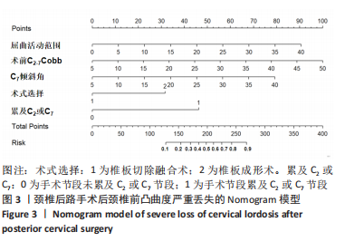
[1] 许春阳,杨晋才,尹鹏,等. 颈椎后路手术治疗脊髓型颈椎病的应用进展[J]. 中华骨与关节外科杂志,2022,15(4):296-301.
[2] 李觅,赵承斌,张捍军,等. 颈前路扩大纵向减压范围植骨融合及钢板内固定治疗脊髓型颈椎病[J]. 中国组织工程研究,2019,23(16): 2473-2478.
[3] 江泽华,张学利,朱如森,等. 颈前路间盘切除植骨融合钛板内固定和颈后路单开门椎管成形治疗多节段脊髓型颈椎病:重建后的稳定性[J]. 中国组织工程研究,2017,21(27):4306-4311.
[4] 王国旗,徐韬,盛伟斌,等. 不同入路治疗多节段脊髓型颈椎病:疗效与安全性的Meta分析[J]. 中国组织工程研究,2014,18(4):637-644.
[5] EDWARDS CC, HELLER JG, MURAKCAMI H. Corpectomy versus laminoplasty formultilevel cervicalmyelopathy: an independentmatched cohortanalysis. Spine. 2002;11:1168-1175.
[6] GU Y, WANG C, HU J, et al. Association Between the Cervical Extensor Musculature and the Demographic Features,Symptoms and Sagittal balance in Patients with Multilevel Cervical Spondylotic Myelopathy. World Neurosurg. 2022. doi: 10.1016/j.wneu.2022.10.014.
[7] MAEDA T, ARIZONO T, SAITO T, et al. Cervical alignment, range of motion, and instability after cervical laminoplasty. Clin Orthop Relat Res. 2002;401:132-138.
[8] SODEYAMA T, GOTO S, MOCHIZUKI M, et al. Effect of decompression enlargement laminoplasty for posterior shifting of the spinal cord. Spine. 1999;24(15):1531-1532.
[9] WHITE AA 3RD, JOHNSON RM, PANJABI MM, et al. Biomechanical analysis of clinical stability in the cervical spine. Clin Orthop Relat Res. 1975;109:85-96.
[10] 李程, 郭开今, 李强,等. Arch钛板与侧块螺钉固定治疗颈椎后纵韧带骨化症疗效比较[J]. 实用骨科杂志,2017,23(10):873-878.
[11] XU R, HAMAN SP, EBRAHEIM NA, et al. The anatomic relation of lateral mass screws to the spinal nerves. A comparison of the Magerl, Anderson, and an techniques. Spine. 1999;24(19):2057-2061.
[12] ZHAO Y, QIU GX, ZHANG JG, et al. Relationship between Cervical Curvature Index(Ishihara) and Cervical Spine Angle(C2-7). J Pract Orthop. 2004;6(3):223-226.
[13] ONO K, MURATA S, MATSUSHITA M, et al. Cervical Lordosis Ratio as a Novel Predictor for the Loss of Cervical Lordosis After Laminoplasty. Neurospine. 2021;18(2):311-318.
[14] LEE SU, SON DW, LEE JS, et al. Does Extension Dysfunction Affect Postoperative Loss of Cervical Lordosis in Patients Who Undergo Laminoplasty? Spine. 2019;44(8):E456-E464.
[15] BABA H, UCHIDA K, MAEZAWA Y, et al. Lordotic alignment and posterior migration of the spinal cord following en bloc open-door laminoplasty for cervical myelopathy: a magnetic resonance imaging study. J Neurol. 1996;243(9):626-632.
[16] HOU SB, SUN XZ, LIU FY, et al. Relationship of Change in Cervical Curvature after Laminectomy with Lateral Mass Screw Fixation to Spinal Cord Shift and Clinical Efficacy. J Neurol Surg Part A. 2022;83(2):129-134.
[17] LIZUKA H, SHIMIZU T, TATENO K, et al. Extensor musculature of the cervical spine after laminoplasty: morphologic evaluation by coronal view of the magnetic resonance image. Spine. 2001;26(20):2220-2226.
[18] 张为,董玉昌,申勇,等. 保留颈半棘肌肌止的椎板成形术的临床应用[J]. 中国矫形外科杂志,2006,14(13):980-982.
[19] LIN S, ZHOU F, SUN Y, et al. The severity of operative invasionto the posterior muscular-ligament complex influences cervical sagittal balance after open-door laminoplasty. Eur Spine J. 2015;24(1):127-135.
[20] KIM T, LEE SY, KIM YC, et al. T1 slope as a predictor of kyphotic alignment change after laminoplasty in patients with cervical myelopathy. Spine. 2013;38(16):E992-E997.
[21] ZHANG JT, LI JQ, NIU RJ, et al. Predictors of cervical lordosis loss after laminoplasty in patients with cervical spondylotic myelopathy. Eur Spine J. 2017;26(4):1205-1210.
[22] LIN T, WANG Z, CHEN G, et al. Is Cervical Sagittal Balance Related to the Progression of Patients with Cervical Spondylotic Myelopathy? World Neurosurg. 2020;137:e52-e67.
[23] LI XY, KONG C, SUN XY, et al. Influence of the Ratio of C2–C7 Cobb Angle to T1 Slope on Cervical Alignment After Laminoplasty. World Neurosurg. 2019;S1878-8750(19)30052-X.
[24] CHUGH A, PATEL M, GERGES C, et al. Use of C7 Slope as a Surrogate Marker for T1 Slope: A Radiographic Study in Patients with and without Cervical Deformity. World Neurosurg. 2020;143:E516-E522.
[25] 房昕,吴恺,刘品端,等. C7倾斜角与ACDF术后矢状位参数的关系研究[J]. 颈腰痛杂志,2021,42(1):126-128.
[26] WOODROFFE RW, HELLAND L, HOLLATZ C, et al. Impact of the Inclusion of C2 in Posterior Cervical Fusions for Cervical Myelopathy on Sagittal Cervical Alignment. Clin Spine Surg. 2020;33(4):E141-E146.
[27] 吴炳轩,刘宝戈,崔维,等. C2及C7倾斜角与退变性颈椎节段后凸矢状位力线及临床功能相关性研究[J]. 中国脊柱脊髓杂志,2021, 31(12):1098-1105.
[28] MAO JP, TIAN W, LIU B, et al. Short-term clinical outcome of modified expansive open-door cervical laminoplasty preserving posterior extensor musculature inserted into C2 and C7 spinous process. Zhonghua Yi Xue Za Zhi. 2010;90(5):337-341.
[29] NORI S, SHIRAISHI T, AOYAMA R, et al. Extremely high preoperative C7 slope limits compensatory cervical lordosis after muscle-preserving selective laminectomy. Eur Spine J. 2018;27(8):2029-2037.
[30] 刘晓伟,陈德玉,王新伟,等. 颈椎后纵韧带骨化症患者K线对两种颈后路手术疗效的影响[J]. 中国脊柱脊髓杂志,2013,23(1):6-10.
[31] 谭俊铭,叶晓健,袁文,等. 三种颈前路融合术后颈椎前柱高度和Cobb角比较[J]. 中国修复重建外科杂志,2006,20(4):367-371. |