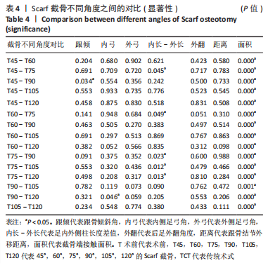
[1] FLORES DV, MEJÍA GÓMEZ C, FERNÁNDEZ HERNANDO M, et al. Adult Acquired Flatfoot Deformity: Anatomy, Biomechanics, Staging, and Imaging Findings. Radiographics. 2019;39(5):1437-1460.
[2] MOLINA-GARCIA P, MOLINA-MOLINA A, SMEETS A, et al. Effects of integrative neuromuscular training on the gait biomechanics of children with overweight and obesity. Scand J Med Sci Sports. 2022;32(7):1119-1130.
[3] 张新语,邢新阳,霍洪峰.矫正鞋垫的设计原理与生物力学功能[J].中国组织工程研究,2020,24(23):3744-3750.
[4] 何晓宇,王朝强,周之平,等.三维有限元方法构建足部健康骨骼与常见疾病模型及生物力学分析[J].中国组织工程研究,2020,24(9): 1410-1415.
[5] OHUCHI H, CHAVEZ JS, ALVAREZ CAD. Changes in calcaneal pitch and heel fat pad thickness in static weight bearing radiographs while wearing shoes with arch support and heel cup orthotics. Asia Pac J Sports Med Arthrosc Rehabil Technol. 2019;17:21-24.
[6] FELLAS A, SINGH-GREWAL D, CHAITOW J, et al. Effect of preformed foot orthoses in reducing pain in children with juvenile idiopathic arthritis: a multicentre randomized clinical trial. Rheumatology (Oxford). 2022;61(6):2572-2582.
[7] EVANS D. Calcaneo-valgus deformity. J Bone Joint Surg Br. 1975;57(3): 270-278.
[8] KOUTSOGIANNIS E. Treatment of mobile flat foot by displacement osteotomy of the calcaneus. J Bone Joint Surg Br. 1971;53(1):96-100.
[9] ZAW H, CALDER JD. Operative management options for symptomatic flexible adult acquired flatfoot deformity: a review. Knee Surg Sports Traumatol Arthrosc. 2010;18(2):135-142.
[10] FEUERSTEIN CA, WEIL L JR, WEIL LS SR, et al. The calcaneal scarf osteotomy: surgical correction of the adult acquired flatfoot deformity and radiographic results. Foot Ankle Spec. 2013;6(5):367-371.
[11] 李晓东,江汉,江毅,等.成人获得性扁平足的手术治疗进展[J].实用骨科杂志,2015,21(7):626-630.
[12] 朱珊,王植,郭林.双下肢负重位三维CT检查技术与步态分析结合初步应用进展[J].陕西医学杂志,2021,50(6):766-768,封3.
[13] LIGON SC, LISKA R, STAMPFL J, et al. Polymers for 3D Printing and Customized Additive Manufacturing. Chem Rev. 2017;117(15):10212-10290.
[14] 陈立翔,娄博,王焕.基于有限元法的数字化建模在拇外翻研究中的应用[J].医用生物力学,2022,37(5):972-977.
[15] HO M, NGUYEN J, HEALES L, et al. The biomechanical effects of 3D printed and traditionally made foot orthoses in individuals with unilateral plantar fasciopathy and flat feet. Gait Posture. 2022;96: 257-264.
[16] OKA K, MURASE T, MORITOMO H, et al. Accuracy of corrective osteotomy using a custom-designed device based on a novel computer simulation system. J Orthop Sci. 2011;16(1):85-92.
[17] 陈亮,桑莉莉,高大伟,等.基于3D打印个性化手术导航模块的微创锥形截骨治疗桡骨上段旋转畸形[J].中华肩肘外科电子杂志, 2018,6(2):151-153.
[18] 钟世镇.数字医学在不同学科中的探索应用[J].中华整形外科杂志,2018,34(6):前插2.
[19] 孙卫东,温建民,胡海威,等.拇外翻第1跖骨颈部不同截骨角度截骨端稳定性有限元分析[J].中华损伤与修复杂志(电子版), 2012,7(5):492-496.
[20] MYERSON MS. Adult acquired flatfoot deformity: treatment of dysfunction of the posterior tibial tendon. Instr Course Lect. 1997;46: 393-405.
[21] JOHNSON KA, STROM DE. Tibialis posterior tendon dysfunction. Clin Orthop Relat Res. 1989;(239):196-206.
[22] CONTI MS, ELLIS SJ, CHAN JY, et al. Optimal Position of the Heel Following Reconstruction of the Stage II Adult-Acquired Flatfoot Deformity. Foot Ankle Int. 2015;36(8):919-927.
[23] 俞光荣,陈雁西,杨云峰,等.伴严重疼痛的成人平足症的手术治疗[J].中国矫形外科杂志,2007,15(9):641-644,中插1.
[24] SCHLEUNES SD, CAMPBELL SN, JONES JM, et al. Radiographic Analysis of the Lateral Column Lengthening Procedure in Stage II Adult Acquired Flatfoot Deformity. J Foot Ankle Surg. 2022. doi: 10.1053/j.jfas.2022.04.005. Online ahead of print.
[25] BAXTER JR, DEMETRACOPOULOS CA, PRADO MP, et al. Lateral column lengthening corrects hindfoot valgus in a cadaveric flatfoot model. Foot Ankle Int. 2015;36(6):705-709.
[26] 胡牧,徐向阳,李星辰,等.足外侧柱延长术联合内侧软组织重建术治疗成人ⅡB期获得性扁平足[J].中国骨与关节外科,2014, 7(4):323-327.
[27] CANAVESE F, DIMEGLIO A, BONNEL F. Postoperative CT-scan 3D reconstruction of the calcaneus following lateral calcaneal lengthening osteotomy for flatfoot deformity in children. Is the surgical procedure potentially associated with subtalar joint damage. Foot Ankle Surg. 2018;24(5):453-459.
[28] ARGERAKIS NG, WEIL L JR, WEIL LS SR, et al. The radiographic effects of the scarf bunionectomy on rearfoot alignment. Foot Ankle Spec. 2015;8(2):89-94.
[29] VERMEULEN K, NEVEN E, VANDEPUTTE G, et al. Relationship of the Scarf valgus-inducing osteotomy of the calcaneus to the medial neurovascular structures. Foot Ankle Int. 2011;32(5):S540-544.
[30] WEIL LS JR, ROUKIS TS. The calcaneal scarf osteotomy: operative technique. J Foot Ankle Surg. 2001;40(3):178-182.
[31] SAUNDERS SM, ELLIS SJ, DEMETRACOPOULOS CA, et al. Comparative Outcomes Between Step-Cut Lengthening Calcaneal Osteotomy vs Traditional Evans Osteotomy for Stage IIB Adult-Acquired Flatfoot Deformity. Foot Ankle Int. 2018;39(1):18-27.
[32] LÔBO C, PIRES EA, BORDALO-RODRIGUES M, et al. Imaging of progressive collapsing foot deformity with emphasis on the role of weightbearing cone beam CT. Skeletal Radiol. 2022;51(6):1127-1141.
[33] KIMURA T, THORHAUER ED, KINDIG MW, et al. Evaluation of the Foot Arch in Partial Weightbearing Conditions. Foot Ankle Int. 2022; 43(1):113-122.
[34] KIMURA T, THORHAUER ED, SANGEORZAN BJ, et al. Foot radiographic angle variation as a function of weightbearing magnitude. J Orthop Res. 2022. doi: 10.1002/jor.25283. Online ahead of print.
[35] 张树,王智,张建中.负重CT在足踝疾病诊疗中的应用[J].足踝外科电子杂志,2017,4(2):57-60.
[36] SRIPANICH Y, WEINBERG M, KRÄHENBÜHL N, et al. Change in the First Cuneiform-Second Metatarsal Distance After Simulated Ligamentous Lisfranc Injury Evaluated by Weightbearing CT Scans. Foot Ankle Int. 2020;41(11):1432-1441.
[37] FULLER RM, KIM J, AN TW, et al. Assessment of Flatfoot Deformity Using Digitally Reconstructed Radiographs: Reliability and Comparison to Conventional Radiographs. Foot Ankle Int. 2022; 43(7):983-993.
[38] KIDO M, IKOMA K, IMAI K, et al. Load response of the medial longitudinal arch in patients with flatfoot deformity: in vivo 3D study. Clin Biomech (Bristol, Avon). 2013;28(5):568-573.
[39] 王文成, 张兴飞, 许亚军.数字化技术在踇外翻治疗中的应用[J].中国组织工程研究,2021,25(12):1911-1916.
[40] MALAKOUTIKHAH H, MADENCI E, LATT LD. The impact of ligament tears on joint contact mechanics in progressive collapsing foot deformity: A finite element study. Clin Biomech (Bristol, Avon). 2022; 94:105630.
[41] MALAKOUTIKHAH H, MADENCI E, LATT LD. A computational model of force within the ligaments and tendons in progressive collapsing foot deformity. J Orthop Res. 2022. doi: 10.1002/jor.25380. Online ahead of print. |