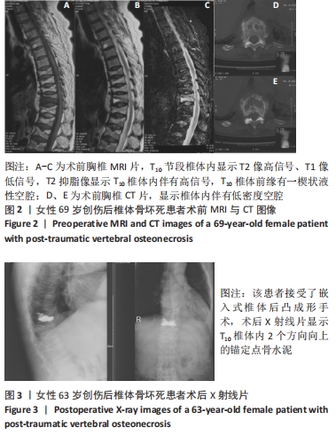
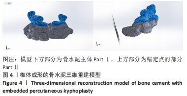
[1] SUN Y, XIONG X, WAN D, et al. Comparison of effectiveness of Vesselplasty and percutaneous kyphoplasty for Kümmell disease. Zhongguo Xiu Fu Chong Jian Wai Ke Za Zhi. 2020;34(12):1539-1544.
[2] 陈伯华,陈其昕,程黎明,等.症状性陈旧性胸腰椎骨质疏松性骨折手术治疗临床指南[J].中华创伤杂志,2020,36(7):577-586.
[3] XU Z, HAO D, DONG L, et al. Surgical options for symptomatic old osteoporotic vertebral compression fractures: a retrospective study of 238 cases. BMC Surg. 2021;21(1):22.
[4] PATIL S, RAWALL S, SINGH D, et al. Surgical patterns in osteoporotic vertebral compression fractures. Eur Spine J. 2013;22(4):883-891.
[5] DAI S, QIN R, SHI X, et al. Percutaneous vertebroplasty versus kyphoplasty for the treatment of neurologically intact osteoporotic Kümmell’s disease. Global Spine J. 2021;21(1): 65.
[6] KIM D, KIM T, PARK K, et al. The proper volume and distribution of cement augmentation on percutaneous vertebroplasty. Korean Neurosurg Soc. 2010;48(2):125-128.
[7] WANG D, LI Y, YIN H, et al. Three-dimensional finite element analysis of optimal distribution model of vertebroplasty. Ann Palliat Med. 2020; 9(3):1062-1072.
[8] WU AM, CHI YL, NI WF. Vertebral compression fracture with intravertebral vacuum cleft sign: pathogenesis, image, and surgical intervention. Asian Spine J. 2013;7(2):148-155.
[9] 郭惠智,梁德,张顺聪,等.腰椎单/双节段骨水泥强化椎弓根螺钉固定对邻近节段影响的有限元分析[J].中国脊柱脊髓杂志,2020, 30(2):159-166.
[10] 陈荣彬,白杰,李勇,等.骨水泥弥散类型影响胸腰段椎体成形后相邻椎体应力的三维有限元分析[J].中国组织工程研究,2020, 24(30):4775-4781.
[11] CHEN C, YUCHI C, GAO Z, et al. Comparative analysis of the biomechanics of the adjacent segments after minimally invasive cervical surgeries versus anterior cervical discectomy and fusion: A finite element study. J Orthop Translat. 2020;23:107-112.
[12] LITTLE J, ADAM C JS. The effect of soft tissue properties on spinal flexibility in scoliosis: biomechanical simulation of fulcrum bending. Spine (Phila Pa 1976). 2009;34(2):E76-82.
[13] 何斌,胡侦明,郝杰,等.后部不同结构切除对胸椎稳定性的影响[J].第三军医大学学报,2012,34(20):2101-4210.
[14] ZHANG W, ZHAO J, LI L, et al. Modelling tri-cortical pedicle screw fixation in thoracic vertebrae under osteoporotic condition: A finite element analysis based on computed tomography. Comput Methods Programs Biomed. 2020;187:105035.
[15] 李楠,张贵林,何达,等.骨水泥的分布与剂量对椎体成形术疗效影响的研究[J].中国骨与关节损伤杂志,2015,30(1):66-68.
[16] 刘长枫,宋文慧,刘昌文,等.经皮椎体成形术骨水泥分布评价及影响因素分析[J].中国脊柱脊髓杂志,2019,29(11):1001-1008.
[17] 卢昌怀,刘志军,张宏波,等.骨水泥量及分布对椎体成形术后相邻椎体生物力学影响的三维有限元分析[J].中国骨质疏松杂志, 2015,21(1):29-33.
[18] ZHANG Z, JING Q, QIAO R, et al. Risk factors analysis of adjacent fractures after percutaneous vertebroplasty for osteoporotic vertebral compression fracture. Zhongguo Xiu Fu Chong Jian Wai Ke Za Zhi. 2021; 35(1):20-25.
[19] 刘昊,迮骁,邹俊,等.经皮椎体后凸成形术的骨水泥分布与临床疗效分析[J].中华骨与关节外科杂志,2017,10(1):8-12.
[20] 包拥政,祝周兴,冯云升,等.骨水泥注射体积与骨质疏松压缩性骨折椎体及邻近椎体应力的关系[J].中国组织工程研究,2015, 19(52):8365-8372.
[21] LEE C, KIM Y, LEE H, et al. Biomechanical effects of hybrid stabilization on the risk of proximal adjacent-segment degeneration following lumbar spinal fusion using an interspinous device or a pedicle screw-based dynamic fixator. J Neurosurg Spine. 2017;27(6):643-649.
[22] PARK J, PARK J, JEON H, et al. Kümmell’s Disease Treated with Percutaneous Vertebroplasty: Minimum 1 Year Follow-Up. Korean J Neurotrauma. 2017;13(2):119-123.
[23] HE X, MENG Y, HUANG Y, et al. Factors Affecting Delayed Union of Vertebral Fractures Following Percutaneous Kyphoplasty. Pain Physician. 2017;20(2):E241-E9.
[24] 李诚,程兆明,张海波.经皮椎体后凸成形术治疗伴周壁破裂的骨质疏松性胸腰椎骨折[J].脊柱外科杂志,2021,19(3):195-197.
[25] ZHANG J, FAN Y, HE X, et al. Bracing after percutaneous vertebroplasty for thoracolumbar osteoporotic vertebral compression fractures was not effective. Clin Interv Aging. 2019;14:265-270.
[26] ZHOU T, LIN H, WANG H, et al. Comparative study on the biomechanics between improved PVP and traditional PKP in the treatment of vertebral peripheral wall damage-type OVCF. Exp Ther Med. 2017; 14(1):575-580.
[27] WANG F, WANG L, MIAO D, et al. Which one is more effective for the treatment of very severe osteoporotic vertebral compression fractures: PVP or PKP? J Pain Res. 2018;11: 2625-2631.
[28] 李永军,梁永辉,韦兴,等.“拖尾征”锚定骨水泥椎体后凸成形术对Kummell′s病的治疗效果[J].中华老年多器官疾病杂志,2020, 19(7):494-498.
[29] HAO D, YANG J, TUO Y, et al. Reliability and application of the new morphological classification system for chronic symptomatic osteoporotic thoracolumbar fracture. J Orthop Surg Res. 2020;15(1):348.
[30] 包拥政,祝周兴,冯云升,等.低弹性模量骨水泥对骨质疏松压缩性骨折椎体及邻近椎体应力的影响:三维有限元分析[J].中国组织工程研究,2016,20(16):2285-2293.
|