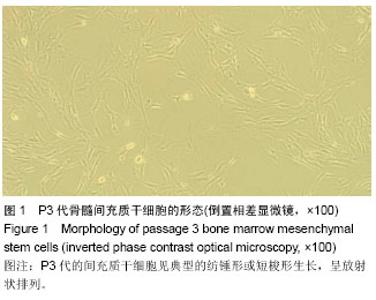
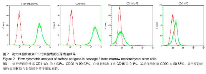
| [1] Minguell JJ, Erices A. Mesenchymal stem cells and the treatment of cardiac disease. Exp Biol Med (Maywood).2006; 231(1):39-49.
[2] Devine SM. Mesenchymal stem cells: will they have a role in the clinic?. J Cell Biochem Suppl.2002,38:73-79.
[3] Pittenger MF, Mackay AM, Beck SC, et al. Multilineage potential of adult human mesenchymal stem cells. Science. 1999;284(5411):143-147.
[4] Menasche P. Current status and future prospects for cell transplantation to prevent congestive heart failure.Semin Thorac Cardiovasc Surg.2008;20(2):131-137.
[5] Lazarus HM, Koc ON, Devine SM, et al.Cotransplantation of HLA-identical sibling culture-expanded mesenchymal stem cells and hematopoietic stem cells in hematologic malignancy patients. Biol Blood Marrow Transplant.2005; 11(5):389-398.
[6] Tambara K, Sakakibara Y, Sakaguchi G, et al. Transplanted skeletal myoblasts can fully replace the infarcted myocardium when they survive in the host in large numbers. Circulation. 2003;108(Suppl 1):I259-I263.
[7] Gao Y, Xu P,Chen L,et al.Prostaglandin E1 encapsulated into lipid nanoparticles improves its anti-inflammatory effect with low side-effect.Int J Pharm. 2010;387(1-2):263-271.
[8] Wu CC, Wu CI, Wang WY, et al.Low concentrations of resveratrol potentiate the antiplatelet effect of prostaglandins. Planta Med.2007;73(5):439-443.
[9] Zhao XS, Pan W, Bekeredjian R, et al. Endogenous endothelin-1 is required for cardiomyocyte survival in vivo. Circulation.2006;114(8):830-837.
[10] 杨焕纳,崔正军,Han Seung-Kyu,等.前列腺素E1对人类脂肪源性基质细胞体外增殖的影响[J].中国组织工程研究与临床康复, 2011,15(14):2517-2520.
[11] Grassel S, Anders S.Cell-based therapy options for osteochondral defects. Autologous mesenchymal stem cells compared to autologous chondrocytes. Orthopade. 2012; 41(5):415-428, 429-430.
[12] Xu N, Liu H, Qu F, et al. Hypoxia inhibits the differentiation of mesenchymal stem cells into osteoblasts by activation of Notch signaling. Exp Mol Pathol. 2013;94(1):33-39.
[13] Fink T, Zachar V. Adipogenic differentiation of human mesenchymal stem cells. Methods Mol Biol.2011;698: 243-251.
[14] Law S,Chaudhuri S.Mesenchymal stem cell and regenerative medicine: regeneration versus immunomodulatory challenges. Am J Stem Cells.2013;2(1):22-38.
[15] Cai B, Li J, Wang J, et al.microRNA-124 regulates cardiomyocyte differentiation of bone marrow-derived mesenchymal stem cells via targeting STAT3 signaling. Stem Cells.2012;30(8):1746-1755.
[16] Zhang Y, Chu Y, Shen W, et al.Effect of 5-azacytidine induction duration on differentiation of human first-trimester fetal mesenchymal stem cells towards cardiomyocyte-like cells. Interact Cardiovasc Thorac Surg.2009;9(6):943-946.
[17] Soma T, Kishimoto J, Fisher D.Isolation of mesenchymal stem cells from human dermis. Methods Mol Biol.2013; 989:265-274.
[18] Lubis A M, Sandhow L, Lubis VK, et al. Isolation and cultivation of mesenchymal stem cells from iliac crest bone marrow for further cartilage defect management. Acta Med Indones, 2011,43(3):178-184.
[19] Conget PA, Minguell JJ. Phenotypical and functional properties of human bone marrow mesenchymal progenitor cells. J Cell Physiol.1999;181(1):67-73.
[20] Gao X, Zhang J, Zhang J, et al.Identification of Rat Respiratory Mucosa Stem cells and comparison of the early neural differentiation potential with the bone marrow mesenchymal stem cells in vitro. Cell Mol Neurobiol. 2014; 34(2):257-268.
[21] 李晓峰,赵劲民,苏伟,等.大鼠骨髓间充质干细胞的培养与鉴定[J]. 中国组织工程研究与临床康复, 2011,15(10):1721-1725.
[22] 杨宇辉,吴继雄.大鼠骨髓间充质干细胞和内皮祖细胞的分离培养及鉴定[J].中国组织工程研究与临床康复, 2009,13(40):7876- 7880.
[23] Liu Y, Wang L, Fatahi R, et al.Isolation of murine bone marrow derived mesenchymal stem cells using Twist2 Cre transgenic mice. Bone.2010;47(5):916-925.
[24] Kitazawa Y, Li X K, Xie L, et al.Bone marrow-derived conventional, but not cloned, mesenchymal stem cells suppress lymphocyte proliferation and prevent graft- versus-host disease in rats. Cell Transplant.2012; 21(2-3): 581-590.
[25] Feng J, Wu G, Liu R, et al. Prostaglandin E1 (PGE1) reduces cardiac-derived TXA2 release in ischaemic arrest in isolated working rat heart. Int J Cardiol. 1996;55(3):265-270.
[26] Bai W, Zheng X, Zhou L, et al. Prostaglandin E1 dose-dependently promotes stability of atherosclerotic plaque in a rabbit model. Can J Physiol Pharmacol. 2012;90(2): 131-139.
[27] Li J, Yang P, Li A, et al. Cardioprotective effect of liposomal prostaglandin E1 on a porcine model of myocardial infarction reperfusion no-reflow. Journal of Zhejiang University Science B.2011;12(8):638-643.
[28] Jia Y,Lin J,Zeng W,et al.Effect of prostaglandin on luteinizing hormone-stimulated proliferation of theca externa cells from chicken prehierarchical follicles. Prostaglandins Other Lipid Mediat.2010;92(1-4):77-84.
[29] 符晓阳,蔡传奇,郑鸿,等.前列腺素E1促进血管内皮细胞增生和迁移及其机制[J].中华医学杂志, 2013,93(3):222-225. |