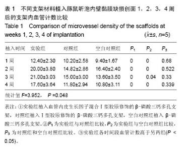
| [1] Bojrab DI,Causse JB,Battista RA,et al.Ossiculoplasty with composite prostheses: Overview and analysis.Otolaryngol Clin North Am.1994;27(4):759-776.
[2] Seebach C,Henrich D,Kähling C,et al.Endothelial progenitor cells and mesenchymal stem cells seeded onto beta-TCP granules enhance early vascularization and bone healing in a critical-sized bone defect in rats.Tissue Eng Part A.2010;16(6): 1961-1970.
[3] 钱成雄,叶毓粦,吴坤南,等.生物陶瓷(β-TCP人工骨)治疗四肢骨折22例临床研究[J]. 中国当代医药,2010,17(10):37-38.
[4] 陈仁吉,丁雯,穆玥,等.β-磷酸三钙用于牙槽嵴裂骨缺损修复的临床研究[J].北京口腔医学,2012,20(3):154-157.
[5] Shinohara T,Gyo K,Saiki T,et al.Ossiculoplasty using hydroxyapatite prostheses: long-term results.Clin Otolaryngol Allied Sci.2000;25(4):287-292.
[6] Chen J,Yang L,Guo L,et al.Sodium hyaluronate as a drug-release system for VEGF 165 improves graft revascularization in anterior cruciate ligament reconstruction in a rabbit model. Exp Ther Med.2012;4(3):430-434.
[7] Weidner N,Folkman J,Pozza F,et al.Tumor angiogenesis:a new significant and independent prognostic indicator in early-stage breast carcinoma. J Natl Cancer Inst. 1992; 84(24):1875-1887.
[8] Brackmann DE,Sheehy JL.Tympanoplasty: ToRps and PoRps.Laryngoscope. 1979;89(1):108-114.
[9] Okuda T,Ioku K,Yonezawa I,et al.The effect of the microstructure of beta-tricalcium phosphate on the metabolism of subsequently formed bone tissue. Biomaterials. 2007;28(16):2612-2621.
[10] Han XY,Zhang FQ,Fu YF,et al.Fabrication and physical properties of porous individual beta-TCP scaffold to reconstruct residual alveolar ridge in dog. Shanghai Kou Qiang Yi Xue.2008;17(1):51-54.
[11] Castilho M,Moseke C,Ewald A,et al.Direct 3D powder printing of biphasic calcium phosphate scaffolds for substitution of complex bone defects. Biofabrication. 2014;6(1):015006.
[12] Doyle H,Lohfeld S,McHugh P.Predicting the Elastic Properties of Selective Laser Sintered PCL/β-TCP Bone Scaffold Materials Using Computational Modelling. Ann Biomed Eng. 2014;42(3):661-677.
[13] Kang Y,Scully A,Young DA,et al.Enhanced mechanical performance and biological evaluation of a PLGA coated beta-TCP composite scaffold for load-bearing applications. Eur Polym J.2011;47(8):1569-1577.
[14] Lou CW,Yao CH,Chen YS,et al.PLA/beta-TCP Complex Tubes: The Mechanical Properties and Applications of Artificial Bone.J Biomater Sci Polym Ed.2011.
[15] Lindhorst D,Tavassol F,Von See C,et al.Effects of VEGF loading on scaffold-confined vascularization.J Biomed Mater Res A.2010;95(3):783-792.
[16] Crouzier T, Sailhan F, Becquart P,et al.The performance of BMP-2 loaded TCP/HAP porous ceramics with a polyelectrolyte multilayer film coating. Biomaterials. 2011;32(30):7543-7554.
[17] Omata K,Matsuno T,Asano K,et al.Enhanced bone regeneration by gelatin-beta-tricalcium phosphate composites enabling controlled release of bFGF. J Tissue Eng Regen Med.2012.
[18] Bai Y,Leng Y,Yin G,et al.Effects of combinations of BMP-2 with FGF-2 and/or VEGF on HUVECs angiogenesis in vitro and CAM angiogenesis in vivo. Cell Tissue Res. 2014. [Epub ahead of print]
[19] Nillesen ST,Geutjes PJ,Wismans R,et al.Increased angiogenesis and blood vessel maturation in acellular collagen-heparin scaffolds containing both FGF2 and VEGF. Biomaterials.2007;28(6):1123-1131.
[20] Kang Y,Kim S,Bishop J,et al.The osteogenic differentiation of human bone marrow MSCs on HUVEC-derived ECM and beta-TCP scaffold.Biomaterials.2012;33(29):6998-7007.
[21] Todo M,Arahira T.In vitro bone formation by mesenchymal stem cells with 3D collagen/beta-TCP composite scaffold.Conf Proc IEEE Eng Med Biol Soc.2013;2013: 409-412.
[22] Sakakibara Y,Nishimura K,Tambara K,et al.Prevascularization with gelatin microspheres containing basic fibroblast growth factor enhances the benefits of cardiomyocyte transplantation.J Thorac Cardiovasc Surg.2002;124(1):50-56.
[23] Springer ML,Chen AS,Kraft PE,et al.VEGF gene delivery to muscle: potential role for vasculogenesis in adults.Mol Cell. 1998;2(5):549-558.
[24] Peng LH,Wei W,Qi XT,et al.Epidermal Stem Cells Manipulated by pDNA-VEGF165/CYD-PEI Nanoparticles Loaded Gelatin/beta-TCP Matrix as a Therapeutic Agent and Gene Delivery Vehicle for Wound Healing. Mol Pharm. 2013; 10(8):3090-3102.
[25] Yang P,Huang X,Wang C,et al.Repair of bone defects using a new biomimetic construction fabricated by adipose-derived stem cells, collagen I, and porous beta-tricalcium phosphate scaffolds. Exp Biol Med (Maywood). 2013;238(12):1331-1343.
[26] Schonfeldt C,Bossmann K.Eine methacrylatein bettung zur histologischen untersuchung entalkter wieauch unentkalkter praparate von zahnen mit ungebendem parodont. Dtsch Zahnatztel Z.1982;35(1):79-89.
[27] 李林.多孔生物陶瓷支架的组织学性能和力学性能表征[D].西南交通大学,2013.
[28] 孟志兵,唐月军,郭军,等.纳米增韧HA-ZrO_2生物陶瓷与骨缺损断面的结合性能观察[J].口腔颌面外科杂志,2013, 23(4):253-256. |