Chinese Journal of Tissue Engineering Research ›› 2014, Vol. 18 ›› Issue (1): 94-99.doi: 10.3969/j.issn.2095-4344.2014.01.016
Previous Articles Next Articles
Location of corneal epithelial stem cells under in vivo and in vitro conditions
Xu Zhong-zhong1, 2, Yu Xiao-fei2, Du Lian-xin2, Li Jing2, Wang Li-ya2
- 1Department of Ophthalmology, the First Affiliated Hospital of Zhengzhou University, Zhengzhou 450052, Henan Province, China
2Henan Eye Institute, Zhengzhou 450003, Henan Province, China
-
Revised:2013-11-11Online:2014-01-01Published:2014-01-01 -
Contact:Wang Li-ya, Professor, Doctoral supervisor, Henan Eye Institute, Zhengzhou 450003, Henan Province, China -
About author:Xu Zhong-zhong, Studying for doctorate, Attending physician, Department of Ophthalmology, the First Affiliated Hospital of Zhengzhou University, Zhengzhou 450052, Henan Province, China; Henan Eye Institute, Zhengzhou 450003, Henan Province, Chi -
Supported by:the National Natural Science Foundation of China, No. 81170831
CLC Number:
Cite this article
Xu Zhong-zhong, Yu Xiao-fei, Du Lian-xin, Li Jing, Wang Li-ya. Location of corneal epithelial stem cells under in vivo and in vitro conditions[J]. Chinese Journal of Tissue Engineering Research, 2014, 18(1): 94-99.
share this article
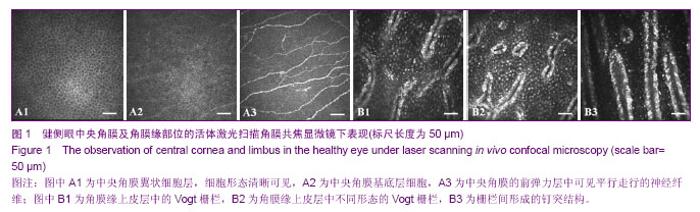
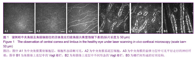
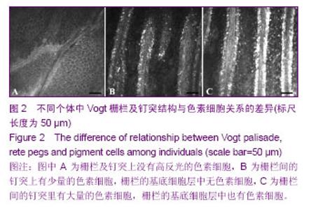
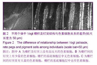
2.1 活体激光扫描角膜共焦显微镜检查 共有24例患者行活体激光扫描角膜共焦显微镜检查,男19例,平均年龄(36.00±12.24)岁;女5例,平均年龄(38.60±5.41)岁。 健侧眼在活体激光扫描角膜共焦显微镜下的表现:中央角膜上皮层各层细胞形态清晰可见,前弹力层可见平行走行的神经纤维(图1 A1-A3);角膜缘上皮层可见乳头状突起的Vogt栅栏及栅栏间形成的钉突结构(图1 B1-B3)。 Vogt栅栏在不同个体间差异很大,形态上多种多样:指状,分支状,圆形及椭圆形等形状;Vogt栅栏及钉突结构与色素细胞的关系也有很大差异:栅栏及钉突上没有高反光的色素细胞(图2A),栅栏间的钉突上有少量的色素细胞,栅栏的基底细胞层中无色素细胞(图2B),栅栏间的钉突里有大量的色素细胞,栅栏的基底细胞层中也有色素细胞(图2C)。 钉突在角膜缘的分布也有不同,在靠近角膜的角膜缘部位很少见到钉突结构,主要集中在角膜缘中间部及近结膜侧角膜缘;角膜缘中间部钉突数量为(5.98±1.92)个/图,近结膜侧角膜缘钉突数量为(3.80±1.10)个/图,其钉突数量低于角膜缘中间部(P < 0.05)。"
"
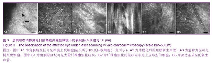
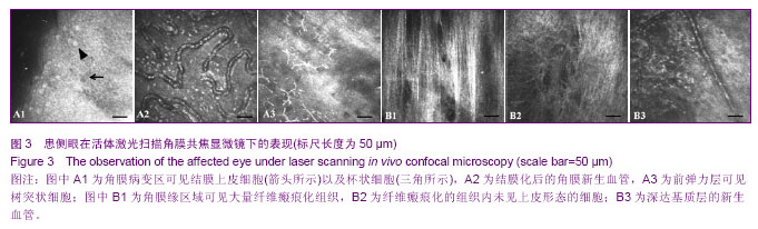
患侧眼在活体激光扫描角膜共焦显微镜下的表现:角膜病变区中典型的上皮细胞形态消失,可见结膜上皮细胞,细胞连接不清晰(图3 A1箭头所示),杯状细胞(图3 A1三角所示),结膜化区域的角膜新生血管(图3A2),前弹力层平行走行的神经纤维消失,上皮层下可见树突状细胞(图3 A3);角膜缘区域Vogt栅栏状结构消失,色素细胞消失,取而代之的是大量纤维瘢痕化组织,角膜缘可见深达基质层的新生血管(图3B1-B3)。"
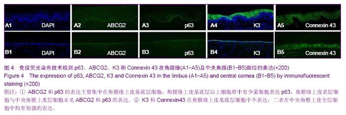
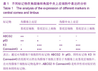
2.2 免疫荧光染色检测p63、ABCG2、K3和Connexin 43的表达 ABCG2和p63的表达主要集中在近结膜侧角膜缘及角膜缘中间部的上皮基底层细胞,角膜缘上皮基底层以上细胞中可见有少量细胞表达p63,角膜中央上皮细胞未见ABCG2和p63的表达(图4);K3和Connexin43在角膜缘上皮基底层以上细胞群中有很强表达,在角膜缘上皮基底层细胞不表达,尤其是Connexin43只在角膜缘基底细胞层的部分细胞中不表达,二者在中央角膜上皮全层细胞中均有很强的表达(图4)。表1为不同标记物在角膜缘及中央角膜上皮层表达结果的强弱分析。"


"
| [1] Davanger M, Evensen A. Role of the pericorneal papillary structure in renewal of corneal epithelium. Nature. 1971; 229(5286):560-561.[2] Schermer A, Galvin S, Sun TT. Differentiation-related expression of a major 64K corneal keratin in vivo and in culture suggests limbal location of corneal epithelial stem cells. J Cell Biol. 1986;103(1):49-62.[3] Chen Z, de Paiva CS, Luo L,et al. Characterization of putative stem cell phenotype in human limbal epithelia.Stem Cells. 2004;22(3):355-366.[4] Majo F, Rochat A, Nicolas M,et al. Oligopotent stem cells are distributed throughout the mammalian ocular surface.Nature. 2008;456(7219):250-254. [5] Sindt CW, Lay B, Bouchard H, et al. Rapid image evaluation system for corneal in vivo confocal microscopy.Cornea. 2013; 32(4):460-465. [6] Hamrah P, Cruzat A, Dastjerdi MH,et al. Unilateral herpes zoster ophthalmicus results in bilateral corneal nerve alteration: an in vivo confocal microscopy study. Ophthalmology. 2013;120(1):40-47.[7] Ledbetter EC, Scarlett JM.In vivo confocal microscopy of the normal equine cornea and limbus.Vet Ophthalmol. 2009;12 Suppl 1:57-64.[8] Priya CG, Prasad T, Prajna NV,et al.Identification of human corneal epithelial stem cells on the basis of high ABCG2 expression combined with a large N/C ratio.Microsc Res Tech. 2013;76(3):242-248.[9] Lim MN, Umapathy T, Baharuddin PJ,et al. Characterization and safety assessment of bioengineered limbal epithelium. Med J Malaysia. 2011;66(4):335-341.[10] Dobrowolski D, Wylega?a E, Orzechowska-Wylega?a B,et al. Culture morphology of the autologous cultivated corneal epithelium.Klin Oczna. 2011;113(7-9):249-253.[11] Turuwhenua JT, Patel DV, McGhee CN. Fully automated montaging of laser scanning in vivo confocal microscopy images of the human corneal subbasal nerve plexus.Invest Ophthalmol Vis Sci. 2012;53(4):2235-2242. [12] Misra S, Craig JP, McGhee CN,et al. Interocular comparison by in vivo confocal microscopy of the 2-dimensional architecture of the normal human corneal subbasal nerve plexus.Cornea. 2012;31(12):1376-1380.[13] Patel DV, Sherwin T, McGhee CN. Laser scanning in vivo confocal microscopy of the normal human corneoscleral limbus. Invest Ophthalmol Vis Sci. 2006;47(7):2823- 2827.[14] Zarei-Ghanavati S, Ramirez-Miranda A, Deng SX. Limbal lacuna: a novel limbal structure detected by in vivo laser scanning confocal microscopy. Ophthalmic Surg Lasers Imaging. 2011;42 Online:e129-131.[15] Petroll WM, Weaver M, Vaidya S,et al. Quantitative 3-dimensional corneal imaging in vivo using a modified HRT-RCM confocal microscope.Cornea. 2013;32(4):e36-43.[16] Salvetat ML, Zeppieri M, Miani F, et al. Comparison between laser scanning in vivo confocal microscopy and noncontact specular microscopy in assessing corneal endothelial cell density and central corneal thickness.Cornea. 2011;30(7): 754-759.[17] Bochert R, Zhivov A, Kraak R,et al. Contribution to comprehension of image formation in confocal microscopy of cornea with Rostock cornea module.Br J Ophthalmol. 2005; 89(10):1351-1355.[18] Shortt AJ, Secker GA, Munro PM,et al. Characterization of the limbal epithelial stem cell niche: novel imaging techniques permit in vivo observation and targeted biopsy of limbal epithelial stem cells.Stem Cells. 2007;25(6):1402-1409.[19] Falke K, Prakasam RK, Guthoff RF,et al. In vivo imaging of limbal epithelium and palisades of Vogt. Klin Monbl Augenheilkd. 2012;229(12):1185-1190.[20] Zhu WQ, Xu JJ, Sun XH,et al. The ocular surface of severe alkali burns patients on confocal microscopy. Zhonghua Yan Ke Za Zhi. 2010;46(1):18-24.[21] Miri A, Alomar T, Nubile M,et al. In vivo confocal microscopic findings in patients with limbal stem cell deficiency.Br J Ophthalmol. 2012;96(4):523-529.[22] Sejpal K, Bakhtiari P, Deng SX. Presentation, diagnosis and management of limbal stem cell deficiency. Middle East Afr J Ophthalmol. 2013;20(1):5-10.[23] Nubile M, Lanzini M, Miri A,et al. In vivo confocal microscopy in diagnosis of limbal stem cell deficiency. Am J Ophthalmol. 2013;155(2):220-232. [24] Lagali N, Edén U, Utheim TP,et al. In vivo morphology of the limbal palisades of vogt correlates with progressive stem cell deficiency in aniridia-related keratopathy. Invest Ophthalmol Vis Sci. 2013;54(8):5333-5342.[25] Ledbetter EC, Scarlett JM. In vivo confocal microscopy of the normal equine cornea and limbus.Vet Ophthalmol. 2009;12 Suppl 1:57-64. [26] Zarei-Ghanavati S, Ramirez-Miranda A, Deng SX. Limbal lacuna: a novel limbal structure detected by in vivo laser scanning confocal microscopy. Ophthalmic Surg Lasers Imaging. 2011;42 Online:e129-131.[27] Fatima S, Zhou S, Sorrentino BP. Abcg2 expression marks tissue-specific stem cells in multiple organs in a mouse progeny tracking model.Stem Cells. 2012;30(2):210-221.[28] de Paiva CS, Chen Z, Corrales RM,et al. ABCG2 transporter identifies a population of clonogenic human limbal epithelial cells.Stem Cells. 2005;23(1):63-73.[29] Budak MT, Alpdogan OS, Zhou M,et al. Ocular surface epithelia contain ABCG2-dependent side population cells exhibiting features associated with stem cells.J Cell Sci. 2005; 118(Pt 8):1715-1724.[30] Chen W, Hara K, Tian Q,et al. Existence of small slow-cycling Langerhans cells in the limbal basal epithelium that express ABCG2. Exp Eye Res. 2007;84(4):626-634.[31] Cancino GI, Yiu AP, Fatt MP,et al. p63 Regulates adult neural precursor and newly born neuron survival to control hippocampal-dependent Behavior. J Neurosci. 2013;33(31): 12569-12585. [32] Pignon JC, Grisanzio C, Geng Y,et al. p63-expressing cells are the stem cells of developing prostate, bladder, and colorectal epithelia. Proc Natl Acad Sci U S A. 2013;110(20): 8105-8110.[33] Nubile M, Curcio C, Dua HS,et al. Pathological changes of the anatomical structure and markers of the limbal stem cell niche due to inflammation. Mol Vis. 2013;19:516-525.[34] Loureiro RR, Cristovam PC, Martins CM,et al. Comparison of culture media for ex vivo cultivation of limbal epithelial progenitor cells. Mol Vis. 2013;19:69-77.[35] Ueno H, Ferrari G, Hattori T,et al. Dependence of corneal stem/progenitor cells on ocular surface innervation.Invest Ophthalmol Vis Sci. 2012;53(2):867-872.[36] Shahdadfar A, Haug K, Pathak M, et al. Ex vivo expanded autologous limbal epithelial cells on amniotic membrane using a culture medium with human serum as single supplement. Exp Eye Res. 2012;97(1):1-9. [37] Márquez-Rosado L, Solan JL, Dunn CA,et al. Connexin43 phosphorylation in brain, cardiac, endothelial and epithelial tissues.Biochim Biophys Acta. 2012;1818(8):1985-1992.[38] Kameritsch P, Pogoda K, Pohl U.Channel-independent influence of connexin 43 on cell migration.Biochim Biophys Acta. 2012;1818(8):1993-2001. [39] Matic M, Evans WH, Brink PR,et al. Epidermal stem cells do not communicate through gap junctions.J Invest Dermatol. 2002;118(1):110-116.[40] Grueterich M, Espana E, Tseng SC. Connexin 43 expression and proliferation of human limbal epithelium on intact and denuded amniotic membrane. Invest Ophthalmol Vis Sci. 2002;43(1):63-71.[41] Chen Z, Evans WH, Pflugfelder SC,et al. Gap junction protein connexin 43 serves as a negative marker for a stem cell-containing population of human limbal epithelial cells. Stem Cells. 2006;24(5):1265-1273. |
| [1] | Lin Qingfan, Xie Yixin, Chen Wanqing, Ye Zhenzhong, Chen Youfang. Human placenta-derived mesenchymal stem cell conditioned medium can upregulate BeWo cell viability and zonula occludens expression under hypoxia [J]. Chinese Journal of Tissue Engineering Research, 2021, 25(在线): 4970-4975. |
| [2] | Pu Rui, Chen Ziyang, Yuan Lingyan. Characteristics and effects of exosomes from different cell sources in cardioprotection [J]. Chinese Journal of Tissue Engineering Research, 2021, 25(在线): 1-. |
| [3] | Zhang Xiumei, Zhai Yunkai, Zhao Jie, Zhao Meng. Research hotspots of organoid models in recent 10 years: a search in domestic and foreign databases [J]. Chinese Journal of Tissue Engineering Research, 2021, 25(8): 1249-1255. |
| [4] | Hou Jingying, Yu Menglei, Guo Tianzhu, Long Huibao, Wu Hao. Hypoxia preconditioning promotes bone marrow mesenchymal stem cells survival and vascularization through the activation of HIF-1α/MALAT1/VEGFA pathway [J]. Chinese Journal of Tissue Engineering Research, 2021, 25(7): 985-990. |
| [5] | Shi Yangyang, Qin Yingfei, Wu Fuling, He Xiao, Zhang Xuejing. Pretreatment of placental mesenchymal stem cells to prevent bronchiolitis in mice [J]. Chinese Journal of Tissue Engineering Research, 2021, 25(7): 991-995. |
| [6] | Liang Xueqi, Guo Lijiao, Chen Hejie, Wu Jie, Sun Yaqi, Xing Zhikun, Zou Hailiang, Chen Xueling, Wu Xiangwei. Alveolar echinococcosis protoscolices inhibits the differentiation of bone marrow mesenchymal stem cells into fibroblasts [J]. Chinese Journal of Tissue Engineering Research, 2021, 25(7): 996-1001. |
| [7] | Fan Quanbao, Luo Huina, Wang Bingyun, Chen Shengfeng, Cui Lianxu, Jiang Wenkang, Zhao Mingming, Wang Jingjing, Luo Dongzhang, Chen Zhisheng, Bai Yinshan, Liu Canying, Zhang Hui. Biological characteristics of canine adipose-derived mesenchymal stem cells cultured in hypoxia [J]. Chinese Journal of Tissue Engineering Research, 2021, 25(7): 1002-1007. |
| [8] | Geng Yao, Yin Zhiliang, Li Xingping, Xiao Dongqin, Hou Weiguang. Role of hsa-miRNA-223-3p in regulating osteogenic differentiation of human bone marrow mesenchymal stem cells [J]. Chinese Journal of Tissue Engineering Research, 2021, 25(7): 1008-1013. |
| [9] | Lun Zhigang, Jin Jing, Wang Tianyan, Li Aimin. Effect of peroxiredoxin 6 on proliferation and differentiation of bone marrow mesenchymal stem cells into neural lineage in vitro [J]. Chinese Journal of Tissue Engineering Research, 2021, 25(7): 1014-1018. |
| [10] | Zhu Xuefen, Huang Cheng, Ding Jian, Dai Yongping, Liu Yuanbing, Le Lixiang, Wang Liangliang, Yang Jiandong. Mechanism of bone marrow mesenchymal stem cells differentiation into functional neurons induced by glial cell line derived neurotrophic factor [J]. Chinese Journal of Tissue Engineering Research, 2021, 25(7): 1019-1025. |
| [11] | Duan Liyun, Cao Xiaocang. Human placenta mesenchymal stem cells-derived extracellular vesicles regulate collagen deposition in intestinal mucosa of mice with colitis [J]. Chinese Journal of Tissue Engineering Research, 2021, 25(7): 1026-1031. |
| [12] | Pei Lili, Sun Guicai, Wang Di. Salvianolic acid B inhibits oxidative damage of bone marrow mesenchymal stem cells and promotes differentiation into cardiomyocytes [J]. Chinese Journal of Tissue Engineering Research, 2021, 25(7): 1032-1036. |
| [13] | Guan Qian, Luan Zuo, Ye Dou, Yang Yinxiang, Wang Zhaoyan, Wang Qian, Yao Ruiqin. Morphological changes in human oligodendrocyte progenitor cells during passage [J]. Chinese Journal of Tissue Engineering Research, 2021, 25(7): 1045-1049. |
| [14] | Wang Zhengdong, Huang Na, Chen Jingxian, Zheng Zuobing, Hu Xinyu, Li Mei, Su Xiao, Su Xuesen, Yan Nan. Inhibitory effects of sodium butyrate on microglial activation and expression of inflammatory factors induced by fluorosis [J]. Chinese Journal of Tissue Engineering Research, 2021, 25(7): 1075-1080. |
| [15] | Wang Xianyao, Guan Yalin, Liu Zhongshan. Strategies for improving the therapeutic efficacy of mesenchymal stem cells in the treatment of nonhealing wounds [J]. Chinese Journal of Tissue Engineering Research, 2021, 25(7): 1081-1087. |
| Viewed | ||||||
|
Full text |
|
|||||
|
Abstract |
|
|||||