Chinese Journal of Tissue Engineering Research
Previous Articles Next Articles
Cytocompatibility of calcium metaphosphate nanoparticles
Wu Yue-heng1, Mai Li-ping1, Chen Peng2, Zhang Ling-min2, Huang Huan-lei1, Zeng Xiang-jun1, Tang Shun-qing2, Xiao Xue-jun1, Yu Xi-yong1
- 1Medical Research Center of Guangdong General Hospital, Guangdong Provincial Cardiovascular Institute, Guangdong Academy of Medical Science, Guangzhou 510100, Guangdong Province, China; 2Institute of Biomedical Engineering, Jinan University, Guangzhou 510632, Guangdong Province, China
-
Received:2013-01-22Revised:2013-06-25Online:2013-09-17Published:2013-09-17 -
Contact:Yu Xi-yong, Professor, Doctoral supervisor, Medical Research Center of Guangdong General Hospital, Guangdong Provincial Cardiovascular Institute, Guangdong Academy of Medical Science, Guangzhou 510100, Guangdong Province, China yuxycn@hotmail.com -
About author:Wu Yue-heng★, Master, Assistant researcher, Medical Research Center of Guangdong General Hospital, Guangdong Provincial Cardiovascular Institute, Guangdong Academy of Medical Science, Guangzhou 510100, Guangdong Province, China edgar_wu@yahoo.com.cn -
Supported by:the National Natural Science Foundation of China, No. 30901468*
CLC Number:
Cite this article
Wu Yue-heng1, Mai Li-ping1, Chen Peng2, Zhang Ling-min, Huang Huan-lei, Zeng Xiang-jun,Tang Shun-qing, Xiao Xue-jun, Yu Xi-yong. Cytocompatibility of calcium metaphosphate nanoparticles[J]. Chinese Journal of Tissue Engineering Research, doi: 10.3969/j.issn.2095-4344.2013.38.015.
share this article
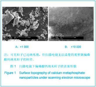
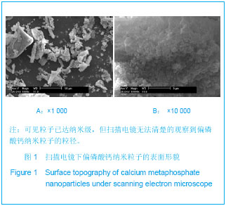
2.1 扫描电镜下偏磷酸钙材料的表面形貌观察 图1为偏磷酸钙纳米粒子的表面形貌,可见粒子已达纳米级,但扫描电镜无法清楚的观察到偏磷酸钙纳米粒子的粒径。"
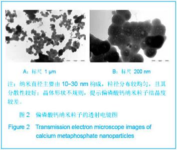
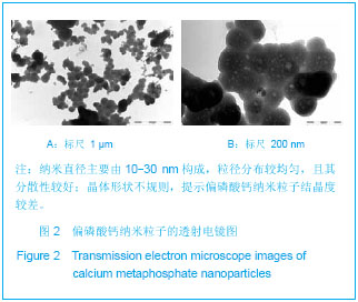
2.2 透射电镜下纳米粒子的表面形貌 图2为偏磷酸钙纳米粒子的透射电镜表面形貌,由图可知,其纳米直径主要由10-30 nm构成,粒径分布较均匀,且其分散性较好;晶体形状不规则,提示偏磷酸钙纳米粒子结晶度较差。"
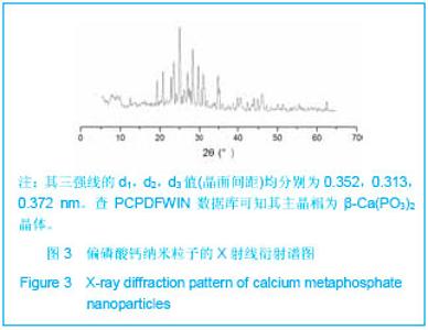
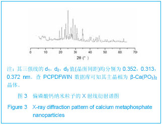
2.3 偏磷酸钙纳米粒子X射线衍射晶相检测结果 样品的X射线衍射图见图3,其三强线的d1,d2,d3值(晶面间距)均分别为0.352,0.313,0.372 nm。查PCPDFWIN数据库可知其主晶相为β-Ca(PO3)2晶体。"
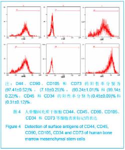
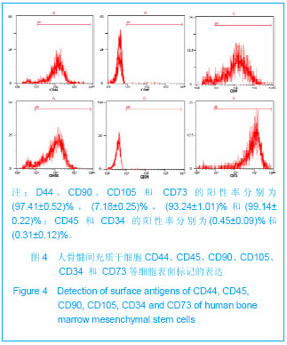
2.4 偏磷酸钙纳米粒子的细胞相容性检测结果 倒置相差显微镜下观察人骨髓间充质干细胞,接种1 d即贴壁,去除悬浮细胞后继续培养3 d贴壁细胞开始增殖。原代细胞呈椭圆型、短梭型、多角型及不规则型等。第3-5代细胞呈均匀一致的长梭型,排列成旋涡状或放射状,细胞生长状态良好。 图4为人骨髓间充质干细胞的流式细胞鉴定结果,CD44、CD90、CD105和 CD73的阳性率分别为(97.41±0.52)%,(7.18±0.25)%,(93.24±1.01)%和(99.14±0.22)%;CD45和CD34的阳性率分别为(0.45±0.09)%和(0.31±0.12)%。"
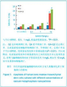
纳米粒子的细胞毒性检测主要考察了偏磷酸钙纳米粒子对人骨髓间充质干细胞凋亡和细胞周期的影响。图5是人骨髓间充质干细胞分别在含高(10 mg/L)、中 (1 mg/L)和低(0.1 mg/L)质量浓度纳米粒子的培养基中培养7 d后,细胞凋亡的测量结果。"
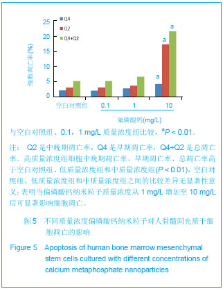
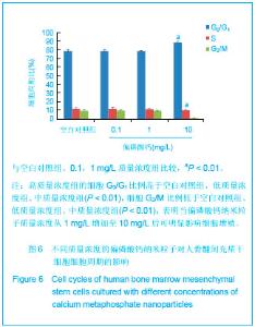
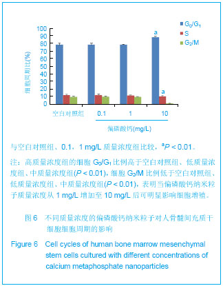
对图5的数据进行方差分析发现,高、中、低质量浓度组及空白对照组间的细胞中晚期凋亡率、早期凋亡率和总凋亡率的比较差异有显著性意义(P < 0.01)。进一步采用S-N-K检验对高、中、低质量浓度组及空白对照组之间分别进行两两比较,结果发现高质量浓度组细胞中晚期凋亡率、早期凋亡率和总凋亡率与空白对照组、低质量浓度组和中质量浓度组之间的比较差异有显著性意义(P < 0.01),空白对照组、低质量浓度组和中质量浓度组之间比较差异无显著性意义。 图6是人骨髓间充质干细胞分别在含高(10 mg/L)、中(1 mg/L) 和低(0.1 mg/L)质量浓度偏磷酸钙纳米粒子的培养基中培养7 d后,细胞周期的测量结果。对图6的数据进行方差分析发现,高、中、低质量浓度组及空白对照组的细胞G0/G1和G2/M期比例比较差异有显著性意义(P < 0.01),高、中、低质量浓度组及空白对照组细胞S期比例的比较差异无显著性意义(P=0.503)。进一步采用S-N-K检验对高、中、低质量浓度组及空白对照组之间细胞G0/G1、G2/M期的比例分别进行两两比较,结果发现,高质量浓度组与空白对照组、低质量浓度组、中质量浓度组之间的细胞G0/G1比例比较差异有显著性意义(P < 0.01),空白对照组、低质量浓度组和中质量浓度组两两之间的G0/G1比例比较差异无显著性意义。高质量浓度组细胞G2/M比例与空白对照组、低质量浓度组、中质量浓度组比较差异有显著性意义(P < 0.01),空白对照组、低质量浓度组和中质量浓度组两两之间的细胞G2/M比例比较差异无显著性意义。"
| [1] Malvindi MA,Brunetti V,Vecchio G,et al.SiO2 nanoparticles biocompatibility and their potential for gene delivery and silencing. Nanoscale.2012;4(2):486-495. [2] Biradar S,Ravichandran P,Gopikrishnan R,et al.Calcium carbonate nanoparticles: synthesis, characterization and biocompatibility.J Nanosci Nanotechnol 2011;11(8): 6868-6874. [3] Tang F,Li L,Chen D.Mesoporous silica nanoparticles: synthesis, biocompatibility and drug delivery.Adv Mater.2012; 24(12):1504-1534. [4] Alarcon EI,Udekwu K,Skog M,et al.The biocompatibility and antibacterial properties of collagen-stabilized,photochemically prepared silver nanoparticles. Biomaterials.2012; 33(19): 4947-4956. [5] Huang X,Zhuang J,Teng X,et al.The promotion of human malignant melanoma growth by mesoporous silica nanoparticles through decreased reactive oxygen species. Biomaterials.2010;31(24):6142-6153. [6] Singh S,Dosani T,Karakoti AS,et al.A phosphate-dependent shift in redox state of cerium oxide nanoparticles and its effects on catalytic properties. Biomaterials.2011;32(28): 6745-6753. [7] Ladewig K,Niebert M,Xu ZP,et al.Efficient siRNA delivery to mammalian cells using layered double hydroxide nanoparticles. Biomaterials.2010;31(7): 821-1829. [8] Labouta HI,Schneider M.Interaction of inorganic nanoparticles with the skin barrier: current status and critical review.Nanomedicine.2013 ;9(1):39-54. [9] Fan K,Cao C,Pan Y,et al.Magnetoferritin nanoparticles for targeting and visualizing tumour tissues.Nat Nanotechnol. 2012;7(7):459-464. [10] Huang HC,Barua S,Sharma G,et al.Inorganic nanoparticles for cancer imaging and therapy.J Control Release.2011; 155(3):344-357. [11] Son SJ,Bai X,Lee SB.Inorganic hollow nanoparticles and nanotubes in nanomedicine Part 1. Drug/gene delivery applications.Drug Discov Today. 2007;12(15-16):650-656. [12] Geidel C,Schmachtel S,Riedinger A,et al.A general synthetic approach for obtaining cationic and anionic inorganic nanoparticles via encapsulation in amphiphilic copolymers. Small.2011;7(20):2929-2934. [13] Pittella F,Zhang M,Lee Y,et al.Enhanced endosomal escape of siRNA-incorporating hybrid nanoparticles from calcium phosphate and PEG-block charge-conversional polymer for efficient gene knockdown with negligible cytotoxicity. Biomaterials.2011;32(11):3106-3114. [14] Yang X,Walboomers XF,van den Dolder J,et al.Non-viral bone morphogenetic protein 2 transfection of rat dental pulp stem cells using calcium phosphate nanoparticles as carriers. Tissue Eng Part A.2008;14(1):71-81. [15] Park EK,Lee YE,Choi JY,et al.Cellular biocompatibility and stimulatory effects of calcium metaphosphate on osteoblastic differentiation of human bone marrow-derived stromal cells.Biomaterials.2004;25(17):3403-3411. [16] Lee YM,Seol YJ,Lim YT,et al.Tissue-engineered growth of bone by marrow cell transplantation using porous calcium metaphosphate matrices.J Biomed Mater Res.2001;54(2) : 216-223. [17] Cho IH,Lee JH,Song YG,et al.Evaluation on the efficacy and safety of calcium metaphosphate coated fixture.J Adv Prosthodont. 2013;5(2):172-178. [18] 江燕萍,吴岳恒,成安衡.多孔偏磷酸钙生物材料的体外加速降解实验[J].中国组织工程研究与临床康复,2008,12(29): 3663-3666. [19] 岳垂源,唐文洁,戴云,等.人骨髓间质干细胞与新型可降解材料生物相容性的实验研究[J].中国病理生理杂志,2005,21(7):1378-1383. [20] 吴岳恒,汤顺清,毛萱,等.组织工程支架材料偏磷酸钙玻璃陶瓷的多孔性能[J]. 中国组织工程研究与临床康复,2007,11(1): 130-132. [21] Lu LX,Zhang XF,Wang YY,et al.Effects of hydroxyapatite-containing composite nanofibers on osteogenesis of mesenchymal stem cells in vitro and bone regeneration in vivo.ACS Appl Mater Inter.2013; 5(2): 319-330. [22] Ning Y,Wei T,Defu C,et al.The research of degradability of a novel biodegradable coralline hydroxyapatite after implanted into rabbit.J Biomed Mater Res B Appl Biomater.2009;88(3): 741-746. [23] Wetherall KM,Pickup DM,Newport RJ,et al.The structure of calcium metaphosphate glass obtained from x-ray and neutron diffraction and reverse Monte Carlo modelling.J Phys Condens Matter.2009;21(3):035109. [24] Kasuga T,Ota Y,Nogami M,et al.Surface modification of calcium metaphosphate fibers. J Mater Sci Mat Med.2000; 11(4):223-225. [25] Salah N,Habib SS,Khan ZH,et al.High-energy ball milling technique for ZnO nanoparticles as antibacterial material.Int J Nanomed.2011;6:863-869. [26] Maksimenko A,Mougin J,Mura S,et al.Polyisoprenoyl gemcitabine conjugates self assemble as nanoparticles, useful for cancer therapy. Cancer Lett.2013;334(2):346-353. [27] Bjerre L,Bunger C,Baatrup A,et al.Flow perfusion culture of human mesenchymal stem cells on coralline hydroxyapatite scaffolds with various pore sizes.J Biomed Mater Res A.2011; 97(3):251-263. [28] Kim JH,Park JS,Yang HN,et al. The use of biodegradable PLGA nanoparticles to mediate SOX9 gene delivery in human mesenchymal stem cells (hMSCs) and induce chondrogenesis. Biomaterials.2011;32(1):268-278. [29] Greulich C,Kittler S,Epple M,et al.Studies on the biocompatibility and the interaction of silver nanoparticles with human mesenchymal stem cells (hMSCs). Langenbecks Arch Surg. 2009;394(3):495-502. [30] Shuang-zhi H,Ping S,Xi-ning P.Culture and identification of human amniotic mesenchymal stem cells. Chin Med Sci J. 2010;25(4):211-214. [31] Xiao Y,Chen J.Proteomics approaches in the identification of molecular signatures of mesenchymal stem cells.Adv Biochem Eng Biotechnol. 2013;129:153-176. [32] Ranera B,Lyahyai J,Romero A,et al.Immunophenotype and gene expression profiles of cell surface markers of mesenchymal stem cells derived from equine bone marrow and adipose tissue.Vet Immunol Immunopathol. 2011; 144(1-2):147-154. [33] Qiu J.Nano-safety studies urged in China.Nature.2012; 489(7416):350. [34] Monem AS,ElBatal HA,Khalil EM,et al.In vivo behavior of bioactive phosphate glass-ceramics from the system P2O5-Na2O-CaO containing TiO2.J Mater Sci Mater Med. 2008;19(3):1097-1108. [35] Hee Soon C,Park SY,Kim S,et al.Effect of different bone substitutes on the concentration of growth factors in platelet-rich plasma.J Biomater Appl.2008;22(6):545-557. [36] Shundo C,Zhang H,Nakanishi T,et al.Cytotoxicity evaluation of magnetite (Fe3O4) nanoparticles in mouse embryonic stem cells.Colloids Surf B Biointerfaces.2012;97:221-225. [37] Park KS,Tae J,Choi B,et al.Characterization, in vitro cytotoxicity assessment,and in vivo visualization of multimodal, RITC-labeled, silica-coated magnetic nanoparticles for labeling human cord blood-derived mesenchymal stem cells.Nanomedicine.2010;6(2):263-276. [38] Tang ZB,Cao JK,Wen N,et al.Posterolateral spinal fusion with nano-hydroxyapatite-collagen/PLA composite and autologous adipose-derived mesenchymal stem cells in a rabbit model.J Tissue Eng Regen Med.2012;6(4):325-336. [39] Phipps MC,Clem WC,Grunda JM,et al.Increasing the pore sizes of bone-mimetic electrospun scaffolds comprised of polycaprolactone, collagen I and hydroxyapatite to enhance cell infiltration.Biomaterials.2012; 33(2):524-534. [40] Dickinson LE,Kusuma S,Gerecht S.Reconstructing the differentiation niche of embryonic stem cells using biomaterials.Macromol Biosci. 2011;11(1):36-49. |
| [1] | Chen Song, He Yuanli, Xie Wenjia, Zhong Linna, Wang Jian. Advantages of calcium phosphate nanoparticles for drug delivery in bone tissue engineering research and application [J]. Chinese Journal of Tissue Engineering Research, 2021, 25(22): 3565-3570. |
| [2] | Gan Zhoujie, Pei Xibo. Enzyme-responsive nanoparticles in tumor therapy: superiority of nanoparticles in accumulation and drug release [J]. Chinese Journal of Tissue Engineering Research, 2021, 25(16): 2562-2568. |
| [3] | Li Xuan, Lu Min, Li Mingxing, Ao Meng, Tang Linmei, Zeng Zhen, Hu Jingwei, Huang Zhiqiang, Xuan Jiqing. In vitro multi-modal imaging of magnetic targeted nanoparticles and their targeting effect on hepatic stellate cells [J]. Chinese Journal of Tissue Engineering Research, 2020, 24(4): 566-571. |
| [4] | Tang Mengmeng, Chen Hechun, Xie Hongchen, Zhang Yu, Tan Xiaoshuang, Sun Yixuan, Huang Yina. Histocompatibility of poly(L-lactide-co-ε-caprolactone)/cross-linked polyvinylpyrrolidone ureteral stent grafted into the rat bladder [J]. Chinese Journal of Tissue Engineering Research, 2020, 24(4): 583-588. |
| [5] | Xiang Haibin, Li Xinxia, Liang Qiuzhen, Song Xinghua. Specific bone-targeting nanoscale drug delivery system: advantages and clinical applicability [J]. Chinese Journal of Tissue Engineering Research, 2020, 24(4): 612-618. |
| [6] | Lei Senlin, Liu Hongyuan, Yang Hongsheng, Xiong Yan, Duan Hong. In vivo biosafety and histocompatibility of absorbable poly-D,L-lactic acid screws implanted with ultrasound-assisted technology [J]. Chinese Journal of Tissue Engineering Research, 2020, 24(34): 5454-5460. |
| [7] | Guo Enhui, Xu Zitong, Liang Yize, Zhou Liang, Lu Zhaoxiang, You Liang, Xia Yujun. Properties of a novel photocrosslinked fish collagen peptide-hyaluronic acid hydrogel [J]. Chinese Journal of Tissue Engineering Research, 2020, 24(28): 4518-4525. |
| [8] | Gao Jianbo, Xia Bing, Li Shengyou, Yang Yujie, Ma Teng, Yu Peng, Luo Zhuojing, Huang Jinghui. Effect of nanoparticles carrying chondroitin sulfate ABC on the migration of Schwann cells in a magnetic field [J]. Chinese Journal of Tissue Engineering Research, 2020, 24(28): 4526-4532. |
| [9] | Zhang Zhongyan, Li Yubo, Qi Tongning, Chang Tao. Three-dimensional printed polylactic acid resin humerus combined with bioactive coating promotes osteoblast adhesion and increases antibacterial ability [J]. Chinese Journal of Tissue Engineering Research, 2020, 24(16): 2485-2492. |
| [10] | Cheng Lei, Jin Jian, Hu Jingguo, Lu Yusong. Inflammatory reaction and lactic acid concentration after implantation of polylactic acid rib nail versus pure titanium embracing fixator in animals#br# [J]. Chinese Journal of Tissue Engineering Research, 2020, 24(16): 2567-2571. |
| [11] | Yuan Bo, Wang Zhiwei, Tang Yifan, Zhou Shengyuan, Chen Xiongsheng, Jia Lianshun. Construction of polycaprolactone-tricalcium phosphate with different mixture ratios using three-dimensional printing technology and its osteoinductivity in vitro [J]. Chinese Journal of Tissue Engineering Research, 2019, 23(6): 821-826. |
| [12] | Wang Xuefeng, Shang Xifu . Curative effects of three filling materials in the treatment of osteoporotic thoracolumbar fractures [J]. Chinese Journal of Tissue Engineering Research, 2019, 23(6): 863-869. |
| [13] | Liu Dan, Min Changqin, Lu Shuai, Chen Yue, Sun Yong. Osseointegration induced by beta-tricalcium phosphate loaded with advanced platelet-rich fibrin [J]. Chinese Journal of Tissue Engineering Research, 2019, 23(6): 888-893. |
| [14] | Cheng Jian, Zhang Jun, Guan Jie, Zeng Junkai, Zhao Xin, Xie Youzhuan. Silver nanoparticle-doped tricalcium phosphate: in vitro and in vivo toxicity in rabbits [J]. Chinese Journal of Tissue Engineering Research, 2019, 23(6): 917-923. |
| [15] | Tian Hongju, Chen Zhongqing. Poly(lactid-co-glycolide) nanoparticles loaded with ropivacaine: preparation and in vivo release in animals [J]. Chinese Journal of Tissue Engineering Research, 2019, 23(6): 924-929. |
| Viewed | ||||||
|
Full text |
|
|||||
|
Abstract |
|
|||||