Chinese Journal of Tissue Engineering Research ›› 2024, Vol. 28 ›› Issue (26): 4178-4183.doi: 10.12307/2024.434
Previous Articles Next Articles
Deficiency of interleukin-9 inhibits osteogenic potential in mice
Wang Yi, Chen Chichi, Zhou Xichao, Shi Qin
- Department of Orthopedics, the First Affiliated Hospital of Soochow University, Institute of Orthopedics of Soochow University, Suzhou 215006, Jiangsu Province, China
-
Received:2023-06-10Accepted:2023-08-01Online:2024-09-18Published:2023-09-28 -
Contact:Shi Qin, MD, Professor, Department of Orthopedics, the First Affiliated Hospital of Soochow University, Institute of Orthopedics of Soochow University, Suzhou 215006, Jiangsu Province, China -
About author:Wang Yi, Department of Orthopedics, the First Affiliated Hospital of Soochow University, Institute of Orthopedics of Soochow University, Suzhou 215006, Jiangsu Province, China -
Supported by:the National Natural Science Foundation of China (General Program), No. 81972059 (to SQ)
CLC Number:
Cite this article
Wang Yi, Chen Chichi, Zhou Xichao, Shi Qin. Deficiency of interleukin-9 inhibits osteogenic potential in mice[J]. Chinese Journal of Tissue Engineering Research, 2024, 28(26): 4178-4183.
share this article
Add to citation manager EndNote|Reference Manager|ProCite|BibTeX|RefWorks

2.1 IL-9-/-小鼠的鉴定 剪取鼠尾组织提取小鼠基因组DNA,使用DNA扩增技术对目的基因(IL-9)进行扩增,根据WT和IL-9-/-小鼠的IL-9基因编码长度不同,最后通过琼脂糖凝胶核酸电泳区分出WT和IL-9-/-小鼠。WT小鼠中IL-9基因的DNA大小为804 bp,IL-9-/-小鼠中IL-9基因的DNA大小为1 118 bp。基因型鉴定结果见图1。"

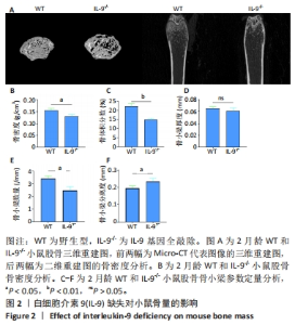
2.2 2月龄WT和IL-9-/-小鼠股骨Micro-CT分析结果 为了探究IL-9缺失对于小鼠骨量的影响,对2月龄WT和IL-9-/-小鼠股骨进行Micro-CT扫描分析。通过小鼠股骨Micro-CT的三维重建图,发现IL-9-/-小鼠骨量明显低于WT小鼠,骨小梁间隙明显增大(图2A),骨密度明显降低(图2B),骨小梁相关参数分析显示,IL-9-/-小鼠骨体积分数、骨小梁数目明显下降,骨小梁分离度明显增大(图2C-F),差异有显著性意义。综上所述,IL-9的缺失导致小鼠出现自发性骨质疏松,骨量显著降低。"

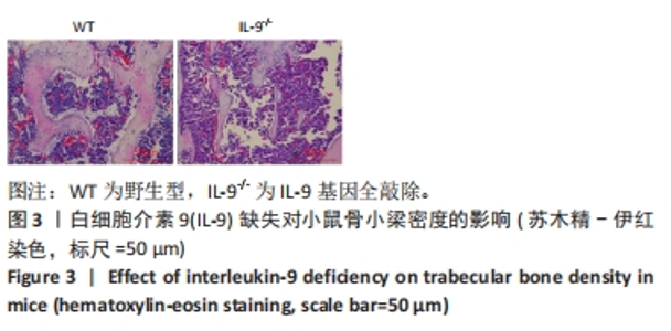
2.3 WT和IL-9-/-小鼠股骨苏木精-伊红染色结果 为了进一步验证IL-9缺失对于小鼠骨量的影响,对WT和IL-9-/-小鼠股骨切片进行苏木精-伊红染色。结果显示,IL-9-/-小鼠骨小梁密度明显降低(图3),这与前面Micro-CT数据得出的结论一致。"

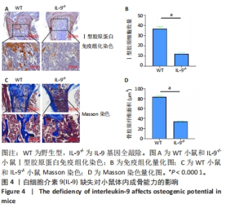
2.4 IL-9-/-小鼠股骨成骨能力减弱 骨重塑是一个骨吸收和骨形成动态平衡的过程。一旦骨重塑失衡,骨吸收速度大于骨形成,就会出现骨质疏松。为了进一步探究IL-9-/-小鼠骨量降低的原因,对WT和IL-9-/-小鼠股骨切片分别进行Ⅰ型胶原蛋白和Masson染色,观察其成骨细胞数量是否存在差异。Ⅰ型胶原蛋白免疫组化染色结果显示,IL-9-/-小鼠股骨远端生长板下方区域Ⅰ型胶原蛋白阳性细胞数目低于WT小鼠;而Masson染色结果说明WT小鼠胶原形成能力较IL-9-/-小鼠更强,差异均有显著性意义(图4)。说明IL-9的缺失抑制了小鼠的成骨分化。"

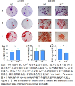
2.5 IL-9的缺失抑制了骨髓间充质干细胞的矿化能力 为了进一步探究IL-9-/-小鼠成骨能力减弱的原因,分别提取了WT和IL-9-/-小鼠原代骨髓细胞进行CFU-F实验。通过结晶紫染色,发现WT和IL-9-/-小鼠骨髓细胞形成的间充质干细胞集落数未见明显差异(图5A)。于是对骨髓细胞进行成骨诱导,检测其成骨分化能力。7 d后,通过碱性磷酸酶染色,发现两者碱性磷酸酶活性未见明显差异(图5B)。21 d后,通过茜素红染色,发现IL-9-/-小鼠来源的原代骨髓间充质干细胞矿化能力明显减弱(图5C)。说明IL-9的缺失显著抑制了骨髓间充质干细胞的矿化能力。"

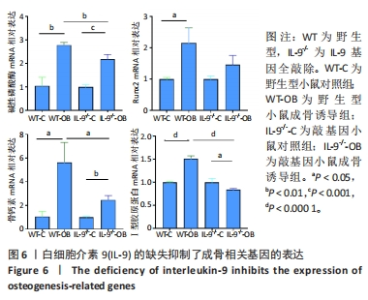
2.6 IL-9的缺失抑制了成骨相关基因的表达 为了进一步探究IL-9对于成骨能力的影响,定量PCR的结果显示,两组小鼠骨髓间充质干细胞经过成骨诱导后,成骨相关基因的表达升高;而野生型小鼠骨髓间充质干细胞的碱性磷酸酶、Runx2、骨钙素和Ⅰ型胶原蛋白表达明显高于IL-9敲基因小鼠,说明IL-9的缺失抑制了小鼠的成骨作用(图6)。"

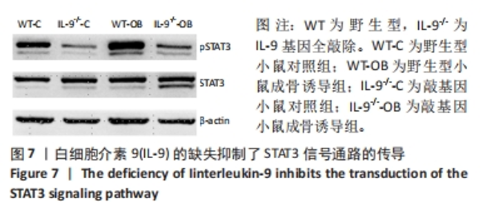
2.7 IL-9的缺失抑制JAK-STAT通路信号传导 为了进一步验证IL-9是否通过JAK-STAT通路进行信号传导,采用Western blot检测STAT3蛋白的表达,结果显示,成骨诱导激活STAT3信号传导,pSTAT3表达明显增多;而IL-9的缺失则抑制此信号通路的传导。说明IL-9通过JAK-STAT3通路调控成骨,与ZHOU等[20]的发现一致(图7)。"

| [1] COMPSTON JE, MCCLUNG MR, LESLIE WD. Osteoporosis. Lancet. 2019;393(10169):364-376. [2] EASTELL R, O’NEILL TW, HOFBAUER LC, et al. Postmenopausal osteoporosis. Nat Rev Dis Primers. 2016;2:16069. [3] Management of Osteoporosis in Postmenopausal Women: The 2021 Position Statement of The North American Menopause Society’’ Editorial Panel. Management of osteoporosis in postmenopausal women: the 2021 position statement of The North American Menopause Society. Menopause. 2021;28(9):973-997. [4] YU F, XIA W. The epidemiology of osteoporosis, associated fragility fractures, and management gap in China. Arch Osteoporos. 2019;14(1):32. [5] ZHU X, BAI W, ZHENG H. Twelve years of GWAS discoveries for osteoporosis and related traits: advances, challenges and applications. Bone Res. 2021;9(1):23. [6] ZEBAZE R, LIBANATI C, MCCLUNG MR, et al. Denosumab Reduces Cortical Porosity of the Proximal Femoral Shaft in Postmenopausal Women With Osteoporosis. J Bone Miner Res. 2016;31(10):1827-1834. [7] CHAVASSIEUX P, CHAPURLAT R, PORTERO-MUZY N, et al. Bone-Forming and Antiresorptive Effects of Romosozumab in Postmenopausal Women With Osteoporosis: Bone Histomorphometry and Microcomputed Tomography Analysis After 2 and 12 Months of Treatment. J Bone Miner Res. 2019;34(9):1597-1608. [8] LOTINUN S, KIVIRANTA R, MATSUBARA T, et al. Osteoclast-specific cathepsin K deletion stimulates S1P-dependent bone formation. J Clin Invest. 2013;123(2):666-681. [9] KRAENZLIN ME, MEIER C. Parathyroid hormone analogues in the treatment of osteoporosis. Nat Rev Endocrinol. 2011;7(11):647-656. [10] REID IR. Short-term and long-term effects of osteoporosis therapies. Nat Rev Endocrinol. 2015; 11(7):418-428. [11] WHITAKER M, GUO J, KEHOE T, et al. Bisphosphonates for osteoporosis--where do we go from here? N Engl J Med. 2012;366(22):2048-2051. [12] FENG X, MCDONALD JM. Disorders of bone remodeling. Annu Rev Pathol. 2011;6:121-145. [13] CHAKRABORTY S, KUBATZKY KF, MITRA DK. An Update on Interleukin-9: From Its Cellular Source and Signal Transduction to Its Role in Immunopathogenesis. Int J Mol Sci. 2019;20(9):2113. [14] GOSWAMI R, KAPLAN MH. A brief history of IL-9. J Immunol. 2011;186(6):3283-3288. [15] NOELLE RJ, NOWAK EC. Cellular sources and immune functions of interleukin-9. Nat Rev Immunol. 2010;10(10):683-687. [16] GENG W, ZHANG W, MA J. IL-9 exhibits elevated expression in osteonecrosis of femoral head patients and promotes cartilage degradation through activation of JAK-STAT signaling in vitro. Int Immunopharmacol. 2018;60:228-234. [17] ZHENG Y, HE Y, XIAO M, et al. Interleukin 9 prevents immune thrombocytopenia in mice via JAK/STAT5 signaling. Exp Cell Res. 2020; 88(1):111801. [18] 邹旺辉,钱楠楠,张萌,等.间充质干细胞临床前研究启示:间充质干细胞的细胞功能与JAK/STAT信号通路的关系[J]. 中国组织工程研究,2022,26(19):3048-3055. [19] SANPAOLO ER, ROTONDO C, CICI D, et al. JAK/STAT pathway and molecular mechanism in bone remodeling. Mol Biol Rep. 2020;47(11):9087-9096. [20] ZHOU S, DAI Q, HUANG X, et al. STAT3 is critical for skeletal development and bone homeostasis by regulating osteogenesis. Nat Commun. 2021;12(1):6891. [21] LEE J, SEONG S, KIM JH, et al. STAT5 is a key transcription factor for IL-3-mediated inhibition of RANKL-induced osteoclastogenesis. Sci Rep. 2016;6:30977. [22] ZHAO Y, XIE L. Unique bone marrow blood vessels couple angiogenesis and osteogenesis in bone homeostasis and diseases. Ann N Y Acad Sci. 2020;1474(1):5-14. [23] DATTA HK, NG WF, WALKER JA, et al. The cell biology of bone metabolism. J Clin Pathol. 2008; 61(5):577-587. [24] KATSIMBRI P. The biology of normal bone remodelling. Eur J Cancer Care (Engl). 2017;26(6). doi: 10.1111/ecc.12740. [25] RAGGATT LJ, PARTRIDGE NC. Cellular and molecular mechanisms of bone remodeling. J Biol Chem. 2010;285(33):25103-25108. [26] SIDDIQUI JA, PARTRIDGE NC. Physiological Bone Remodeling: Systemic Regulation and Growth Factor Involvement. Physiology (Bethesda). 2016;31(3):233-245. [27] BATOON L, MILLARD SM, RAGGATT LJ, et al. Osteal macrophages support osteoclast-mediated resorption and contribute to bone pathology in a postmenopausal osteoporosis mouse model. J Bone Miner Res. 2021; 36(11):2214-2228. [28] MCNAMARA LM. Osteocytes and Estrogen Deficiency. Curr Osteoporos Rep. 2021;19(6):592-603. [29] FöGER-SAMWALD U, KERSCHAN-SCHINDL K, BUTYLINA M, et al. Age Related Osteoporosis: Targeting Cellular Senescence. Int J Mol Sci. 2022;23(5):2701. [30] SCHETT G. Effects of inflammatory and anti-inflammatory cytokines on the bone. Eur J Clin Invest. 2011;41(12):1361-1366. [31] ZUPAN J, JERAS M, MARC J. Osteoimmunology and the influence of pro-inflammatory cytokines on osteoclasts. Biochem Med (Zagreb). 2013;23(1):43-63. [32] RAUBER S, LUBER M, WEBER S, et al. Resolution of inflammation by interleukin-9-producing type 2 innate lymphoid cells. Nat Med. 2017;23(8):938-944. [33] KAR S, GUPTA R, MALHOTRA R, et al. Interleukin-9 Facilitates Osteoclastogenesis in Rheumatoid Arthritis. Int J Mol Sci. 2021;22(19):10397. [34] CHAKRABORTY S, SCHNEIDER J, MITRA DK, et al. Mechanistic insight of interleukin-9 induced osteoclastogenesis. Immunology. 2023;169(3):309-322. |
| [1] | Chen Kaijia, Liu Jingyun, Cao Ning, Sun Jianbo, Zhou Yan, Mei Jianguo, Ren Qiang. Application and prospect of tissue engineering in treatment of osteonecrosis of the femoral head [J]. Chinese Journal of Tissue Engineering Research, 2024, 28(9): 1450-1456. |
| [2] | Guo Sutong, Feng Dehong, Guo Yu, Wang Ling, Ding Yujian, Liu Yi, Qian Zhengying, Li Mingyang. Construction and finite element analysis of normal and osteoporotic hip models [J]. Chinese Journal of Tissue Engineering Research, 2024, 28(9): 1342-1346. |
| [3] | Zhang Xiaoyun, Liu Hua, Chai Yuan, Chen Feng, Zeng Hao, Gao Zhengang, Huang Yourong. Effect of Yishen Gushu Formula on bone metabolic markers and clinical efficacyn in patients with osteoporosis of kidney deficiency and blood stasis type [J]. Chinese Journal of Tissue Engineering Research, 2024, 28(8): 1155-1160. |
| [4] | Dai Yuexing, Zheng Liqin, Wu Minhui, Li Zhihong, Li Shaobin, Zheng Desheng, Lin Ziling. Effect of vessel number on computational fluid dynamics in vascular networks [J]. Chinese Journal of Tissue Engineering Research, 2024, 28(8): 1206-1210. |
| [5] | Pan Xiaolong, Fan Feiyan, Ying Chunmiao, Liu Feixiang, Zhang Yunke. Effect and mechanism of traditional Chinese medicine on inhibiting the aging of mesenchymal stem cells [J]. Chinese Journal of Tissue Engineering Research, 2024, 28(7): 1091-1098. |
| [6] | Ma Shuwei, He Sheng, Han Bing, Zhang Liaoyun. Exosomes derived from mesenchymal stem cells in treatment of animals with acute liver failure: a meta-analysis [J]. Chinese Journal of Tissue Engineering Research, 2024, 28(7): 1137-1142. |
| [7] | Feng Ruiqin, Han Na, Zhang Meng, Gu Xinyi, Zhang Fengshi. Combination of 1% platelet-rich plasma and bone marrow mesenchymal stem cells improves the recovery of peripheral nerve injury [J]. Chinese Journal of Tissue Engineering Research, 2024, 28(7): 985-992. |
| [8] | Qiu Xiaoyan, Li Bixin, Li Jingdi, Fan Chuiqin, Ma Lian, Wang Hongwu. Differentiation of insulin-producing cells from human umbilical cord mesenchymal stem cells infected by MAFA-PDX1 overexpressed lentivirus [J]. Chinese Journal of Tissue Engineering Research, 2024, 28(7): 1000-1006. |
| [9] | Liu Qiwei, Zhang Junhui, Yang Yuan, Wang Jinjuan. Role and mechanism of umbilical cord mesenchymal stem cells on polycystic ovary syndrome [J]. Chinese Journal of Tissue Engineering Research, 2024, 28(7): 1015-1020. |
| [10] | Zhang Min, Peng Jing, Zhang Qiang, Chen Dewang. Mechanical properties of L3/4 laminar decompression and intervertebral fusion in elderly osteoporosis patients analyzed by finite element method [J]. Chinese Journal of Tissue Engineering Research, 2024, 28(6): 847-851. |
| [11] | Xue Xiaofeng, Wei Yongkang, Qiao Xiaohong, Du Yuyong, Niu Jianjun, Ren Lixin, Yang Huifeng, Zhang Zhimin, Guo Yuan, Chen Weiyi. Finite element analysis of osteoporosis in proximal femur after cannulated screw fixation for femoral neck fracture [J]. Chinese Journal of Tissue Engineering Research, 2024, 28(6): 862-867. |
| [12] | Kaiyisaier•Abudukelimu, Maimaitimin•Abulimiti, Li Lei, Yang Xiaokai, Zhang Yukun, Liu Shuai. Effect of lumbar CT values in the diagnosis of osteoporosis in women patients with lumbar degenerative diseases [J]. Chinese Journal of Tissue Engineering Research, 2024, 28(6): 945-949. |
| [13] | Wang Liping, Lian Tianxing, Hu Yongrong, Yang Hongsheng, Zeng Zhimou, Liu Hao, Qu Bo. HU value of chest CT vertebral body in the opportunistic screening of type 2 diabetes mellitus osteoporosis [J]. Chinese Journal of Tissue Engineering Research, 2024, 28(6): 950-954. |
| [14] | Zhang Kefan, Shi Hui. Research status and application prospect of cytokine therapy for osteoarthritis [J]. Chinese Journal of Tissue Engineering Research, 2024, 28(6): 961-967. |
| [15] | Yu Zhaoyu, Tan Lixin, Sun Kai, Lu Yao, Li Yong. Meta-analysis of cement-augmented pedicle screw for thoracolumbar degenerative diseases with osteoporosis [J]. Chinese Journal of Tissue Engineering Research, 2024, 28(5): 813-820. |
| Viewed | ||||||
|
Full text |
|
|||||
|
Abstract |
|
|||||