Chinese Journal of Tissue Engineering Research ›› 2024, Vol. 28 ›› Issue (6): 950-954.doi: 10.12307/2023.795
Previous Articles Next Articles
HU value of chest CT vertebral body in the opportunistic screening of type 2 diabetes mellitus osteoporosis
Wang Liping, Lian Tianxing, Hu Yongrong, Yang Hongsheng, Zeng Zhimou, Liu Hao, Qu Bo
- Department of Orthopedics, First Affiliated Hospital of Chengdu Medical College, Chengdu 610500, Sichuan Province, China
-
Received:2022-11-16Accepted:2023-01-18Online:2024-02-28Published:2023-07-12 -
Contact:Qu Bo, MD, Associate chief physician, Master’s supervisor, Department of Orthopedics, First Affiliated Hospital of Chengdu Medical College, Chengdu 610500, Sichuan Province, China -
About author:Wang Liping, Master candidate, Department of Orthopedics, First Affiliated Hospital of Chengdu Medical College, Chengdu 610500, Sichuan Province, China -
Supported by:Sichuan Provincial Department of Science and Technology Project, No. 21ZDYF3033 (to QB); Special Project for Scientific and Technological Research of Sichuan Administration of Traditional Chinese Medicine, No. 2020LC0037 (to QB); Chengdu Medical College Fund, No. CYZ19-23 (to ZZM); 2021 Postgraduate Innovation Fund, No. YCX2021-28 (to WLP)
CLC Number:
Cite this article
Wang Liping, Lian Tianxing, Hu Yongrong, Yang Hongsheng, Zeng Zhimou, Liu Hao, Qu Bo. HU value of chest CT vertebral body in the opportunistic screening of type 2 diabetes mellitus osteoporosis[J]. Chinese Journal of Tissue Engineering Research, 2024, 28(6): 950-954.
share this article
Add to citation manager EndNote|Reference Manager|ProCite|BibTeX|RefWorks
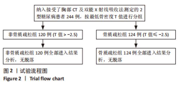
2.1 参与者数量分析 最终共计纳入244例患者,其中根据最低骨密度(股骨颈骨密度T值和髋部骨密度T值两者相比,较低的为最低骨密度)结果将研究对象分为非骨质疏松组(120例)和骨质疏松组(124例)。所有患者均获得完整的临床资料,无脱落。 2.2 试验流程图 见图2。"
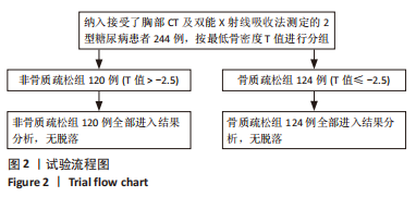
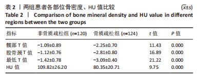
2.3 患者一般情况 共纳入了244例研究对象,根据最低T值分为非骨质疏松组120例及骨质疏松组124例。两组患者性别、年龄、体质量指数、糖化血红蛋白、平均血糖、钙、磷水平、2型糖尿病患病时间、高血压病史、高脂血症病史之间对比差异无显著性意义(P > 0.05),组间具有可比性,见表1。"

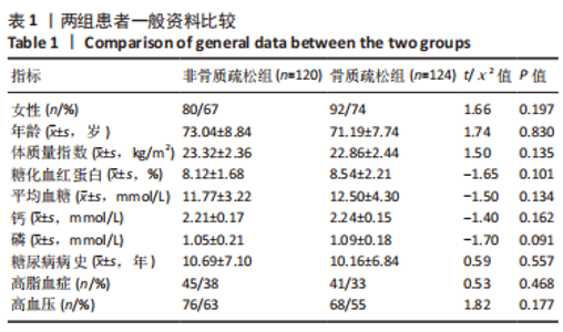
2.4 两组患者骨密度T值与HU值比较 非骨质疏松组的股骨颈T值、髋部T值、最低T值、L1椎体HU值的平均值均大于骨质疏松组(均P < 0.05),差异有显著性意义,见表2所示。"
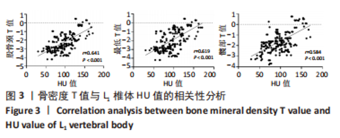
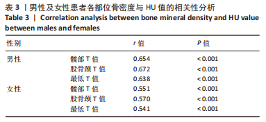
2.5 HU值与骨密度T值的相关性分析 通过采用Pearson相关性分析,L1椎体HU值与髋部T值呈中度正相关(r=0.584,P < 0.01),与股骨颈T值呈中度正相关(r=0.641,P < 0.01),与最低T值呈中度正相关关系(r=0.619,P < 0.01)。其中L1椎体HU值与股骨颈T值相关性最高,见图3。再根据性别及年龄对HU值与骨密度的相关性进行分析,在男性患者中L1椎体HU值与髋部T值呈中度正相关(r=0.654,P < 0.01),与股骨颈T值呈高度正相关(r=0.672,P < 0.01),与最低T值呈中度正相关关系(r=0.638,P < 0.01);而在女性患者中,L1椎体HU值与髋部T值呈中度正相关(r=0.551,P < 0.01),与股骨颈T值呈中度正相关(r=0.570,P < 0.01),与最低T值呈中度正相关关系(r=0.541,P < 0.01)。结果表明,不同性别患者的L1椎体HU值均与股骨颈T值相关性最高,见表3所示。"
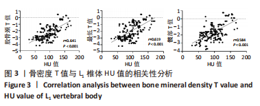
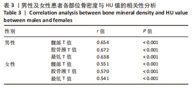
"
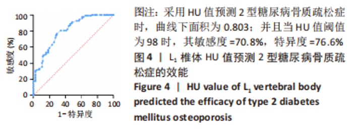
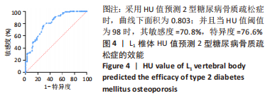
2.6 HU值预测T2DMOP的效能 通过受试者工作特征曲线发现L1椎体HU值在评估T2DMOP时,其曲线下面积为0.803(P < 0.05),表明L1椎体HU值评估T2DMOP有一定的准确性;当约登指数(约登指数=灵敏度+特异度-1)取最大值0.474时,阈值为98,敏感度为70.8%,特异性为76.6%;表明当HU值取98时,其筛查T2DMOP患者的敏感度为70.8%,具有良好的敏感性,见图4。"
| [1] SI Y, WANG C, GUO Y, et al. Prevalence of osteoporosis in patients with type 2 diabetes mellitus in the Chinese mainland: A protocol of systematic review and meta-analysis. Medicine (Baltimore). 2020;99(16):e19762. [2] LI Y, TENG D, SHI X, et al. Prevalence of diabetes recorded in mainland China using 2018 diagnostic criteria from the American Diabetes Association: national cross sectional study. BMJ. 2020;369:m997. [3] PORTAL-NÚÑEZ S, ARDURA JA, LOZANO D, et al. Adverse Effects of Diabetes Mellitus on the Skeleton of Aging Mice. J Gerontol A Biol Sci Med Sci. 2016;71(3): 290-299. [4] SLEEMAN A, CLEMENTS JN. Abaloparatide: A new pharmacological option for osteoporosis. Am J Health Syst Pharm. 2019;76(3):130-135. [5] GREGSON CL, ARMSTRONG DJ, BOWDEN J, et al. UK clinical guideline for the prevention and treatment of osteoporosis. Arch Osteoporos. 2022;17(1):58. [6] 陈明月,张雪丽,汤光宇. 基于CT的机会性筛查评估骨质疏松症的研究进展[J]. 国际医学放射学杂志,2022,45(4):459-465. [7] 夏维波,章振林,林华,等. 原发性骨质疏松症诊疗指南(2017)[J]. 中国骨质疏松杂志,2019,25(3):281-309. [8] 中国健康促进基金会基层医疗机构骨质疏松症诊断与治疗专家共识委员会. 基层医疗机构骨质疏松症诊断和治疗专家共识(2021)[J]. 中国骨质疏松杂志, 2021,27(7):937-944. [9] 杜雪平,黄凯,孙艳格,等. 北京市社区医务人员骨质疏松症相关知识认知状况调查[J]. 中国全科医学,2013,16(18):1646-1647. [10] SCHREIBER JJ, ANDERSON PA, ROSAS HG, et al. Hounsfield Units for Assessing Bone Mineral Density and Strength: A Tool for Osteoporosis Management. J Bone Joint Surg. 2011;93(11):1057-1063. [11] SALZMANN SN, OKANO I, JONES C, et al. Preoperative MRI-based Vertebral Bone Quality (VBQ) score assessment in patients undergoing lumbar spinal fusion. Spine J. 2022;22(8):1301-1308. [12] PAIVA LC, FILARDI S, PINTO-NETO AM, et al. Impact of degenerative radiographic abnormalities and vertebral fractures on spinal bone density of women with osteoporosis. Sao Paulo Med J. 2002;120(1):9-12. [13] YU EW, THOMAS BJ, BROWN JK, et al. Simulated increases in body fat and errors in bone mineral density measurements by DXA and QCT. J Bone Miner Res. 2012; 27(1):119-124. [14] 包洁,邹达,李危石. 椎体CT值评估腰椎退变患者骨密度的研究进展[J]. 中国脊柱脊髓杂志,2020,30(8):745-750. [15] ZOU D, MUHEREMU A, SUN Z, et al. Computed tomography Hounsfield unit–based prediction of pedicle screw loosening after surgery for degenerative lumbar spine disease. J Neurosurg Spine. 2020;32(5):716-721. [16] 郭立新. 2021年糖尿病领域年度重大进展回顾[J]. 中华糖尿病杂志,2022, 14(1):1-8. [17] 王盼,吴科锋,崔燎.不同阶段2型糖尿病诱发骨质疏松症的致病机制研究进展[J]. 中国骨质疏松杂志,2020,26(4):619-624. [18] NAPOLI N, SCHWARTZ AV, PALERMO L, et al. Risk Factors for Subtrochanteric and Diaphyseal Fractures: The Study of Osteoporotic Fractures. J Clin Endocrinol Metab. 2013;98(2):659-667. [19] FAN Y, WEI F, LANG Y, et al. Diabetes mellitus and risk of hip fractures: a meta-analysis. Osteoporos Int. 2016;27(1):219-228. [20] 叶紫梦玮,戴璇,刘亚鸽,等. 糖尿病性骨质疏松症的临床诊断方法探讨[J]. 中国骨质疏松杂志,2021,27(7):1005-1010. [21] 符桑,文章新,陈蓉,等. 跟骨定量超声在中老年2型糖尿病并发骨质疏松症预测诊断中的应用[J]. 中华骨质疏松和骨矿盐疾病杂志,2021,14(4):352-359. [22] de LIEFDE II, van der KLIFT M, de LAET CE, et al. Bone mineral density and fracture risk in type-2 diabetes mellitus: the Rotterdam Study. Osteoporos Int. 2005;16(12):1713-1720. [23] AHERN DP, MCDONNELL JM, RIFFAULT M, et al. A meta-analysis of the diagnostic accuracy of Hounsfield units on computed topography relative to dual-energy X-ray absorptiometry for the diagnosis of osteoporosis in the spine surgery population. Spine J. 2021;21(10):1738-1749. [24] NA MK, WON YD, KIM CH, et al. Opportunistic osteoporosis screening via the measurement of frontal skull Hounsfield units derived from brain computed tomography images. PLoS One. 2018;13(5):e197336. [25] 刘云,李培岭,郭永杰,等. 中老年骨质疏松症患者腰1-3椎体骨密度值与CT值相关性研究[J]. 风湿病与关节炎,2021,10(6):29-31. [26] ZHANG D, WU Y, LUO S, et al. Characteristics of Lumbar Bone Density in Middle-Aged and Elderly Subjects: A Correlation Study between T-Scores Determined by the DEXA Scan and Hounsfield Units from CT. J Health Eng. 2021;2021:1-7. [27] PERRIER-CORNET J, OMOROU AY, FAUNY M, et al. Opportunistic screening for osteoporosis using thoraco-abdomino-pelvic CT-scan assessing the vertebral density in rheumatoid arthritis patients. Osteoporos Int. 2019;30(6):1215-1222. [28] 陈金春,黄建华,黄建武,等. 绝经后骨质疏松症骨密度T值与腰椎椎体CT值的相关性[J]. 中医正骨,2009,21(9):1-3. [29] 李宏军. 新型冠状病毒肺炎影像学辅助诊断指南[J]. 中国医学影像技术, 2020,36(3):321-331. [30] 庞惠荧,林俊杰,孙珊. 探讨腰椎CT值评估2型糖尿病患者骨质疏松的临床运用[J]. 疾病监测与控制,2021,15(6):457-459. [31] SCHREIBER JJ, ANDERSON PA, HSU WK. Use of computed tomography for assessing bone mineral density. Neurosurg Focus. 2014;37(1):E4. [32] LI YL, WONG KH, LAW MW, et al. Opportunistic screening for osteoporosis in abdominal computed tomography for Chinese population. Arch Osteoporos. 2018;13(1):76. [33] GAUSDEN EB, NWACHUKWU BU, SCHREIBER JJ, et al. Opportunistic Use of CT Imaging for Osteoporosis Screening and Bone Density Assessment: A Qualitative Systematic Review. J Bone Joint Surg Am. 2017;99(18):1580-1590. [34] RATHINAVELU S, GUIDRY-ELIZONDO C, BANU J. Molecular Modulation of Osteoblasts and Osteoclasts in Type 2 Diabetes. J Diabetes Res. 2018;2018:1-11. [35] PRITCHARD JM, GIANGREGORIO LM, ATKINSON SA, et al. Association of larger holes in the trabecular bone at the distal radius in postmenopausal women with type 2 diabetes mellitus compared to controls. Arthritis Care Res. 2012;64(1):83-91. [36] LEE S, CHUNG CK, OH SH, et al. Correlation between Bone Mineral Density Measured by Dual-Energy X-Ray Absorptiometry and Hounsfield Units Measured by Diagnostic CT in Lumbar Spine. J Korean Neurosurg Soc. 2013;54(5):384. [37] BUCKENS CF, DIJKHUIS G, DE KEIZER B, et al. Opportunistic screening for osteoporosis on routine computed tomography? An external validation study. Eur Radiol. 2015;25(7):2074-2079. [38] PICKHARDT PJ, POOLER BD, LAUDER T, et al. Opportunistic Screening for Osteoporosis Using Abdominal Computed Tomography Scans Obtained for Other Indications. Ann Int Med. 2013;158(8):588. [39] ZOU D, LI W, DENG C, et al. The use of CT Hounsfield unit values to identify the undiagnosed spinal osteoporosis in patients with lumbar degenerative diseases. Eur Spine J. 2019;28(8):1758-1766. [40] MOONEY J, MORGAN S, BROCKINGTON D, et al. Inter-rater reliability and correlation of L1 Hounsfield unit measurements with DXA scores. J Clin Densitom. 2022;25(4):668-673. |
| [1] | Zhang Min, Peng Jing, Zhang Qiang, Chen Dewang. Mechanical properties of L3/4 laminar decompression and intervertebral fusion in elderly osteoporosis patients analyzed by finite element method [J]. Chinese Journal of Tissue Engineering Research, 2024, 28(6): 847-851. |
| [2] | Xue Xiaofeng, Wei Yongkang, Qiao Xiaohong, Du Yuyong, Niu Jianjun, Ren Lixin, Yang Huifeng, Zhang Zhimin, Guo Yuan, Chen Weiyi. Finite element analysis of osteoporosis in proximal femur after cannulated screw fixation for femoral neck fracture [J]. Chinese Journal of Tissue Engineering Research, 2024, 28(6): 862-867. |
| [3] | Kaiyisaier•Abudukelimu, Maimaitimin•Abulimiti, Li Lei, Yang Xiaokai, Zhang Yukun, Liu Shuai. Effect of lumbar CT values in the diagnosis of osteoporosis in women patients with lumbar degenerative diseases [J]. Chinese Journal of Tissue Engineering Research, 2024, 28(6): 945-949. |
| [4] | Yin Linwei, Huang Xiarong, Qu Mengjian, Yang Lu, Wang Jinling, Jia Feiyang, Liao Yang, Zhou Jun. Effects of treadmill exercise on osteoporosis and wnt/beta-catenin signal pathway in aged rats [J]. Chinese Journal of Tissue Engineering Research, 2024, 28(2): 231-236. |
| [5] | He Lijun, Qi Xiaojuan. Adipose-derived mesenchymal stem cells overexpressing bone morphogenetic protein 2 promote alveolar bone defect repair in osteoporosis rats [J]. Chinese Journal of Tissue Engineering Research, 2024, 28(1): 32-37. |
| [6] | Li Xiaomin, Tian Xiangdong, Tan Yetong, Zhu Guangyu, Wang Rongtian, Wang Jian, Xue Zhipeng, Ma Sheng, Hu Yuanyi, Huang Ye, Ding Tiansong. Changes of lower limb force line and knee function after high tibial osteotomy in osteoporotic medial ventricular knee osteoarthritis [J]. Chinese Journal of Tissue Engineering Research, 2023, 27(9): 1325-1329. |
| [7] | Wu Tianliang, Tao Xiuxia, Xu Hongguang. Influence of different bone mineral densities on cage subsidence after stand-alone oblique lateral interbody fusion: three-dimensional finite element analysis [J]. Chinese Journal of Tissue Engineering Research, 2023, 27(9): 1352-1358. |
| [8] | Jiang Xiaocheng, Shi Lu, Wang Yinbin, Li Qiujiang, Xi Chuangzhen, Ma Zefeng, Cai Lijun. Systematical evaluation of bone fusion rate after interbody fusion in patients with osteoporosis and lumbar degenerative disease treated with teriparatide [J]. Chinese Journal of Tissue Engineering Research, 2023, 27(9): 1427-1433. |
| [9] | Sun Jiajia, Zhu Haidi, Lu Yun, Zhang Kai. Comparison of bone metabolism markers between type 2 diabetes mellitus and non-type 2 diabetes mellitus patients with hip fracture [J]. Chinese Journal of Tissue Engineering Research, 2023, 27(8): 1156-1160. |
| [10] | Wang Ji, Zhang Min, Yang Zhongya, Zhang Long. A review of physical activity intervention in type 2 diabetes mellitus with sarcopenia [J]. Chinese Journal of Tissue Engineering Research, 2023, 27(8): 1272-1277. |
| [11] | Long Yanming, Xie Mengsheng, Huang Jiajie, Xue Wenli, Rong Hui, Li Xiaojie. Casein kinase 2-interaction protein-1 regulates the osteogenic ability of bone marrow mesenchymal stem cells in osteoporosis rats [J]. Chinese Journal of Tissue Engineering Research, 2023, 27(6): 878-882. |
| [12] | Wang Jinling, Huang Xiarong, Qu Mengjian, Huang Fujin, Yin Lingwei, Zhong Peirui, Liu Jin, Sun Guanghua, Liao Yang, Zhou Jun. Effects of exercise training on bone mass and bone microstructure in aged osteoporotic rats [J]. Chinese Journal of Tissue Engineering Research, 2023, 27(5): 676-682. |
| [13] | Zhang Min, Zhang Xiaoming, Liu Tongbin. Application potential of naringin in bone tissue regeneration [J]. Chinese Journal of Tissue Engineering Research, 2023, 27(5): 787-792. |
| [14] | Cheng Yunzhong, Liu Yuzeng, Hai Yong, Guan Li, Pan Aixing, Zhang Xinuo, Tao Luming, Li Yue. Bibliometric and visual analysis of the research status and development trend of cortical bone trajectory screws [J]. Chinese Journal of Tissue Engineering Research, 2023, 27(4): 513-519. |
| [15] | Liu Hao, Yang Hongsheng, Zeng Zhimou, Wang Liping, Yang Kunhai, Hu Yongrong, Qu Bo. Lumbar MRI vertebral bone quality score to evaluate the severity of osteoporosis in postmenopausal women [J]. Chinese Journal of Tissue Engineering Research, 2023, 27(4): 606-611. |
| Viewed | ||||||
|
Full text |
|
|||||
|
Abstract |
|
|||||