Chinese Journal of Tissue Engineering Research ›› 2023, Vol. 27 ›› Issue (5): 676-682.doi: 10.12307/2022.1020
Previous Articles Next Articles
Effects of exercise training on bone mass and bone microstructure in aged osteoporotic rats
Wang Jinling1, 2, 3, Huang Xiarong1, 2, 3, Qu Mengjian1, 2, 3, Huang Fujin1, 2, 3, Yin Lingwei1, 2, 3, Zhong Peirui1, 2, 3, Liu Jin1, 2, 3, Sun Guanghua1, 2, 3, Liao Yang1, 2, 3, Zhou Jun1, 2, 3
- 1Rehabilitation Medicine Center, 2Department of Rehabilitation, 3Rehabilitation Laboratory, the First Affiliated Hospital, Hengyang Medical School, University of South China, Hengyang 421001, Hunan Province, China
-
Received:2022-01-15Accepted:2022-03-07Online:2023-02-18Published:2022-07-21 -
Contact:Zhou Jun, MD, Chief physician, Master’s supervisor, 1Rehabilitation Medicine Center, 2Department of Rehabilitation, 3Rehabilitation Laboratory, the First Affiliated Hospital, Hengyang Medical School, University of South China, Hengyang 421001, Hunan Province, China -
About author:Wang Jinling, Master candidate, 1Rehabilitation Medicine Center, 2Department of Rehabilitation, 3Rehabilitation Laboratory, the First Affiliated Hospital, Hengyang Medical School, University of South China, Hengyang 421001, Hunan Province, China -
Supported by:Natural Science Foundation of Hunan Province, No. 2020JJ4085 (to ZJ); Key Project of Hunan Provincial Health Commission, No. 202103060198 (to ZJ); Key Project of South China University, No. USCKF201902K02 (to ZJ)
CLC Number:
Cite this article
Wang Jinling, Huang Xiarong, Qu Mengjian, Huang Fujin, Yin Lingwei, Zhong Peirui, Liu Jin, Sun Guanghua, Liao Yang, Zhou Jun. Effects of exercise training on bone mass and bone microstructure in aged osteoporotic rats[J]. Chinese Journal of Tissue Engineering Research, 2023, 27(5): 676-682.
share this article
Add to citation manager EndNote|Reference Manager|ProCite|BibTeX|RefWorks
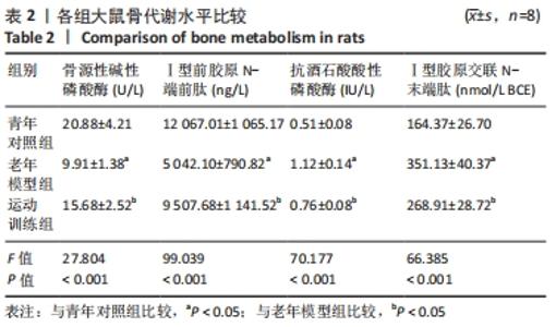
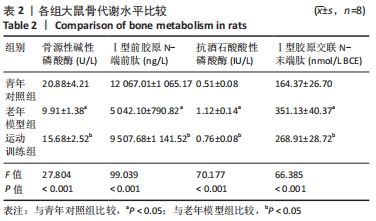
2.1 实验动物数量分析 实验共选用24只SD大鼠,分为3组,实验过程中大鼠均无死亡。 2.2 血清骨代谢指标比较 如表2所示,与青年对照组大鼠比较,老年模型组大鼠血清中骨源性碱性磷酸酶和Ⅰ型前胶原N-端前肽水平降低,而抗酒石酸酸性磷酸酶和Ⅰ型胶原交联N-末端肽水平升高,差异有显著性意义(P < 0.05);与老年模型组相比较,运动训练组大鼠血清中骨源性碱性磷酸酶和Ⅰ型前胶原N-端前肽水平升高,而抗酒石酸酸性磷酸酶和Ⅰ型胶原交联N-末端肽水平降低,差异有显著性意义(P < 0.05)。"
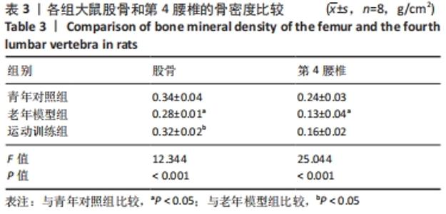
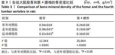
2.3 骨密度的比较 如表3所示,与青年对照组大鼠比较,老年模型组大鼠股骨和第4腰椎的骨密度下降,差异有显著性意义(P < 0.05);经运动干预后,运动训练组大鼠股骨骨密度高于老年模型组,差异有显著性意义(P < 0.05);而运动训练组大鼠第4腰椎骨密度高于老年模型组,但差异无显著性意义(P > 0.05)。"
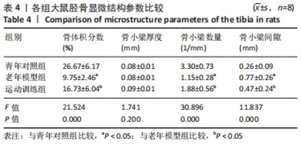
2.4 骨微结构的变化 Micro-CT对3组大鼠胫骨和第5腰椎骨微结构进行检测,并对其中显微结果参数进行分析。如表4所示,老年模型组大鼠胫骨的骨体积分数、骨小梁数量均低于青年对照组,骨小梁间隙均高于青年对照组,运动训练组大鼠胫骨的骨体积分数、骨小梁数量均高于老年模型组,骨小梁间隙低于老年模型组,以上差异均有显著性意义(P < 0.05)。与青年对照组比较,老年模型组大鼠胫骨的骨小梁厚度差异无显著性意义(P > 0.05);与老年模型组比较,运动训练组大鼠胫骨的骨小梁厚度差异无显著性意义(P > 0.05)。"


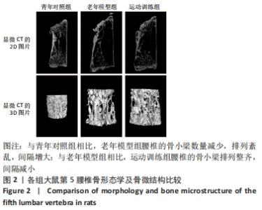
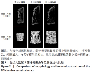
如表5所示,老年模型组大鼠第5腰椎的骨体积分数、骨小梁数量、骨小梁厚度均低于青年对照组,骨小梁间隙均高于青年对照组,运动训练组大鼠第5腰椎的骨小梁间隙低于老年模型组,以上差异均有显著性意义(P < 0.05),运动训练组大鼠第5腰椎的骨体积分数、骨小梁厚度、骨小梁数量均高于老年模型组,但差异无显著性意义(P > 0.05)。 "
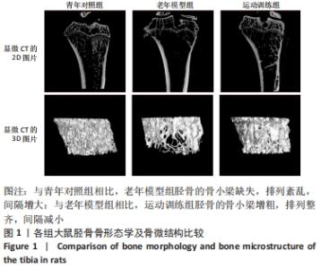
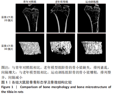
MicroCT扫描结果显示,与青年对照组相比,老年模型组大鼠骨小梁组织断裂明显,结构紊乱,骨髓腔内空洞较多;与老年模型组相比,运动训练组骨小梁数量变多,厚度变厚,组织完整,排列整齐,见图1,图2。 "
"
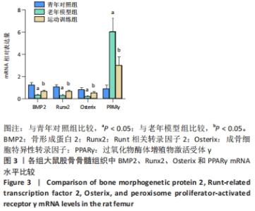
2.5 BMP2、Runx2、Osterix和PPARγ mRNA表达比较 如图3所示,与青年对照组大鼠相比较,老年模型组大鼠BMP2、Runx2与Osterix mRNA表达均减少,差异有显著性意义(P < 0.05);而经过运动训练后,运动训练组老年大鼠BMP2、Runx2与Osterix mRNA表达均增加,差异有显著性意义(P < 0.05);相反,与青年对照组大鼠相比较,老年模型组大鼠PPARγ mRNA表达增加,差异有显著性意义(P < 0.05);而经过运动训练后,运动训练组老年大鼠PPARγ mRNA表达减少,差异有显著性意义(P < 0.05)。"


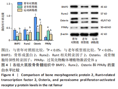
2.6 BMP2、Runx2、Osterix和PPARγ蛋白表达水平 如图4所示,各组大鼠BMP2、Runx2、Osterix 、PPARγ 蛋白表达的趋势与mRNA表达一致。与青年对照组大鼠相比较,老年模型组大鼠BMP2、Runx2与Osterix表达均减少,差异有显著性意义(P < 0.05);而经过运动训练后,运动训练组大鼠BMP2、Runx2与Osterix表达均增加,差异有显著性意义(P < 0.05);相反,与青年对照组大鼠相比较,老年模型组大鼠PPARγ表达增加,差异有显著性意义(P < 0.05);而经过运动训练后,运动训练组大鼠PPARγ表达均减少,差异有显著性意义(P < 0.05)。"

| [1] 中华医学会骨质疏松和骨矿盐疾病分会. 原发性骨质疏松症诊疗指南(2017)[J].中华骨质疏松和骨矿盐疾病杂志,2017,10(5):413-444. [2] QADIR A, LIANG S, WU Z, et al. Senile Osteoporosis: The Involvement of Differentiation and Senescence of Bone Marrow Stromal Cells. Int J Mol Sci. 2020;21(1):349. [3] 丁超,孙强.老年性骨质疏松症相关问题研究进展[J].中国骨质疏松杂志,2016,22(3):372-375. [4] COMPSTON JE, MCCLUNG MR, LESLIE WD. Osteoporosis. Lancet. 2019; 393(10169):364-376. [5] WANG L, YU W, YIN X, et al. Prevalence of Osteoporosis and Fracture in China: The China Osteoporosis Prevalence Study. JAMA Netw Open. 2021;4(8):e2121106. [6] 贺丽英,孙蕴,要文娟,等.2010-2016年中国老年人骨质疏松症患病率Meta分析 [J].中国骨质疏松杂志,2016,22(12):1590-1596. [7] CHIEN MY, WU YT, HSU AT, et al. Efficacy of a 24-week aerobic exercise program for osteopenic postmenopausal women. Calcif Tissue Int. 2000;67(6):443-448. [8] YEH JK, ALOIA JF, TIERNEY JM, et al. Effect of treadmill exercise on vertebral and tibial bone mineral content and bone mineral density in the aged adult rat: determined by dual energy X-ray absorptiometry. Calcif Tissue Int. 1993;52(3):234-238. [9] HOWE TE, SHEA B, DAWSON LJ, et al. Exercise for preventing and treating osteoporosis in postmenopausal women. Cochrane Database Syst Rev. 2011;(7):CD000333. [10] PAGNOTTI GM, STYNER M, UZER G, et al. Combating osteoporosis and obesity with exercise: leveraging cell mechanosensitivity. Nat Rev Endocrinol. 2019;15(6):339-355. [11] NIH CONSENSUS DEVELOPMENT PANEL ON OSTEOPOROSIS PREVENTION, DIAGNOSIS, AND THERAPY. Osteoporosis prevention, diagnosis, and therapy. JAMA. 2001;285(6):785-795. [12] 王春燕,何成奇.骨质疏松症治疗中的运动疗法[J].中国组织工程研究,2013,17(37):6657-6663. [13] LANE NE. Epidemiology, etiology, and diagnosis of osteoporosis. Am J Obstet Gynecol. 2006;194(2 Suppl):S3-11. [14] LIN Z, HE H, WANG M, et al. MicroRNA-130a controls bone marrow mesenchymal stem cell differentiation towards the osteoblastic and adipogenic fate. Cell Prolif. 2019;52(6):e12688. [15] WEN J, BAO M, TANG M, et al. Low magnitude vibration alleviates age-related bone loss by inhibiting cell senescence of osteogenic cells in naturally senescent rats. Aging (Albany NY). 2021;13(8):12031-12045. [16] HADJIDAKIS DJ, ANDROULAKIS II. Bone remodeling. Ann N Y Acad Sci. 2006;1092:385-396. [17] YU S, GUO J, SUN Z, et al. BMP2-dependent gene regulatory network analysis reveals Klf4 as a novel transcription factor of osteoblast differentiation. Cell Death Dis. 2021;12(2):197. [18] ZHAO C, GU Y, WANG Y, et al. miR-129-5p Promotes Osteogenic Differentiation of BMSCs and Bone Regeneration via Repressing Dkk3. Stem Cells Int. 2021;2021:7435605. [19] ZHU F, LIU Z, REN Y. Mechanism of melatonin combined with calcium carbonate on improving osteoporosis in aged rats. Exp Ther Med. 2018;16(1):192-196. [20] 周静媛,刘肖珩,沈阳,等.骨质疏松症动物模型的构建及实验方法研究进展[J].生物医学工程研究,2021,40(1):83-87. [21] GU Q, ZHAO L, MA YP, et al. Contribution of mitochondrial function to exercise-induced attenuation of renal dysfunction in spontaneously hypertensive rats. Mol Cell Biochem. 2015;406(1-2):217-225. [22] BEDFORD TG, TIPTON CM, WILSON NC, et al. Maximum oxygen consumption of rats and its changes with various experimental procedures. J Appl Physiol Respir Environ Exerc Physiol. 1979;47(6): 1278-1283. [23] LUO D, REN H, ZHANG H, et al. The protective effects of triptolide on age-related bone loss in old male rats. Biomed Pharmacother. 2018; 98:280-285. [24] CHEN K, YANG YH, JIANG SD, et al. Decreased activity of osteocyte autophagy with aging may contribute to the bone loss in senile population. Histochem Cell Biol. 2014;142(3):285-295. [25] ZHU X, LUO J, CHEN X, et al. Expression characteristic and significance of interleukin-6, nuclear factor kappa beta, and bone formation markers in rat models of osteoporosis. Transl Res. 2008;152(1):18-23. [26] KOIVULA MK, RISTELI L, RISTELI J. Measurement of aminoterminal propeptide of type I procollagen (PINP) in serum. Clin Biochem. 2012; 45(12):920-927. [27] 董伟,冯晓洁,梁永强,等.双膦酸盐对破骨细胞分化及抗酒石酸酸性磷酸酶的影响[J].中国组织工程研究,2014,18(38): 6069-6073. [28] HERRMANN M, SEIBEL MJ. The amino- and carboxyterminal cross-linked telopeptides of collagen type I, NTX-I and CTX-I: a comparative review. Clin Chim Acta. 2008;393(2):57-75. [29] EYRE DR. Bone biomarkers as tools in osteoporosis management. Spine (Phila Pa 1976). 1997;22(24 Suppl):17S-24S. [30] DUQUE G, TROEN BR. Understanding the mechanisms of senile osteoporosis: new facts for a major geriatric syndrome. J Am Geriatr Soc. 2008;56(5):935-941. [31] 张翔,颜卫文,王雅纯.红景天甙联合碳酸钙对老年雌性大鼠骨量、骨代谢和氧化应激水平影响[J].中国骨质疏松杂志,2020,26(10): 1441-1445. [32] JAIN RK, VOKES T. Dual-energy X-ray Absorptiometry. J Clin Densitom. 2017;20(3):291-303. [33] GREGSON CL, ARMSTRONG DJ, BOWDEN J, et al. UK clinical guideline for the prevention and treatment of osteoporosis. Arch Osteoporos. 2022;17(1):58. [34] KANIS JA. Assessment of fracture risk and its application to screening for postmenopausal osteoporosis: synopsis of a WHO report. WHO Study Group. Osteoporos Int. 1994;4(6):368-381. [35] YUAN Y, ZHANG L, TONG X, et al. Mechanical Stress Regulates Bone Metabolism Through MicroRNAs. J Cell Physiol. 2017;232(6):1239-1245. [36] LLOYD SA, LANG CH, ZHANG Y, et al. Interdependence of muscle atrophy and bone loss induced by mechanical unloading. J Bone Miner Res. 2014;29(5):1118-1130. [37] MATSUBARA T, KIDA K, YAMAGUCHI A, et al. BMP2 regulates Osterix through Msx2 and Runx2 during osteoblast differentiation. J Biol Chem. 2008;283(43):29119-29125. [38] HALLORAN D, DURBANO HW, NOHE A. Bone Morphogenetic Protein-2 in Development and Bone Homeostasis. J Dev Biol. 2020;8(3):19. [39] LIN W, ZHU X, GAO L, et al. Osteomodulin positively regulates osteogenesis through interaction with BMP2. Cell Death Dis. 2021; 12(2):147. [40] CHOI Y, KIM MH, NAM YK, et al. Palmul-Tang, a Korean Medicine, Promotes Bone Formation via BMP-2 Pathway in Osteoporosis. Front Pharmacol. 2021;12:643482. [41] KATAGIRI T, YAMAGUCHI A, KOMAKI M, et al. Bone morphogenetic protein-2 converts the differentiation pathway of C2C12 myoblasts into the osteoblast lineage. J Cell Biol. 1994;127(6 Pt 1):1755-1766. [42] GOMATHI K, AKSHAYA N, SRINAATH N, et al. Regulation of Runx2 by post-translational modifications in osteoblast differentiation. Life Sci. 2020;245:117389. [43] PHIMPHILAI M, ZHAO Z, BOULES H, et al. BMP signaling is required for RUNX2-dependent induction of the osteoblast phenotype. J Bone Miner Res. 2006;21(4):637-646. [44] NAKASHIMA K, ZHOU X, KUNKEL G, et al. The novel zinc finger-containing transcription factor osterix is required for osteoblast differentiation and bone formation. Cell. 2002;108(1):17-29. [45] WOELLER CF, LIM SA, ROZTOCIL E, et al. Neonatal hyperoxia impairs adipogenesis of bone marrow-derived mesenchymal stem cells and fat accumulation in adult mice. Free Radic Biol Med. 2021;167:287-298. [46] KO FC, KOBELSKI MM, ZHANG W, et al. Phosphate restriction impairs mTORC1 signaling leading to increased bone marrow adipose tissue and decreased bone in growing mice. J Bone Miner Res. 2021;36(8): 1510-1520. [47] CAO J, OU G, YANG N, et al. Impact of targeted PPARγ disruption on bone remodeling. Mol Cell Endocrinol. 2015;410:27-34. [48] SHEN GS, ZHOU HB, ZHANG H, et al. The GDF11-FTO-PPARγ axis controls the shift of osteoporotic MSC fate to adipocyte and inhibits bone formation during osteoporosis. Biochim Biophys Acta Mol Basis Dis. 2018;1864(12):3644-3654. [49] ZHOU RP, DENG MT, CHEN LY, et al. Shp2 regulates chlorogenic acid-induced proliferation and adipogenic differentiation of bone marrow-derived mesenchymal stem cells in adipogenesis. Mol Med Rep. 2015; 11(6):4489-4495. [50] 徐众华,莫雨晴,周驰.基于BMP/Runx2/Osx信号通路研究淫羊藿总黄酮改善绝经后骨质疏松模型大鼠的作用机制 [J].中国药房, 2020,31(19):2333-2338. [51] 卜淑敏,杨涵. 5种干预疗法对去卵巢骨质疏松大鼠骨髓脂肪细胞数目和PPARγ2蛋白表达水平的影响[J].首都体育学院学报,2016, 28(3):270-273. [52] CHEN T, YANG T, ZHANG W, et al. The therapeutic potential of mesenchymal stem cells in treating osteoporosis. Biol Res. 2021; 54(1):42. [53] HU Y, XU R, CHEN CY, et al. Extracellular vesicles from human umbilical cord blood ameliorate bone loss in senile osteoporotic mice. Metabolism. 2019;95:93-101. [54] LIAO HW, HUANG TH, CHANG YH, et al. Exercise Alleviates Osteoporosis in Rats with Mild Chronic Kidney Disease by Decreasing Sclerostin Production. Int J Mol Sci. 2019;20(8):2044. [55] 黄晓,李建华.跑台运动对骨质疏松大鼠骨组织Wnt/β-catenin信号通路的影响[J].中华物理医学与康复杂志,2018,40(10):727-732. [56] FELBER K, ELKS PM, LECCA M, et al. Expression of osterix Is Regulated by FGF and Wnt/β-Catenin Signalling during Osteoblast Differentiation. PLoS One. 2015;10(12):e0144982. [57] LUO Y, ZHANG Y, MIAO G, et al. Runx1 regulates osteogenic differentiation of BMSCs by inhibiting adipogenesis through Wnt/β-catenin pathway. Arch Oral Biol. 2019;97:176-184. [58] MURUGANANDAN S, IONESCU AM, SINAL CJ. At the Crossroads of the Adipocyte and Osteoclast Differentiation Programs: Future Therapeutic Perspectives. Int J Mol Sci. 2020;21(7):2277. |
| [1] | Zhong Yizheng, Huang Peizhen, Cai Qunbin, Zheng Liqin, He Xingpeng, Dong Hang. Microstructural indexes that determine the trabecular bone maximum stress of micro-finite element models [J]. Chinese Journal of Tissue Engineering Research, 2023, 27(9): 1313-1318. |
| [2] | Cao Sheng, Kong Lingwei, Xu Kun, Sun Zhijie. Correlation of cervical sagittal force line parameters with degenerative segment and Pfirrmann classification in patients with cervical intervertebral disc degeneration [J]. Chinese Journal of Tissue Engineering Research, 2023, 27(9): 1319-1324. |
| [3] | Ke Yuqi, Chen Changjian, Wu Hao, Zheng Lianjie. Comparison of 12-month follow-up results of primary total hip arthroplasty between modified direct anterior approach and direct anterior approach [J]. Chinese Journal of Tissue Engineering Research, 2023, 27(9): 1377-1382. |
| [4] | Zhang Lichuang, Gao Huali, Wang Jingchao, Lin Huijun, Wu Chonggui, Ma Yinghui, Huang Yunfei, Fang Xue, Zhai Weitao. Effect of tendon manipulation with equal emphasis on muscles and bones on accelerating the functional rehabilitation of quadriceps femoris after total knee arthroplasty [J]. Chinese Journal of Tissue Engineering Research, 2023, 27(9): 1383-1389. |
| [5] | Du Xueting, Zhang Xiaodong, Chen Yanjun, Wang Mei, Chen Wubiao, Huang Wenhua. Application of compressed sensing technology in two-dimensional magnetic resonance imaging of the ankle joint [J]. Chinese Journal of Tissue Engineering Research, 2023, 27(9): 1396-1402. |
| [6] | You Zhengqiu, Zhang Zhongzu, Wang Qunbo. Early symptomatic intervertebral disc pseudocysts after discectomy detected on MRI [J]. Chinese Journal of Tissue Engineering Research, 2023, 27(9): 1403-1409. |
| [7] | Li Chao, Zhang Peipei, Xu Mengting, Li Linlin, Ding Jiangtao, Liu Xihua, Bi Hongyan. Respiratory training improves morphological changes of the multifidus muscle in patients with chronic nonspecific lower back pain assessed by musculoskeletal ultrasound [J]. Chinese Journal of Tissue Engineering Research, 2023, 27(9): 1417-1421. |
| [8] | He Yinhao, Li Xiaosheng, Chen Hongwen, Chen Tiezhu. 3D printed porous tantalum metal in the treatment of developmental dysplasia of the hip: current status and application prospect [J]. Chinese Journal of Tissue Engineering Research, 2023, 27(9): 1455-1461. |
| [9] | Jiang Xiaocheng, Shi Lu, Wang Yinbin, Li Qiujiang, Xi Chuangzhen, Ma Zefeng, Cai Lijun. Systematical evaluation of bone fusion rate after interbody fusion in patients with osteoporosis and lumbar degenerative disease treated with teriparatide [J]. Chinese Journal of Tissue Engineering Research, 2023, 27(9): 1427-1433. |
| [10] | Liu Xiaolin, Mu Xinyue, Ma Ziyu, Liu Shutai, Wang Wenlong, Han Xiaoqian, Dong Zhiheng. Effect of hydrogel-loaded simvastatin microspheres on osteoblast proliferation and differentiation [J]. Chinese Journal of Tissue Engineering Research, 2023, 27(7): 998-1003. |
| [11] | Zhu Lin, Gu Weiping, Wang Can, Chen Gang. Biomechanical analysis of All-on-Four and pterygomaxillary implants under different maxillary bone conditions [J]. Chinese Journal of Tissue Engineering Research, 2023, 27(7): 985-991. |
| [12] | Jiang Yifang, Cai Qimin, Chu Zhengyi, Qin Min, Shen Yurong, Gu Yuanping. Simulation analysis of stress distribution of NRT FILES in curved root canals [J]. Chinese Journal of Tissue Engineering Research, 2023, 27(7): 1038-1042. |
| [13] | Xu Yan, Li Ping, Lai Chunhua, Zhu Peijun, Yang Shuo, Xu Shulan. Piezoelectric materials for vascularized bone regeneration [J]. Chinese Journal of Tissue Engineering Research, 2023, 27(7): 1126-1132. |
| [14] | Wu Yujie, Wan Xiaofang, Wei Mianxing, Peng Shiyuan, Xu Xiaomei. Correlation between autophagy and the Hippo-YAP protein pathway in periodental ligament cells on the pressure side of a mouse model of orthodontic tooth movement [J]. Chinese Journal of Tissue Engineering Research, 2023, 27(5): 683-689. |
| [15] | Yang Heng, Zheng Liying, Liu Zhili, Wang Qinzhang, Hao Zhiqiang, Wang Jingshen, Wang Yuan, Li Yongle, Tan Minghui, Zou Xiaofeng, Zhang Guoxi, Huang Ruohui, Jiang Bo, Qian Biao. Physiological function of the testis in rats undergoing different parts of vasectomy [J]. Chinese Journal of Tissue Engineering Research, 2023, 27(5): 720-725. |
| Viewed | ||||||
|
Full text |
|
|||||
|
Abstract |
|
|||||