Chinese Journal of Tissue Engineering Research ›› 2023, Vol. 27 ›› Issue (5): 683-689.doi: 10.12307/2023.086
Previous Articles Next Articles
Correlation between autophagy and the Hippo-YAP protein pathway in periodental ligament cells on the pressure side of a mouse model of orthodontic tooth movement
Wu Yujie1, 2, Wan Xiaofang1, 2, Wei Mianxing1, 2, Peng Shiyuan1, 2, Xu Xiaomei1, 2
- 1Department of Orthodontics, The Affiliated Stomatology Hospital of Southwest Medical University, Luzhou 646000, Sichuan Province, China; 2Oral & Maxillofacial Reconstruction and Regeneration Laboratory, Southwest Medical University, Luzhou 646000, Sichuan Province, China
-
Received:2022-02-28Accepted:2022-04-24Online:2023-02-18Published:2022-07-22 -
Contact:Xu Xiaomei, MD, Chief physician, Associate professor, Department of Orthodontics, The Affiliated Stomatology Hospital of Southwest Medical University, Luzhou 646000, Sichuan Province, China; Oral & Maxillofacial Reconstruction and Regeneration Laboratory, Southwest Medical University, Luzhou 646000, Sichuan Province, China -
About author:Wu Yujie, Master candidate, Department of Orthodontics, The Affiliated Stomatology Hospital of Southwest Medical University, Luzhou 646000, Sichuan Province, China; Oral & Maxillofacial Reconstruction and Regeneration Laboratory, Southwest Medical University, Luzhou 646000, Sichuan Province, China -
Supported by:Science and Technology Strategic Cooperation Project of Luzhou Municipal People’s Government & Southwest Medical University, No. 2020LZXNYDZ06 (to XXM); Sichuan Provincial Department of Science and Technology Planning Project, No. 2021YJ0151 (to XXM)
CLC Number:
Cite this article
Wu Yujie, Wan Xiaofang, Wei Mianxing, Peng Shiyuan, Xu Xiaomei. Correlation between autophagy and the Hippo-YAP protein pathway in periodental ligament cells on the pressure side of a mouse model of orthodontic tooth movement[J]. Chinese Journal of Tissue Engineering Research, 2023, 27(5): 683-689.
share this article
Add to citation manager EndNote|Reference Manager|ProCite|BibTeX|RefWorks
2.1 实验动物数量分析 实验共纳入C57BL/6小鼠54只,分为9组,实验期间无动物死亡,无加力装置脱落,全部进入结果分析。 2.2 牙移动距离测量 正畸牙移动的距离随正畸力作用时间的延长而增加。与空白组相比,正畸力施加12 h组测量到磨牙出现(0.020±0.007) mm移动(P < 0.01),加力1 d组可测量出明显的牙移动距离(0.030±0.005) mm(P < 0.01),加力7 d牙移动距离最远(0.110±0.020) mm(P < 0.01),见图2。"
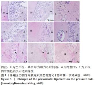
2.3 压力侧牙周膜形态改变 观察发现,加力30 min至4 h组压力侧牙周膜没有发生明显的变化。加力12 h组开始观察到随加力时间延长,压力侧牙周膜内细胞及纤维排列由规则有序逐渐变紊乱,牙周膜逐渐被压缩,至3 d组压力侧牙周膜间隙最窄,7 d组牙周膜间隙稍恢复;加力1 d组压力区出现透明样变化,至7 d组透明样变达最大面积,见图3。"
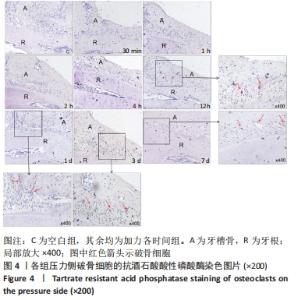
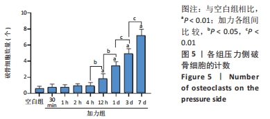
2.4 TRAP染色 阳性染色破骨细胞的胞质呈紫红色,见图4。空白组和加力30 min至4 h组压力侧偶尔可见到破骨细胞的出现;加力12 h组压力侧破骨细胞开始增多(P < 0.01),随后随加力时间的增加而增加,至加力7 d其数量达最大值(P < 0.01),见图5。"

"
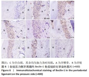
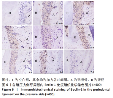
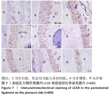
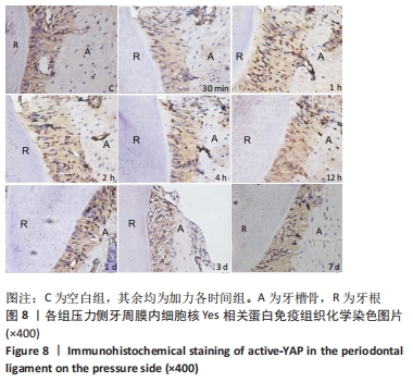
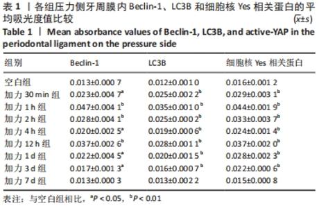
2.5 Belin-1、LC3B以及细胞核YAP的免疫组化染色 免疫组织化学染色可见:压力侧牙周膜内Beclin-1和LC3B主要在细胞质中表达,细胞核YAP即active-YAP主要在细胞核内表达,三者的阳性染色皆为棕色,见图6-8。"

"

"
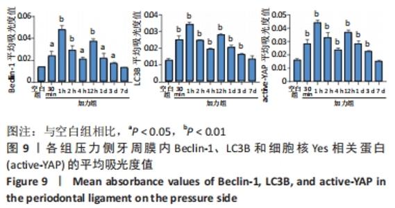
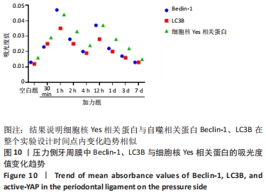
Beclin-1和LC3B在实验组压力侧牙周膜中免疫组化染色的变化趋势类似。加力30 min 时压力侧Beclin-1、LC3B的平均吸光度值均高于空白组(P < 0.05),加力1 h二者平均吸光度值均最高(P < 0.01);加力1-4 h期间逐渐减弱,但4 h组二者的平均吸光度值仍然比空白组高(P < 0.05);加力12 h二者的平均吸光度值明显增强,达又一峰值(P < 0.01);随后回落,至加力7 d它们的表达水平与空白组接近(P > 0.05),见图9及表1。 "

"
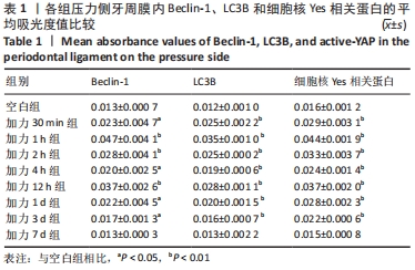
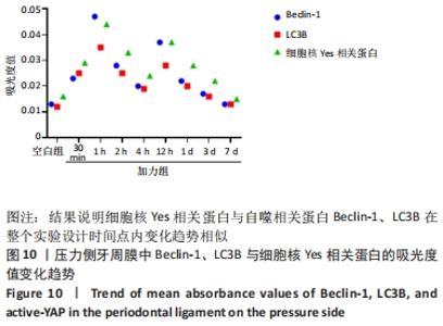
加力30 min组压力侧牙周膜内可见active-YAP的平均吸光度值增加(P < 0.01);加力1 h其平均吸光度值最强(P < 0.01);加力1-4 h组逐渐减少,至4 h组active-YAP的平均吸光度值达最弱,但仍强于空白组(P < 0.01);加力12 h组再次增强(P < 0.01),随后逐渐减弱,至加力7 d组与空白组无差异(P > 0.05),见图9及表1。 2.6 Beclin-1、LC3B与active-YAP表达变化的相关性分析 实验结果中压力侧牙周膜中Beclin-1和LC3B的表达变化趋势与active-YAP趋势相似,表达强度都在加力1 h达峰值。随后逐渐降低,于加力4 h降至谷值,加力12 h它们的表达增强达又一峰值,而后回落,于7 d都降至基础水平,见图10。 "
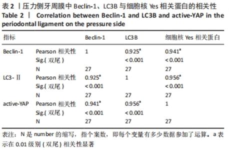
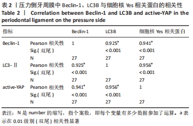
Pearson相关性分析:可见Beclin-1与active-YAP的表达变化呈高度正相关,相关系数r=0.941,P < 0.01;LC3B与active-YAP的表达变化也呈高度正相关,相关系数r=0.956,P < 0.01,见表2。 "
| [1] XU J, ZHAO X, ZENG J, et al. Role of autophagy in the periodontal ligament reconstruction during orthodontic tooth movement in rats. J Dent Sci. 2020;15(3):351-363. [2] COBBAUT M, KARAGIL S, BRUNO L, et al. Dysfunctional Mechanotransduction through the YAP/TAZ/Hippo Pathway as a Feature of Chronic Disease. Cells. 2020;9(1):151. [3] GAO Y, LUO C, RUI T, et al. Autophagy inhibition facilitates wound closure partially dependent on the YAP/IL-33 signaling in a mouse model of skin wound healing. FASEB J. 2021;35(10):e21920. [4] MAYR A, MARCINIAK J, EGGERS B, et al. Autophagy Induces Expression of IL-6 in Human Periodontal Ligament Fibroblasts Under Mechanical Load and Overload and Effects Osteoclastogenesis in vitro. Front Physiol. 2021;12:716441. [5] RAVANAN P, SRIKUMAR IF, TALWAR P. Autophagy: the spotlight for cellular stress responses. Life Sci. 2017;188:53-67. [6] GALATI S, BONI C, GERRA MC, et al. Autophagy: A Player in response to Oxidative Stress and DNA Damage. Oxid Med Cell Longev. 2019; 2019:5692958. [7] CHEN L, MO S, HUA Y. Compressive force-induced autophagy in periodontal ligament cells downregulates osteoclastogenesis during tooth movement. J Periodontol. 2019;90(10):1170-1181. [8] LI W, ZHAO J, SUN W, et al. Osteocytes promote osteoclastogenesis via autophagy-mediated RANKL secretion under mechanical compressive force. Arch Biochem Biophys. 2020;694:108594. [9] KAZAMA T. Electron microscopic study of periodontal tissues in the pressure side by intermittent forces. Tsurumi Shigaku. 1989;15:87-108. [10] LI Y, JACOX LA, COATS S, et al. Roles of autophagy in orthodontic tooth movement. Am J Orthod Dentofacial Orthop. 2021;159(5):582-593. [11] 尹圆圆,马华钰,李昕怡,等.小鼠正畸牙移动中牙周组织自噬相关基因表达的初步研究[J].国际口腔医学杂志,2020,47(6):627-634. [12] MAEJIMA Y, ZABLOCKI D, NAH J, et al. The role of the Hippo pathway in autophagy in the heart [published online ahead of print, 2022 Feb 12]. Cardiovasc Res. 2022; cvac014. [13] ZHOU J, HU M, HE M, et al. TNFAIP3 Interacting Protein 3 Is an Activator of Hippo-YAP Signaling Protecting Against Hepatic Ischemia/Reperfusion Injury. Hepatology. 2021;74(4):2133-2153. [14] TANG F, GAO R, JEEVAN-RAJ B, et al. LATS1 but not LATS2 represses autophagy by a kinase-independent scaffold function. Nat Commun. 2019;10(1):5755. [15] DUPONT S. Role of YAP/TAZ in cell-matrix adhesion-mediated signalling and mechanotransduction. Exp Cell Res. 2016;343(1):42-53. [16] PAVEL M, PARK SJ, FRAKE RA, et al. α-Catenin levels determine direction of YAP/TAZ response to autophagy perturbation. Nat Commun. 2021; 12(1):1703. [17] LIU Z, ZENG W, WANG S, et al. A potential role for the Hippo pathway protein, YAP, in controlling proliferation, cell cycle progression, and autophagy in BCPAP and KI thyroid papillary carcinoma cells. Am J Transl Res. 2017;9(7):3212-3223. [18] KILKENNY C, BROWNE W, CUTHILL IC, et al. Animal research: reporting in vivo experiments: the ARRIVE guidelines. Br J Pharmacol. 2010; 160(7):1577-1579. [19] NAKAMURA T, YAMASHITA M, IKEGAMI K, et al. Autophagy facilitates type I collagen synthesis in periodontal ligament cells. Sci Rep. 2021;11(1):1291. [20] WESTOVER L, FAULKNER G, FLORES-MIR C, et al. Non-invasive evaluation of periodontal ligament stiffness during orthodontic tooth movement. Angle Orthod. 2019;89:228e34. [21] JIANG N, HE D, MA Y, et al. Force-Induced Autophagy in Periodontal Ligament Stem Cells Modulates M1 Macrophage Polarization via AKT Signaling. Front Cell Dev Biol. 2021;9:666631. [22] HUANG Y, LIU H, GUO R, et al. Long Non-coding RNA FER1L4 Mediates the Autophagy of Periodontal Ligament Stem Cells Under Orthodontic Compressive Force via AKT/FOXO3 Pathway. Front Cell Dev Biol. 2021; 9:631181. [23] DAS J, AGARWAL T, CHAKRABORTY S, et al. Compressive stress-induced autophagy promotes invasion of HeLa cells by facilitating protein turnover in vitro. Exp Cell Res. 2019;381(2):201-207. [24] 吕佳岭,徐洁,曾锦,等.正畸牙压力区牙周膜细胞自噬相关蛋白Beclin-1与微管相关蛋白2轻链3的表达[J].华西口腔医学杂志, 2019,37(2):168-173. [25] HUANG L, YIN X, CHEN J, et al. Lithium chloride promotes osteogenesis and suppresses apoptosis during orthodontic tooth movement in osteoporotic model via regulating autophagy. Bioact Mater. 2021; 6(10):3074-3084. [26] JABER FA, KHAN NM, ANSARI MY, et al. Autophagy plays an essential role in bone homeostasis. J Cell Physiol. 2019;234(8):12105-12115. [27] TONG X, ZHANG C, WANG D, et al. Suppression of AMP-activated protein kinase reverses osteoprotegerin-induced inhibition of osteoclast differentiation by reducing autophagy. Cell Prolif. 2020;53(1):e12714. [28] SHI J, WANG L, ZHANG H, et al. Glucocorticoids: Dose-related effects on osteoclast formation and function via reactive oxygen species and autophagy. Bone. 2015;79:222-232. [29] LI J, SUN Z, LIN Y, et al. Syndecan 4 contributes to osteoclast differentiation induced by RANKL through enhancing autophagy. Int Immunopharmacol. 2021;91:107275. [30] TOTARO A, ZHUANG Q, PANCIERA T, et al. Cell phenotypic plasticity requires autophagic flux driven by YAP/TAZ mechanotransduction. Proc Natl Acad Sci USA. 2019;116(36):17848-17857. [31] SHEN Y, PAN Y, GUO S, et al. The roles of mechanosensitive ion channels and associated downstream MAPK signaling pathways in PDLC mechanotransduction. Mol Med Rep. 2020;21(5):2113-2122. [32] JIANG Y, GUAN Y, LAN Y, et al. Mechanosensitive Piezo1 in Periodontal Ligament Cells Promotes Alveolar Bone Remodeling During Orthodontic Tooth Movement. Front Physiol. 2021;12:767136. [33] BENHAM-PYLE BW, PRUITT BL, NELSON WJ. Cell adhesion. Mechanical strain induces E-cadherin-dependent Yap1 and β-catenin activation to drive cell cycle entry. Science. 2015;348(6238):1024-1027. [34] MENG Z, QIU Y, LIN KC, et al. RAP2 mediates mechanoresponses of the Hippo pathway. Nature. 2018;560(7720):655-660. [35] DUPONT S, MORSUT L, ARAGONA M, et al. Role of YAP/TAZ in mechanotransduction. Nature. 2011;474(7350):179-183. [36] NGUYEN MT, LEE W. MiR-141-3p regulates myogenic differentiation in C2C12 myoblasts via CFL2-YAP-mediated mechanotransduction. BMB Rep. 2022;55(2):104-109. [37] PANCIERA T, AZZOLIN L, CORDENONSI M, et al. Mechanobiology of YAP and TAZ in physiology and disease. Nat Rev Mol Cell Biol. 2017;18:758-770. [38] SUN B, WEN Y, WU X, et al. Expression pattern of YAP and TAZ during orthodontic tooth movement in rats. J Mol Histol. 2018;49(2):123-131. [39] WANG D, HE J, HUANG B, et al. Emerging role of the Hippo pathway in autophagy. Cell Death Dis. 2020;11(10):880. [40] PAVEL M, RENNA M, PARK SJ, et al. Contact inhibition controls cell survival and proliferation via YAP/TAZ-autophagy axis. Nat Commun. 2018;9(1):2961. [41] WANG C, ZHU X, FENG W, et al. Verteporfin inhibits YAP function through up-regulating 14-3-3δ sequestering YAP in the cytoplasm. Am J Cancer Res. 2015;6(1):27-37. |
| [1] | Li Long, Li Guangdi, Shi Hao, Deng Keqi. Circular RNA as a competing endogenous RNA is involved in the regulation of osteoarthritis [J]. Chinese Journal of Tissue Engineering Research, 2023, 27(5): 751-757. |
| [2] | Li Zhichao, Tan Guoqing, Su Hui, Xu Zhanwang, Xue Haipeng. Regulatory role of non-coding RNAs as potential therapeutic targets in spinal cord injury [J]. Chinese Journal of Tissue Engineering Research, 2023, 27(5): 758-764. |
| [3] | Tao Xin, Xu Yi, Song Zhiwen, Liu Jinbo. Hippo signaling pathway in the regulation of spinal cord injury [J]. Chinese Journal of Tissue Engineering Research, 2023, 27(4): 619-625. |
| [4] | Wang Huanhuan, Wang Qing, Tang Peng, Zhang Rui, Min Hongwei. Effects of extracorporeal shock wave on the proliferation and autophagy of chondrocytes from osteoarthritis rats [J]. Chinese Journal of Tissue Engineering Research, 2023, 27(2): 252-257. |
| [5] | Lan Qian, Gu Yangcong, Xiao Xin, Bi Xueting, Li Na. Human periodontal ligament stem cells-derived exosomes interfere with the proliferation and differentiation of MC3T3-E1 cells [J]. Chinese Journal of Tissue Engineering Research, 2023, 27(1): 54-58. |
| [6] | Luo Xiaoling, Zhang Li, Yang Maohua, Xu Jie, Xu Xiaomei. Effect of naringenin on osteogenic differentiation of human periodontal ligament stem cells [J]. Chinese Journal of Tissue Engineering Research, 2022, 26(7): 1051-1056. |
| [7] | Cui Xing, Sun Xiaoqi, Zheng Wei, Ma Dexin. Huangqin Decoction regulates autophagy to intervene with intestinal acute graft-versus-host disease in mice [J]. Chinese Journal of Tissue Engineering Research, 2022, 26(7): 1057-1062. |
| [8] | Xu Yixin, Wang Yixin, Li Yongming. Autophagy level of the mandible in nasal obstruction rats [J]. Chinese Journal of Tissue Engineering Research, 2022, 26(35): 5633-5638. |
| [9] | Fan Jilin, Zhu Tingting, Tian Xiaoling, Liu Sijia, Su Jing, Zhang Shiliang. Effect and mechanism of non-coding RNA regulating autophagy in myocardial ischemia-reperfusion injury [J]. Chinese Journal of Tissue Engineering Research, 2022, 26(35): 5716-5723. |
| [10] | Ma Hailong, Zhao Zhenqun, Liu Wanlin, Sun Jun. Microtubule-associated protein 1 light chain 3 is involved in the occurrence and development of steroid-induced avascular necrosis of the femoral head in a rabbit model [J]. Chinese Journal of Tissue Engineering Research, 2022, 26(32): 5167-5172. |
| [11] | Song Na, Liu Guanjuan, Luo Shanshan, Huo Hua, Cheng Yuting, Hong Wei, Liao Jian. Autophagy and inflammation-induced alveolar bone metabolism [J]. Chinese Journal of Tissue Engineering Research, 2022, 26(32): 5217-5222. |
| [12] | Li Zulong, Rong Zhen, Sun Hua, Jiang Ruiyuan, Zhong Xiaoting, Mo Chunmei. Mechanism underlying the effect of Fuhebeihua Recipe on the proliferation of CD133+HepG2 hepatoma stem cells and related autophagy proteins [J]. Chinese Journal of Tissue Engineering Research, 2022, 26(31): 4988-4995. |
| [13] | Wei Hewei, Zheng Weipeng, Liu Zhijun, Zhao Guoyuan, Fang Weihua, Chen Sheng, Liao Zhihao, Wan Lei. Expression of autophagy in rotator cuff tendon stem cells induced by oxidative stress [J]. Chinese Journal of Tissue Engineering Research, 2022, 26(31): 4954-4961. |
| [14] | Li Xiheng, Li Xinyue, Mao Tianjiao, Yang Wanqi, Tang Liang, Li Jiang. Cannabidiol promotes proliferation and osteogenic differentiation of human periodontal ligament stem cells [J]. Chinese Journal of Tissue Engineering Research, 2022, 26(30): 4867-4872. |
| [15] | Wu Mengxin, Liang Wenhong, Yang Kun, Han Yingying. Influencing factors of periodontal ligament stem cells promoting periodontal tissue regeneration [J]. Chinese Journal of Tissue Engineering Research, 2022, 26(30): 4912-4920. |
| Viewed | ||||||
|
Full text |
|
|||||
|
Abstract |
|
|||||