Chinese Journal of Tissue Engineering Research ›› 2024, Vol. 28 ›› Issue (15): 2315-2322.doi: 10.12307/2024.372
Previous Articles Next Articles
Polypyrrole-chitosan conductive composite hydrogel promotes recovery of cardiac function after ischemia-reperfusion injury
Wang Xinzhu1, Wang Qi1, Lang Limin2, He Sheng2
- 1Center for Regenerative Medicine, Shanxi Key Laboratory of Birth Defects and Cell Regeneration, Shanxi Medical University, Taiyuan 030001, Shanxi Province, China; 2Department of Radiology, First Hospital of Shanxi Medical University, Taiyuan 030001, Shanxi Province, China
-
Received:2023-03-02Accepted:2023-06-05Online:2024-05-28Published:2023-09-19 -
Contact:He Sheng, MD, Chief physician, Department of Radiology, First Hospital of Shanxi Medical University, Taiyuan 030001, Shanxi Province, China -
About author:Wang Xinzhu, Master, Center for Regenerative Medicine, Shanxi Key Laboratory of Birth Defects and Cell Regeneration, Shanxi Medical University, Taiyuan 030001, Shanxi Province, China -
Supported by:National Natural Science Foundation of China, No. 81900279 (to HS); China Postdoctoral Science Foundation, No. 2021M691991 (to HS)
CLC Number:
Cite this article
Wang Xinzhu, Wang Qi, Lang Limin, He Sheng. Polypyrrole-chitosan conductive composite hydrogel promotes recovery of cardiac function after ischemia-reperfusion injury[J]. Chinese Journal of Tissue Engineering Research, 2024, 28(15): 2315-2322.
share this article
Add to citation manager EndNote|Reference Manager|ProCite|BibTeX|RefWorks
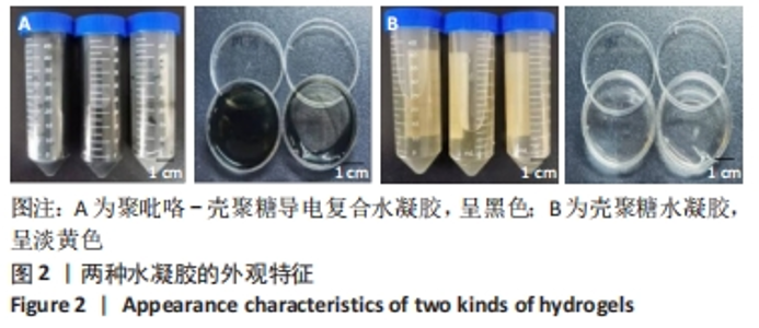
2.1 聚吡咯-壳聚糖导电复合水凝胶的大体结构 首先吡咯单体在水系统中聚合成聚吡咯,然后使用FeCl3作为氧化剂将聚吡咯接合到非导电天然材料壳聚糖主链上,最终合成呈黑色液体流动形式的聚吡咯-壳聚糖复合材料,后加入适量4%戊二醛交联为黑色水凝胶状,见图2A所示。未与聚吡咯耦合的单纯壳聚糖材料呈现偏黄色流动液体形态,加入4%戊二醛交联成淡黄色水凝胶,见图2B所示。"

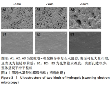
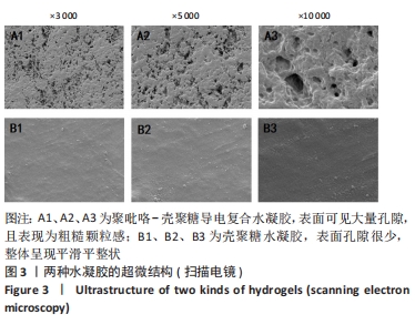
2.2 聚吡咯-壳聚糖导电复合水凝胶的微观特性 扫描电镜下可见,聚吡咯-壳聚糖导电复合水凝胶表面可见大量孔隙,且表现为粗糙颗粒感;壳聚糖水凝胶表面孔隙很少,整体呈现平滑平整状,见图3。聚吡咯-壳聚糖导电复合水凝胶的多孔性可以为承载细胞、输送治疗药物增加优势,并且有益于细胞的黏附、生长和生物因子的缓释。"

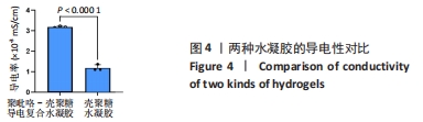
2.3 聚吡咯-壳聚糖导电复合水凝胶的导电性 聚吡咯-壳聚糖导电复合水凝胶的导电率为(3.19±0.03)×10-3 mS/cm,图4证明聚吡咯-壳聚糖导电复合水凝胶的导电性要高于壳聚糖水凝胶的导电性。聚吡咯-壳聚糖导电复合水凝胶这种优良的导电性能将会对心肌组织电生理的调节发挥重要作用。"

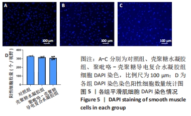
2.4 聚吡咯-壳聚糖导电复合水凝胶的生物相容性 将3组细胞进行DAPI细胞核染色比较细胞的生长情况,在倒置荧光显微镜下观察并进行拍照[16],见图5所示,与对照组相比,在两组水凝胶材料上生长的细胞均保持细胞完整性,3组细胞数量无明显差异,说明聚吡咯-壳聚糖导电复合水凝胶不影响细胞的正常生长,具有良好的生物相容性。"
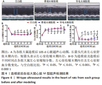
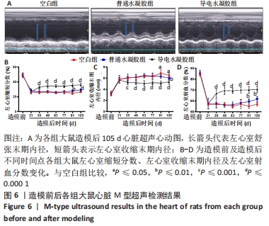
2.5 聚吡咯-壳聚糖导电复合水凝胶的体内实验结果 2.5.1 实验动物数量分析 30只大鼠全部进入结果分析。 2.5.2 超声心动图检查结果 与造模前相比,缺血-再灌注损伤后各组大鼠左心室收缩末期内径增大、左心室缩短分数和射血分数明显减小;经过不同治疗后,空白组和普通水凝胶组大鼠心脏收缩功能无明显好转,导电水凝胶组大鼠心脏收缩功能明显改善,见图6。可能是聚吡咯-壳聚糖导电复合水凝胶促进了心脏收缩功能的恢复。"
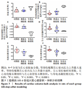
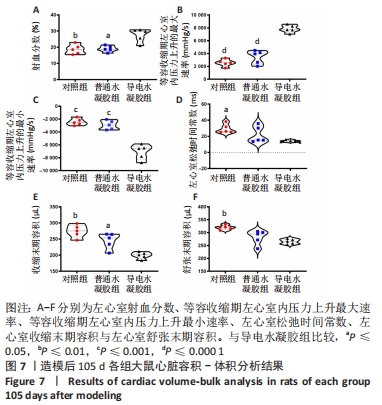
2.5.3 心脏压力-容积分析结果 与空白组、普通水凝胶组相比,导电水凝胶组大鼠等容收缩期左心室内压力上升最大速率增大、等容收缩期左心室内压力上升最小速率减小,左心室收缩末期和舒张末期容积减少,左心室射血分数增加,等容舒张期心室压延迟时间缩短,见图7。进一步说明聚吡咯-壳聚糖导电复合水凝胶具有改善心脏收缩功能的作用。"
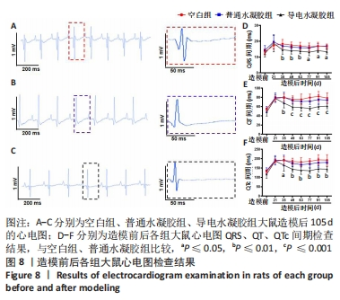
2.5.4 心电图检测结果 缺血-再灌注损伤前,各组大鼠心电图显示心脏电活动正常。缺血-再灌注损伤后,所有大鼠的QRS、QT间期延长约50%以上,即缺血-再灌注损伤破坏了心脏正常的电信号传播。造模后105 d,与空白组、普通水凝胶组相比,导电水凝胶组大鼠心电图表现出近20%-30%的QRS间期缩短和50%-60%的QT/QTc间期缩短,见图8。结果表明,使用聚吡咯-壳聚糖导电复合水凝胶可改善缺血-再灌注损伤大鼠心脏的电传导功能。"
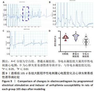
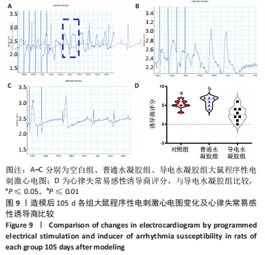
2.5.5 心脏程序性电刺激检测结果 程序性电刺激是诱导心律失常的标准临床方法。图9显示了各组大鼠程序性电刺激后心电图变化及诱导评分,与空白组和普通水凝胶组相比,导电水凝胶组大鼠心律失常评分明显降低,提示心律失常易感性较低。说明聚吡咯-壳聚糖导电复合水凝胶可以降低心律失常的发生。"
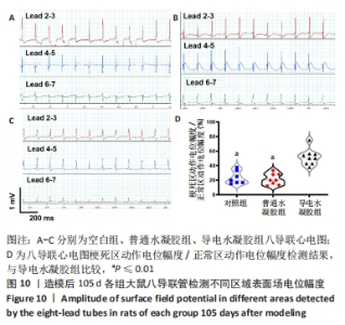
2.5.6 八导联心电图检测结果 由图10可见,与空白组和普通水凝胶组相比,导电水凝胶组大鼠心脏梗死瘢痕区具有最高的表面场电位幅度比(梗死瘢痕区电位幅度/正常区电位幅度),说明聚吡咯-壳聚糖导电复合水凝胶促进了梗死瘢痕区心肌的电传导。"
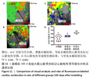
2.5.7 心脏荧光标记电传导速度检测结果 如图11所示,从心尖部向心底部电传导(箭头所示),当传导区域距离相同时,与空白组和普通凝胶组相比,导电水凝胶组传导所需时间更短(15.8 ms);3组电传导速度分析结果显示,导电水凝胶组纵向传导速度大于其他两组(P≤0.05,P≤0.001),表明聚吡咯-壳聚糖导电复合水凝胶极大改善了缺血-再灌注损伤后大鼠的心脏电信号传导。"


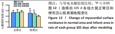
2.5.8 各组心脏表面电阻检测结果 如图12所示,与空白组和普通水凝胶组比较,导电水凝胶组大鼠心肌梗死瘢痕区表面电阻降低(即增大了梗死瘢痕区的电导性)(P < 0.05),正常区表面电阻改变不明显(P > 0.05)。"

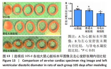
2.5.9 各组大鼠心脏标本环大体观察结果 如图13所示,各组大鼠心脏标本环中均有白色梗死区且有不同程度的左心室壁变薄,导电水凝胶组显示存在黑色材料,导电水凝胶组大鼠左心室舒张末期内径低于空白组、普通水凝胶组(P ≤0.01)。"
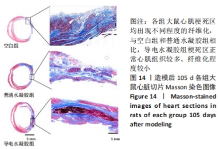
2.5.10 各组大鼠心脏组织Masson染色结果 如图14所示,各组大鼠心肌梗死区均出现不同程度的纤维化,与空白组和普通水凝胶组相比,导电水凝胶组梗死区正常心肌组织较多、纤维化程度较小。证明聚吡咯-壳聚糖导电复合水凝胶材料可降低缺血-再灌注损伤后的心室重构。"

| [1] HERR DJ, SINGH T, DHAMMU T, et al. Regulation of metabolism by mitochondrial enzyme acetylation in cardiac ischemia-reperfusion injury. Biochim Biophys Acta Mol Basis Dis. 2020;1866(6):165728. [2] MARINESCU MC, LAZAR AL, MARTA MM, et al. Non-Coding RNAs: Prevention, Diagnosis, and Treatment in Myocardial Ischemia-Reperfusion Injury. Int J Mol Sci. 2022;23(5):2728. [3] BRISTOW MR, SAXON LA, BOEHMER J, et al. Cardiac-resynchronization therapy with or without an implantable defibrillator in advanced chronic heart failure. N Engl J Med. 2004;350(21):2140-2150. [4] CLELAND JG, DAUBERT JC, ERDMANN E, et al. The effect of cardiac resynchronization on morbidity and mortality in heart failure. N Engl J Med. 2005;352(15):1539-1549. [5] HE S, WU J, LI SH, et al. The conductive function of biopolymer corrects myocardial scar conduction blockage and resynchronizes contraction to prevent heart failure. Biomaterials. 2020;258:120285. [6] GABETTI S, SILEO A, MONTRONE F, et al. Versatile electrical stimulator for cardiac tissue engineering-Investigation of charge-balanced monophasic and biphasic electrical stimulations. Front Bioeng Biotechnol. 2022;10:1031183. [7] CUI Z, NI NC, WU J, et al. Polypyrrole-chitosan conductive biomaterial synchronizes cardiomyocyte contraction and improves myocardial electrical impulse propagation. Theranostics. 2018;8(10):2752-2764. [8] MASSOUMI B, ABBASIAN M, JAHANBAN-ESFAHLAN R, et al. A novel bio-inspired conductive, biocompatible, and adhesive terpolymer based on polyaniline, polydopamine, and polylactide as scaffolding biomaterial for tissue engineering application. Int J Biol Macromol. 2020;147:1174-1184. [9] LEE M, KIM MC, LEE JY. Nanomaterial-Based Electrically Conductive Hydrogels for Cardiac Tissue Repair. Int J Nanomedicine. 2022;17: 6181-6200. [10] WANJARE M, KAWAMURA M, HU C, et al. Vascularization of Engineered Spatially Patterned Myocardial Tissue Derived From Human Pluripotent Stem Cells in vivo. Front Bioeng Biotechnol. 2019;7:208. [11] BEAUCHAMP P, JACKSON CB, OZHATHIL LC, et al. 3D Co-culture of hiPSC-Derived Cardiomyocytes With Cardiac Fibroblasts Improves Tissue-Like Features of Cardiac Spheroids. Front Mol Biosci. 2020;7:14. [12] LI Y, QIU X. Bioelectricity-coupling patches for repairing impaired myocardium. Wiley Interdiscip Rev Nanomed Nanobiotechnol. 2022;14(4):e1787. [13] LI Y, WEI L, LAN L, et al. Conductive biomaterials for cardiac repair: A review. Acta Biomater. 2022;139:157-178. [14] QUE W, HAN C, ZHAO X, et al. An ECG generative model of myocardial infarction. Comput Methods Programs Biomed. 2022;225:107062. [15] FAVERE K, VAN FRAEYENHOVE J, JACOBS G, et al. Cardiac electrophysiology studies in mice via the transjugular route: a comprehensive practical guide. Am J Physiol Heart Circ Physiol. 2022;323(4):H763-h773. [16] 姜增誉,阎长平,李健丁,等.新型导电复合材料聚吡咯壳聚糖对新生大鼠心室肌细胞钙信号传导的影响及可能机制[J].中西医结合心脑血管病杂志,2018,16(12): 1650-1655. [17] KOLETSI D, ILIADI A, TZANETAKIS GN, et al. Cardiovascular Disease and Chronic Endodontic Infection. Is There an Association? A Systematic Review and Meta-Analysis. Int J Environ Res Public Health. 2021;18(17):9111. [18] WANG Y, LI G, YANG L, et al. Development of Innovative Biomaterials and Devices for the Treatment of Cardiovascular Diseases. Adv Mater. 2022;34(46):e2201971. [19] HE M, WANG D, XU Y, et al. Nitric Oxide-Releasing Platforms for Treating Cardiovascular Disease. Pharmaceutics. 2022;14(7):1345. [20] AGGARWAL M, AGGARWAL B, RAO J. Integrative Medicine for Cardiovascular Disease and Prevention. Med Clin North Am. 2017;101(5):895-923. [21] DE GREGORIO C. Physical Training and Cardiac Rehabilitation in Heart Failure Patients. Adv Exp Med Biol. 2018;1067:161-181. [22] 郎丽敏,何生,姜增誉,等.导电复合材料在心肌梗死组织工程治疗领域的应用进展[J].中国组织工程研究,2021,25(22):3584-3590. [23] MCCARTNEY SL, PATEL C, DEL RIO JM. Long-term outcomes and management of the heart transplant recipient. Best Pract Res Clin Anaesthesiol. 2017;31(2):237-248. [24] LI Y, WEI L, LAN L, et al. Conductive biomaterials for cardiac repair: A review. Acta Biomater. 2022;139:157-178. [25] CHO S, DISCHER DE, LEONG KW, et al. Challenges and opportunities for the next generation of cardiovascular tissue engineering. Nat Methods. 2022;19(9):1064-1071. [26] MADONNA R, VAN LAAKE LW, BOTKER HE, et al. ESC Working Group on Cellular Biology of the Heart: position paper for Cardiovascular Research: tissue engineering strategies combined with cell therapies for cardiac repair in ischaemic heart disease and heart failure. Cardiovasc Res. 2019;115(3):488-500. [27] ASHTARI K, NAZARI H, KO H, et al. Electrically conductive nanomaterials for cardiac tissue engineering. Adv Drug Deliv Rev. 2019;144:162-179. [28] PHUTANE P, TELANGE D, AGRAWAL S, et al. Biofunctionalization and Applications of Polymeric Nanofibers in Tissue Engineering and Regenerative Medicine. Polymers (Basel). 2023;15(5):1202. [29] GHOVVATI M, KHARAZIHA M, ARDEHALI R, et al. Recent Advances in Designing Electroconductive Biomaterials for Cardiac Tissue Engineering. Adv Healthc Mater. 2022; 11(13):e2200055. [30] MOND HG, HELLAND JR, STOKES K, et al. The electrode-tissue interface: the revolutionary role of steroid-elution. Pacing Clin Electrophysiol. 2014;37(9):1232-1249. [31] BLACKBURN NJ, SOFRENOVIC T, KURAITIS D, et al. Timing underpins the benefits associated with injectable collagen biomaterial therapy for the treatment of myocardial infarction. Biomaterials. 2015;39:182-192. [32] CUPA J, STREBEL I, BADERTSCHER P, et al. Diagnostic and prognostic value of QRS duration and QTc interval in patients with suspected myocardial infarction. Cardiol J. 2018;25(5):601-610. [33] JASTRZĘBSKI M, MOSKAL P, HUYBRECHTS W, et al. Left bundle branch-optimized cardiac resynchronization therapy (LOT-CRT): Results from an international LBBAP collaborative study group. Heart Rhythm. 2022;19(1):13-21. [34] THEUNS D, VAN BOVEN N, SCHAER BA, et al. Predicting Early Mortality Among Implantable Defibrillator Patients Treated With Cardiac Resynchronization Therapy. J Card Fail. 2019; 25(10):812-818. [35] BEHAR JM, JACKSON T, HYDE E, et al. Optimized Left Ventricular Endocardial Stimulation Is Superior to Optimized Epicardial Stimulation in Ischemic Patients With Poor Response to Cardiac Resynchronization Therapy: A Combined Magnetic Resonance Imaging, Electroanatomic Contact Mapping, and Hemodynamic Study to Target Endocardial Lead Placement. JACC Clin Electrophysiol. 2016;2(7):799-809. [36] MORSINK M, SEVERINO P, LUNA-CERON E, et al. Effects of electrically conductive nano-biomaterials on regulating cardiomyocyte behavior for cardiac repair and regeneration. Acta Biomater. 2022;139:141-156. [37] MOUSAVI A, VAHDAT S, BAHEIRAEI N, et al. Multifunctional Conductive Biomaterials as Promising Platforms for Cardiac Tissue Engineering. ACS Biomater Sci Eng. 2021;7(1):55-82. [38] UL HAQ A, CAROTENUTO F, DE MATTEIS F, et al. Intrinsically Conductive Polymers for Striated Cardiac Muscle Repair. Int J Mol Sci. 2021;22(16):8550. [39] UL HAQ A, CAROTENUTO F, DI NARDO P, et al. Extrinsically Conductive Nanomaterials for Cardiac Tissue Engineering Applications. Micromachines (Basel). 2021;12(8):914. [40] WANG L, LIU Y, YE G, et al. Injectable and conductive cardiac patches repair infarcted myocardium in rats and minipigs. Nat Biomed Eng. 2021;5(10):1157-1173. |
| [1] | Yang Yifeng, Ye Nan, Wang Lin, Guo Shuaicheng, Huang Jian. Signaling pathway of dexmedetomidine against ischemia-reperfusion injury [J]. Chinese Journal of Tissue Engineering Research, 2024, 28(9): 1464-1469. |
| [2] | Lou Guo, Zhang Yan, Fu Changxi. Role of endothelial nitric oxide synthase in exercise preconditioning-induced improvement of myocardial ischemia-reperfusion injury [J]. Chinese Journal of Tissue Engineering Research, 2024, 28(8): 1283-1288. |
| [3] | Yue Yun, Wang Peipei, Yuan Zhaohe, He Shengcun, Jia Xusheng, Liu Qian, Li Zhantao, Fu Huiling, Song Fei, Jia Menghui. Effects of croton cream on JNK/p38 MAPK signaling pathway and neuronal apoptosis in cerebral ischemia-reperfusion injury rats [J]. Chinese Journal of Tissue Engineering Research, 2024, 28(8): 1186-1192. |
| [4] | Yang Yifeng, Huang Jian, Ye Nan, Wang Lin. Ischemia-reperfusion injury in total knee arthroplasty [J]. Chinese Journal of Tissue Engineering Research, 2024, 28(6): 955-960. |
| [5] | Wang Wu, Fan Xiaolei, Xie Jie, Hu Yihe, Zeng Min. Hydroxyapatite-polyvinyl alcohol/collagen-chitosan-gelatin composite hydrogel for repairing rabbit osteochondral defect [J]. Chinese Journal of Tissue Engineering Research, 2024, 28(5): 682-689. |
| [6] | Zhang Ya, Mu Qiuju, Wang Zilin, Liu Hongjie, Zhu Lili. Hydrogel loaded with platelet-rich plasma promotes wound healing in diabetic rats [J]. Chinese Journal of Tissue Engineering Research, 2024, 28(5): 690-696. |
| [7] | Shen Ziqing, Xia Tian, Shan Yibo, Zhu Ruijun, Wan Haoxin, Ding Hao, Pan Shu, Zhao Jun. Vascularized tracheal substitutes constructed by exosome-load hydrogel-modified 3D printed scaffolds [J]. Chinese Journal of Tissue Engineering Research, 2024, 28(5): 697-705. |
| [8] | Zhu Liwei, Wang Jiangyue, Bai Ding. Application value of nanocomposite gelatin methacryloyl hydrogels in different bone defect environments [J]. Chinese Journal of Tissue Engineering Research, 2024, 28(5): 753-758. |
| [9] | Chen Xiaofang, Zheng Guoshuang, Li Maoyuan, Yu Weiting. Preparation and application of injectable sodium alginate hydrogels [J]. Chinese Journal of Tissue Engineering Research, 2024, 28(5): 789-794. |
| [10] | Dai Jing, Liu Shasha, Shen Mingjing. Exosome-loaded injectable hydrogel for repairing bone defects around implants [J]. Chinese Journal of Tissue Engineering Research, 2024, 28(3): 347-354. |
| [11] | Gu Mingxi, Wang Changcheng, Tian Fengde, An Ning, Hao Ruihu, Guo Lin. Preparation and in vitro evaluation of a three-dimensional porous cartilage scaffold made of silk fibroin/gelatin/chitosan [J]. Chinese Journal of Tissue Engineering Research, 2024, 28(3): 366-372. |
| [12] | Cao Sheng, Kong Lingwei, Xu Kun, Sun Zhijie. Effect of gelatin methacryloyl hydrogel loaded with salvianolic acid B on intervertebral disc degeneration [J]. Chinese Journal of Tissue Engineering Research, 2024, 28(3): 380-386. |
| [13] | Bi Yujie, Ma Dujun, Peng Liping, Zhou Ziqiong, Zhao Jing, Zhu Houjun, Zhong Qiuhui, Yang Yuxin. Strategy and significance of Chinese medicine combined with medical hydrogel for disease treatment [J]. Chinese Journal of Tissue Engineering Research, 2024, 28(3): 419-425. |
| [14] | Chen Pinrui, Pei Xibo, Xue Yiyuan. Function and advantages of magnetically responsive hydrogel in bone tissue engineering [J]. Chinese Journal of Tissue Engineering Research, 2024, 28(3): 452-457. |
| [15] | Long Zhirui, Huang Lei, Xiao Fang, Wang Lin, Wang Xiaobei. Characteristics of hydrogel microspheres in bone tissue engineering [J]. Chinese Journal of Tissue Engineering Research, 2024, 28(3): 472-478. |
| Viewed | ||||||
|
Full text |
|
|||||
|
Abstract |
|
|||||