Chinese Journal of Tissue Engineering Research ›› 2024, Vol. 28 ›› Issue (10): 1497-1504.doi: 10.12307/2024.374
Previous Articles Next Articles
Various arginine configurations-modified chitosan hydrogels promote skin wound repair
Deng Jing, Li Tinghua, Zhu Hai, Yang Xiao, Cao Jun, Zhu Xiangdong
- College of Biomedical Engineering, Sichuan University, Chengdu 610064, Sichuan Province, China
-
Received:2023-05-13Accepted:2023-06-15Online:2024-04-08Published:2023-08-17 -
Contact:Zhu Xiangdong, Researcher, College of Biomedical Engineering, Sichuan University, Chengdu 610064, Sichuan Province, China -
About author:Deng Jing, Master candidate, College of Biomedical Engineering, Sichuan University, Chengdu 610064, Sichuan Province, China -
Supported by:Science and Technology Plan Project of Sichuan Province (Key Research and Development Project), No. 2020YFS0038 (to YX)
CLC Number:
Cite this article
Deng Jing, Li Tinghua, Zhu Hai, Yang Xiao, Cao Jun, Zhu Xiangdong. Various arginine configurations-modified chitosan hydrogels promote skin wound repair[J]. Chinese Journal of Tissue Engineering Research, 2024, 28(10): 1497-1504.
share this article
Add to citation manager EndNote|Reference Manager|ProCite|BibTeX|RefWorks
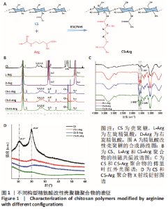
2.1 壳聚糖-精氨酸聚合物的合成及表征结果 如图1A所示,不同构型精氨酸可通过酰胺化反应键合到壳聚糖的侧链。核磁氢谱结果显示,壳聚糖-精氨酸聚合物中不仅出现了壳聚糖的特征吸收峰(δ=2.0×10⁻⁶),还在δ=1.5×10⁻⁶以及δ=3.0×10⁻⁶-3.2×10⁻⁶处均出现了精氨酸的特征吸收峰,以上结果说明精氨酸成功接枝到壳聚糖链上,见图1B。进一步,通过将精氨酸在a和d处-CH2特征吸收峰面积和壳聚糖的-CH3特征吸收峰面积进行比较,可以计算得到精氨酸的接枝率。CS-L-Arg、CS-D-Arg和CS-DL-Arg的接枝率分别为8.7%,9.2%和6.4%。 红外图谱结果如图1C所示,壳聚糖-精氨酸聚合物在 1 642 cm-1和1 549 cm-1 2个位置出现了新的吸收峰,分别是酰胺Ⅰ带C=O的伸缩振动吸收峰和酰胺Ⅱ带C-N伸缩振动和N-H变形振动的组合,说明了有新的酰胺键形成。同时,1 587 cm-1左右的N-H 弯曲振动峰被新生成的酰胺Ⅱ带覆盖,进一步证明了壳聚糖聚合物中的氨基和精氨酸分子中的羧基成功进行了酰胺化反应。 X射线衍射分析结果如图1D所示,未改性壳聚糖在衍射角为10.7°和19.8°处有2个明显的衍射峰,改性后壳聚糖的X衍射图谱有了明显改变,壳聚糖在10.7°处的衍射峰几乎完全消失,在19.8°处的衍射峰位置和强度都有变化,衍射角向大角度方向略有偏移,同时峰强度显著降低,这表明精氨酸的引入明显降低了壳聚糖的结晶度。"

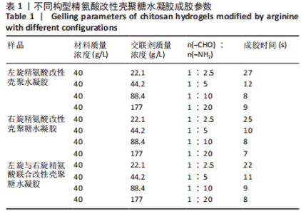
2.2 精氨酸改性壳聚糖水凝胶的构建及理化性能表征结果 首先考察壳聚糖-精氨酸聚合物和交联剂浓度对水凝胶理化性能的影响,分别配制质量分数4%聚合物与不同质量浓度的交联剂溶液,于室温条件下通过席夫碱反应成胶并记录成胶时间。结果如表1所示,随着交联剂质量浓度的增加,水凝胶成胶时间逐渐减少,同时水凝胶的强度增加、韧性变差。鉴于壳聚糖-精氨酸聚合物质量分数为4%、交联剂质量浓度为44.2 mg/mL形成的水凝胶强度、韧性等更适用于创面修复,故后续实验以该参数制备水凝胶进行评价。"

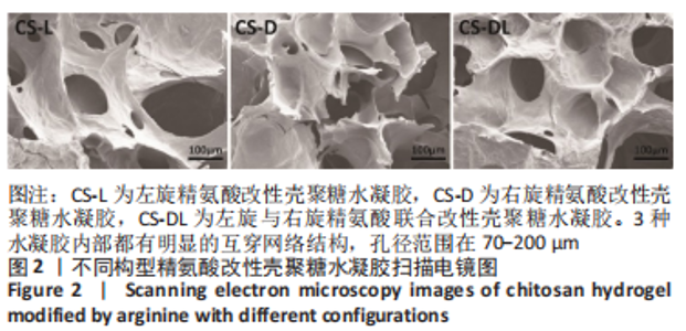
将构建的水凝胶冻干后用扫描电镜观察内部形貌,结果如图2所示,3种水凝胶内部都有明显的互穿网络结构,孔径范围在70-200 μm。水凝胶的溶胀检测结果显示,CS-L、CS-D和CS-DL水凝胶溶胀平衡时的吸水溶胀率分别为2 135%,2 288%和2 068%,见图3A,其中CS-DL水凝胶的接枝率略低,这可能源于该组聚合物的接枝率更低进而影响了分子间氢键作用。对水凝胶的降解能力进行了评价,结果如图3B所示,CS-D、CS-L和CS-DL水凝胶均呈现初期降解速度较快,之后开始缓慢降解的趋势,14 d的降解率分别为70.86%,67.99%和68.22%。"


"

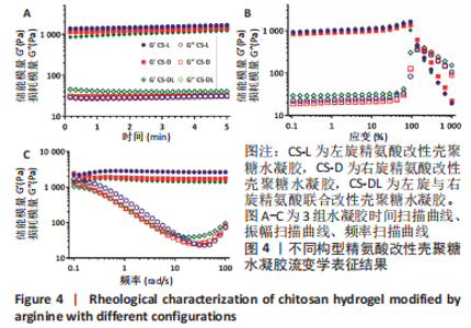
进一步考察3组水凝胶的流变学特征,结果如图4所示。由时间扫描曲线可知,3组水凝胶的储能模量(G’)均大于损耗模量(G”),表现为黏弹性固体行为特性,说明成功构建了水凝胶。同时,测量时间内3组水凝胶的储能模量和损耗模量基本保持稳定,G’约为1 000 Pa,G”为200-300 Pa。由振幅扫描曲线可知,在低应变区间(70%)以下时,3组水凝胶的G’和G”都保持稳定,此区间为水凝胶的线性黏弹区。当应变大于70%时,随着应变的增加,水凝胶的G’逐渐变小,G”逐渐变大,直至出现两条曲线相交,超过这个临界点后G’ < G”,说明水凝胶的结构已经被破坏,由黏弹性固体转变为液体状态。CS-L、CS-D、CS-DL水凝胶的转变临界值分别为158%,193%和132%。由频率扫描曲线可知,无论是在高频或者低频范围内,3组水凝胶的G’都始终大于G”,表明水凝胶形态一直维持,具有较好的稳定性。"

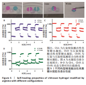
由图5A照片可知,水凝胶切成两半后重新贴附在一起,10 min过后用镊子可以轻松夹起水凝胶,两半水凝胶明显愈合为一块水凝胶,证明了制备的水凝胶具有良好的自愈合性能。进一步通过动态交替时间扫描定量表征水凝胶自愈合性能,结果如图5-D所示。当施加200%大应变时,3组水凝胶都是G’ < G”,这表明凝胶结构此时已经被破坏;但当应变恢复1%时,3组凝胶的G’和G”均很快都恢复至接近初始值,表明凝胶结构重新形成。在3次循环之后,3组水凝胶的储能模量和损耗模量几乎保持不变,证明了水凝胶具有良好的自愈合性能。"

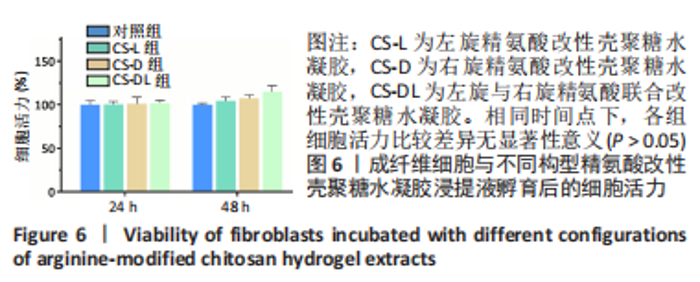
2.3 精氨酸改性壳聚糖水凝胶的生物相容性评价 采用CCK-8法评价3组水凝胶的细胞相容性,结果如图6所示。在培养24 h时,3组水凝胶浸提液的细胞存活率和对照组没有明显区别,待培养时间延长到48 h时,3组水凝胶浸提液中细胞存活率均高于对照组。细胞存活率结果说明,水凝胶对L929细胞没有毒性,具有良好的细胞生物相容性。"

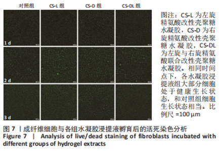
进一步,通过活死染色评价L929细胞和3组水凝胶浸提液共培养后的细胞状态,结果如图7所示。3组水凝胶与细胞培养1,2,3 d后,大部分细胞处于健康生长状态,和对照组细胞生长状态相当,证明3组水凝胶材料具有良好的细胞相容性。"


此外,通过溶血实验考察了构建的3种水凝胶的血液相容性,结果如图8所示。阳性对照组离心后明显看到上清液变为红色,说明红细胞已经被损坏破裂,血红蛋白逸出呈红色。然而,与各壳聚糖水凝胶进行共孵育后,上清液为淡黄色,与阴性对照生理盐水组一致。3组水凝胶的溶血率都远小于生物材料溶血安全临界标准(< 5%),说明3组水凝胶都具有良好的血液相容性。"


2.4 精氨酸改性壳聚糖水凝胶促成纤维细胞迁移 由图9A显微镜成像可知,孵育48 h后,3种水凝胶孵育组相较于对照组划痕剩余面积都更小,其中CS-D水凝胶组细胞迁移最快,说明构建的3种水凝胶均促进L929细胞的迁移。随后统计了各组未迁移划痕面积以及迁移率,结果如图9B、C所示。孵育48 h 后,对照组细胞划痕迁移率为69.1%,CS-L组、CS-D组和CS-DL组细胞划痕迁移率分别为81.9%,89.1%,84.3%,均明显高于对照组(P < 0.05)。综上,不同构型精氨酸改性壳聚糖水凝胶都能促进细胞迁移,进而促进细胞划痕的愈合。"


2.5 精氨酸改性壳聚糖水凝胶促巨噬细胞产生NO的能力 结果如图10所示,RAW264.7细胞在和水凝胶浸提液孵育24 h后,对照组产生了1.4 μmol/L的NO,而CS-L、CS-D和CS-DL组分别产生了5.17,1.87和1.69 μmol/L的NO。可以看出,只有CS-L组具有明显诱导巨噬细胞的NO生成的能力,这与研究表明的左旋精氨酸为NO天然供体一致[29]。"


2.6 精氨酸改性壳聚糖水凝胶促巨噬细胞分型 由图11可知,RAW264.7巨噬细胞与3组水凝胶浸提液共孵育后,无论是CD86 蛋白(M1 型巨噬细胞标志物)还是CD206蛋白(M2型巨噬细胞标志物)的平均荧光强度值都比对照组高(P < 0.05),并且CS-D组的平均荧光强度值最高(P < 0.05)。以上结果说明,3种水凝胶具有促巨噬细胞极化的能力,其中CS-D水凝胶的促巨噬细胞极化能力最强。"

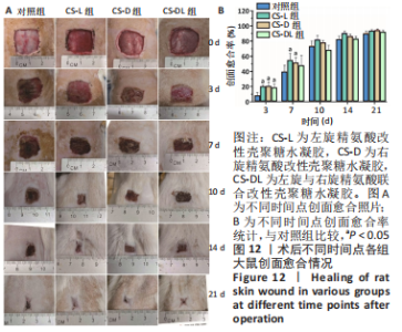
2.7 精氨酸改性壳聚糖水凝胶促大鼠全层缺损创面愈合能力评价 2.7.1 实验动物数量分析 36只大鼠全部进入结果分析。 2.7.2 各组大鼠创面愈合情况 术后不同时间的创面愈合照片如图12A,相比对照组,3种水凝胶促创面修复的能力更强。进一步对创面愈合率统计,如图12B所示,CS-L组和CS-D组术后各个时间点的创面愈合率都要高于对照组,特别是第3天和第7天明显高于对照组(P < 0.05)。术后21 d,CS-L组、CS-D组和CS-DL组创面愈合率分别为94.0%,94.5%,92.3%,而对照组创面愈合率不到90%。综上说明了不同构型精氨酸改性壳聚糖水凝胶都具有促进创面愈合的作用。"

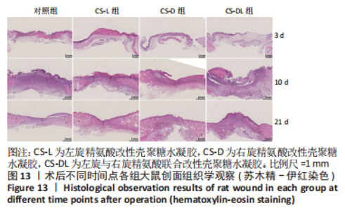
2.7.3 各组大鼠创面组织形态学观察 苏木精-伊红染色显示,术后3 d,各组大鼠创面组织中都有大量炎性细胞浸润,伴有新生血管以及成纤维细胞;术后10 d,对照组创面中仍有较多的炎性细胞浸润,相较而言,各实验组炎症情况有所改善,特别是CS-D组炎性细胞减少较为明显;术后21 d,各组创面上皮修复良好,CS-L组和CS-D组基本没有炎性细胞浸润,对照组还有少量炎性细胞浸润,见图13。"

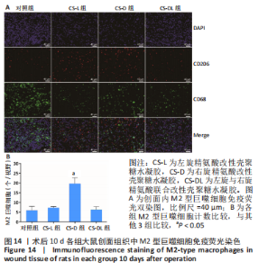
2.7.4 各组大鼠创面组织巨噬细胞表型极化分析 术后10 d,各组大鼠创面组织中M2型巨噬细胞免疫荧光染色结果如图14所示。对照组、CS-L组、CS-D组和CS-DL组创面组织中的M2巨噬细胞数目分别为(6.00±1.63),(7.33±0.47),(19.67±2.49),(6.33±1.25)个/视野,其中CS-D组创面中M2型巨噬细胞数量最多(P < 0.05)。"

| [1] KOEHLER J, BRANDL FP, GOEPFERICH AM. Hydrogel wound dressings for bioactive treatment of acute and chronic wounds. Eur Polym J. 2018;100:1-11. [2] WINTER GD. Formation of the scab and the rate of epithelization of superficial wounds in the skin of the young domestic pig. Nature. 1962;193:293-294. [3] DONG RN, GUO BL. Smart wound dressings for wound healing. Nano Today. 2021;41:22. [4] GHOMI ER, KHALILI S, KHORASANI SN, et al. Wound dressings: Current advances and future directions. J Appl Polym Sci. 2019;136(27):12. [5] FARAHANI M, SHAFIEE A. Wound Healing: From Passive to Smart Dressings. Adv Healthc Mater. 2021;10(16):32. [6] GALLASTEGUI A, SPESIA MB, DELL’ERBA IE, et al. Controlled release of antibiotics from photopolymerized hydrogels: Kinetics and microbiological studies. Mater Sci Eng C-Mater Biol Appl. 2019;102:896-905. [7] SU JJ, LI JK, LIANG JH, et al. Hydrogel Preparation Methods and Biomaterials for Wound Dressing. Life-Basel. 2021;11(10):22. [8] LIANG YP, HE JH, GUO BL. Functional Hydrogels as Wound Dressing to Enhance Wound Healing. ACS Nano. 2021;15(8):12687-12722. [9] CHEN SJ, TIAN HR, MAO JL, et al. Preparation and application of chitosan-based medical electrospun nanofibers. Int J Biol Macromol. 2023;226:410-422. [10] TORKAMAN S, RAHMANI H, ASHORI A, et al. Modification of chitosan using amino acids for wound healing purposes: A review. Carbohydr Polym. 2021;258:8. [11] QU J, ZHAO X, LIANG YP, et al. Antibacterial adhesive injectable hydrogels with rapid self-healing, extensibility and compressibility as wound dressing for joints skin wound healing. Biomaterials. 2018;183:185-199. [12] LI H, ZHOU XJ, LUO L, et al. Bio-orthogonally crosslinked catechol-chitosan hydrogel for effective hemostasis and wound healing. Carbohydr Polym. 2022;281:11. [13] HRISTINA K, LANGERHOLC T, TRAPECAR M. Novel metabolic roles of L-arginine in body energy metabolism and possible clinical applications. J Nutr Health Aging. 2014;18(2):213-218. [14] HUSSEIN Y, EL-FAKHARANY EM, KAMOUN EA, et al. Electrospun PVA/hyaluronic acid/L-arginine nanofibers for wound healing applications: Nanofibers optimization and in vitro bioevaluation. Int J Biol Macromol. 2020;164:667-676. [15] YU J, ZHANG RL, CHEN BH, et al. Injectable Reactive Oxygen Species-Responsive Hydrogel Dressing with Sustained Nitric Oxide Release for Bacterial Ablation and Wound Healing. Adv Funct Mater. 2022;32(33):11. [16] PELUFFO RD. L-Arginine currents in rat cardiac ventricular myocytes. J Physiol-London. 2007; 580(3):925-936. [17] LIM HK, RAHIM AB, LEO VI, et al. Polyamine Regulator AMD1 Promotes Cell Migration in Epidermal Wound Healing. J Invest Dermatol. 2018;138(12):2653-2665. [18] ITO D, ITO H, IDETA T, et al. Systemic and topical administration of spermidine accelerates skin wound healing. Cell Commun Signal. 2021;19(1):12. [19] LING ZX, DENG J, ZHANG ZR, et al. Spatiotemporal manipulation of L-arginine release from bioactive hydrogels initiates rapid skin wound healing accompanied with repressed scar formation. Appl Mater Today. 2021;24:16. [20] MARIANI E, LISIGNOLI G, BORZI RM, et al. Biomaterials: Foreign Bodies or Tuners for the Immune Response? Int J Mol Sci. 2019;20(3):42. [21] VISHWAKARMA A, BHISE NS, EVANGELISTA MB, et al. Engineering Immunomodulatory Biomaterials To Tune the Inflammatory Response. Trends Biotechnol. 2016;34(6): 470-482. [22] JULIER Z, PARK AJ, BRIQUEZ PS, et al. Promoting tissue regeneration by modulating the immune system. Acta Biomater. 2017;53:13-28. [23] LYNCH RI, LAVELLE EC. Immuno-modulatory biomaterials as anti-inflammatory therapeutics. Biochem Pharmacol. 2022;197:17. [24] WYNN TA, VANNELLA KM. Macrophages in Tissue Repair, Regeneration, and Fibrosis. Immunity. 2016;44(3):450-462. [25] QIAN YN, ZHENG YJ, JIN J, et al. Immunoregulation in Diabetic Wound Repair with a Photoenhanced Glycyrrhizic Acid Hydrogel Scaffold. Adv Mater. 2022;34(29):15. [26] GREEN DW, LEE JM, KIM EJ, et al. Chiral Biomaterials: From Molecular Design to Regenerative Medicine. Adv Mater Interfaces. 2016;3(6):13. [27] DUAN YY, ZHENG HH, LI ZH, et al. Unsaturated polyurethane films grafted with enantiomeric polylysine promotes macrophage polarization to a M2 phenotype through PI3K/Akt1/mTOR axis. Biomaterials. 2020;246:14. [28] GRIFFIN DR, ARCHANG MM, KUAN CH, et al. Activating an adaptive immune response from a hydrogel scaffold imparts regenerative wound healing. Nat Mater. 2021;20(4):560-569. [29] ZHU JW, TIAN J, YANG C, et al. L-Arg-Rich Amphiphilic Dendritic Peptide as a Versatile NO Donor for NO/Photodynamic Synergistic Treatment of Bacterial Infections and Promoting Wound Healing. Small. 2021;17(32):18. [30] WILKINSON HN, HARDMAN MJ. Wound healing: cellular mechanisms and pathological outcomes. Open Biol. 2020;10(9):14. [31] RAZIYEVA K, KIM Y, ZHARKINBEKOV Z, et al. Immunology of Acute and Chronic Wound Healing. Biomolecules. 2021;11(5):25. [32] HE YX, LI Y, SUN YD, et al. A double-network polysaccharide-based composite hydrogel for skin wound healing. Carbohydr Polym. 2021;261:11. [33] ZHANG M, YANG M, WOO MW, et al. High-mechanical strength carboxymethyl chitosan-based hydrogel film for antibacterial wound dressing. Carbohydr Polym. 2021;256:9. [34] ZOU FX, WANG YS, ZHENG YD, et al. A novel bioactive polyurethane with controlled degradation and L-Arg release used as strong adhesive tissue patch for hemostasis and promoting wound healing. Bioact Mater. 2022;17:471-487. [35] AHMED R, AUGUSTINE R, CHAUDHRY M, et al. Nitric oxide-releasing biomaterials for promoting wound healing in impaired diabetic wounds: State of the art and recent trends. Biomed Pharmacother. 2022;149:12707. [36] FENG ZJ, SU Q, ZHANG CN, et al. Bioinspired Nanofibrous Glycopeptide Hydrogel Dressing for Accelerating Wound Healing: A Cytokine-Free, M2-Type Macrophage Polarization Approach. Adv Funct Mater. 2020;30(52):13. [37] EMING SA, KRIEG T, DAVIDSON JM. Inflammation in wound repair: Molecular and cellular mechanisms. J Invest Dermatol. 2007;127(3):514-525. [38] MAO JY, CHEN L, CAI ZW, et al. Advanced Biomaterials for Regulating Polarization of Macrophages in Wound Healing. Adv Funct Mater. 2022;32(12):25. [39] LIU GW, MA HX, QIU L, et al. Phenotypic and functional switch of macrophages induced by regulatory CD4(+)CD25(+) T cells in mice. Immunol Cell Biol. 2011;89(1):130-142. [40] ZHANG W, XIA SZ, WENG TT, et al. Antibacterial coaxial hydro-membranes accelerate diabetic wound healing by tuning surface immunomodulatory functions. Mater Today Bio. 2022;16:18. [41] YUAN Y, FAN DD, SHEN SH, et al. An M2 macrophage-polarized anti-inflammatory hydrogel combined with mild heat stimulation for regulating chronic inflammation and impaired angiogenesis of diabetic wounds. Chem Eng J. 2022;433:18. |
| [1] | Wang Weiqing, Zhou Yue. Chronic inflammation regulates adipose tissue fibrosis [J]. Chinese Journal of Tissue Engineering Research, 2024, 28(8): 1307-1312. |
| [2] | Zeng Fanzhuo, Li Yuxin, Sun Jiachen, Gu Xinyang, Wen Shan, Tian He, Mei Xifan. Efficient strategies for microglia replacement in spinal cord injury models [J]. Chinese Journal of Tissue Engineering Research, 2024, 28(7): 1007-1014. |
| [3] | Chen Xiaofang, Zheng Guoshuang, Li Maoyuan, Yu Weiting. Preparation and application of injectable sodium alginate hydrogels [J]. Chinese Journal of Tissue Engineering Research, 2024, 28(5): 789-794. |
| [4] | Wang Wu, Fan Xiaolei, Xie Jie, Hu Yihe, Zeng Min. Hydroxyapatite-polyvinyl alcohol/collagen-chitosan-gelatin composite hydrogel for repairing rabbit osteochondral defect [J]. Chinese Journal of Tissue Engineering Research, 2024, 28(5): 682-689. |
| [5] | Zhang Ya, Mu Qiuju, Wang Zilin, Liu Hongjie, Zhu Lili. Hydrogel loaded with platelet-rich plasma promotes wound healing in diabetic rats [J]. Chinese Journal of Tissue Engineering Research, 2024, 28(5): 690-696. |
| [6] | Shen Ziqing, Xia Tian, Shan Yibo, Zhu Ruijun, Wan Haoxin, Ding Hao, Pan Shu, Zhao Jun. Vascularized tracheal substitutes constructed by exosome-load hydrogel-modified 3D printed scaffolds [J]. Chinese Journal of Tissue Engineering Research, 2024, 28(5): 697-705. |
| [7] | Zhu Liwei, Wang Jiangyue, Bai Ding. Application value of nanocomposite gelatin methacryloyl hydrogels in different bone defect environments [J]. Chinese Journal of Tissue Engineering Research, 2024, 28(5): 753-758. |
| [8] | Yang Yuqing, Chen Zhiyu. Role and application of early transient presence of M1 macrophages in bone tissue engineering [J]. Chinese Journal of Tissue Engineering Research, 2024, 28(4): 594-601. |
| [9] | Xing Hao, Meng Qingfeng, Chang Zhengqi. Mechanism of negative pressure wound therapy in the auxiliary treatment of bone and soft tissue infection [J]. Chinese Journal of Tissue Engineering Research, 2024, 28(4): 621-626. |
| [10] | Dai Jing, Liu Shasha, Shen Mingjing. Exosome-loaded injectable hydrogel for repairing bone defects around implants [J]. Chinese Journal of Tissue Engineering Research, 2024, 28(3): 347-354. |
| [11] | Gu Mingxi, Wang Changcheng, Tian Fengde, An Ning, Hao Ruihu, Guo Lin. Preparation and in vitro evaluation of a three-dimensional porous cartilage scaffold made of silk fibroin/gelatin/chitosan [J]. Chinese Journal of Tissue Engineering Research, 2024, 28(3): 366-372. |
| [12] | Cao Sheng, Kong Lingwei, Xu Kun, Sun Zhijie. Effect of gelatin methacryloyl hydrogel loaded with salvianolic acid B on intervertebral disc degeneration [J]. Chinese Journal of Tissue Engineering Research, 2024, 28(3): 380-386. |
| [13] | Bi Yujie, Ma Dujun, Peng Liping, Zhou Ziqiong, Zhao Jing, Zhu Houjun, Zhong Qiuhui, Yang Yuxin. Strategy and significance of Chinese medicine combined with medical hydrogel for disease treatment [J]. Chinese Journal of Tissue Engineering Research, 2024, 28(3): 419-425. |
| [14] | Chen Pinrui, Pei Xibo, Xue Yiyuan. Function and advantages of magnetically responsive hydrogel in bone tissue engineering [J]. Chinese Journal of Tissue Engineering Research, 2024, 28(3): 452-457. |
| [15] | Long Zhirui, Huang Lei, Xiao Fang, Wang Lin, Wang Xiaobei. Characteristics of hydrogel microspheres in bone tissue engineering [J]. Chinese Journal of Tissue Engineering Research, 2024, 28(3): 472-478. |
| Viewed | ||||||
|
Full text |
|
|||||
|
Abstract |
|
|||||