Chinese Journal of Tissue Engineering Research ›› 2024, Vol. 28 ›› Issue (16): 2534-2541.doi: 10.12307/2024.225
Previous Articles Next Articles
Aerobic exercise modulates mitochondrial quality control system to reverse cardiac pathological remodeling in aging rats
Tang Liang1, Wang Hexia1, Wang Qingbo2, Pi Yihua2, Zhang Yan2
- 1College of Physical Education and Arts Humanities, China University of Petroleum, Beijing 102249, China; 2Guangxi University of Chinese Medicine, Nanning 530021, Guangxi Zhuang Autonomous Region, China
-
Received:2022-12-19Accepted:2023-03-02Online:2024-06-08Published:2023-07-29 -
Contact:Zhang Yan, Master, Associate professor, Guangxi University of Chinese Medicine, Nanning 530021, Guangxi Zhuang Autonomous Region, China -
About author:Tang Liang, Master, Associate professor, College of Physical Education and Arts Humanities, China University of Petroleum, Beijing 102249, China -
Supported by:Guangxi Educational Science “13th Five-Year Plan” Project, No. 2017C386 (to ZY)
CLC Number:
Cite this article
Tang Liang, Wang Hexia, Wang Qingbo, Pi Yihua, Zhang Yan. Aerobic exercise modulates mitochondrial quality control system to reverse cardiac pathological remodeling in aging rats[J]. Chinese Journal of Tissue Engineering Research, 2024, 28(16): 2534-2541.
share this article
Add to citation manager EndNote|Reference Manager|ProCite|BibTeX|RefWorks
2.1 实验动物数量分析 实验过程中,由于意外死亡、拒跑等原因,最终样本量为n=39(青年安静组n=15、老年安静组n=13、老年运动组n=11)。 2.2 各组大鼠体质量、心脏质量和心脏指数比较 与青年安静组比较,老年安静组和老年运动组大鼠体质量、心脏质量、心脏质量指数、左心室质量和左心室质量指数增加(P < 0.05);与老年安静组比较,老年运动组大鼠体质量下降(P < 0.05),各心脏质量参数无明显变化(P > 0.05),见表1。"
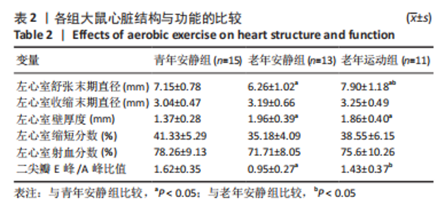
2.3 各组大鼠心脏结构与功能比较 与青年安静组比较,老年安静组大鼠左心室舒张末期直径下降(P < 0.05),左心室壁厚度增加(P < 0.05),二尖瓣E峰/A峰比值下降(P < 0.05),左心室收缩末期直径、左心室缩短分数和左心室射血分数无明显变化(P > 0.05);与青年安静组比较,老年运动组左心室舒张末期直径和左心室壁厚度增加(P < 0.05),左心室收缩末期直径、左心室缩短分数、左心室射血分数和二尖瓣E峰/A峰比值无明显变化(P > 0.05);与老年安静组比较,老年运动组大鼠左心室舒张末期直径和二尖瓣E峰/A峰比值升高(P < 0.05),左心室收缩末期直径、左心室壁厚度、左心室缩短分数和左心室射血分数无明显变化(P > 0.05),见表2。"
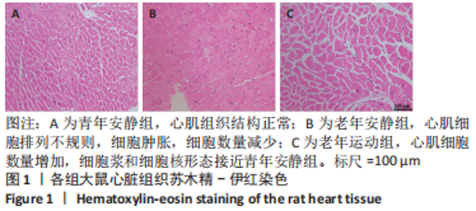
2.4 各组大鼠心脏病理组织学、心肌细胞凋亡和心肌线粒体超微结构观察 心脏苏木精-伊红染色显示,青年安静组大鼠心肌组织结构正常;老年安静组大鼠心肌细胞排列不规则,细胞肿胀,细胞数量减少;老年运动组大鼠心肌细胞数量增加,细胞浆和细胞核形态接近青年安静组,见图1。与青年安静组比较,老年安静组和老年运动组大鼠心肌细胞横截面积升高(P < 0.05);与老年安静组比较,老年运动组心肌细胞横截面积无明显变化(P > 0.05),见图2A。各组心肌细胞计数比较显示,与青年安静组比较,老年安静和老年运动组大鼠心肌细胞计数减少(P < 0.05);与老年安静组比较,老年运动组心肌细胞计数增加(P < 0.05),见图2B。"

"
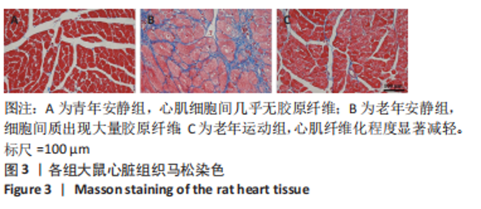
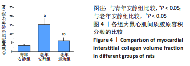
心脏马松染色显示,青年安静组大鼠心肌细胞间几乎无胶原纤维;老年安静组大鼠心肌细胞间质出现大量胶原纤维;老年运动组大鼠心肌纤维化程度显著减轻,见图3。与青年安静组比较,老年安静组和老年运动组大鼠心肌胶原容积分数增加(P < 0.05);与老年安静组比较,老年运动组大鼠心肌胶原容积分数降低(P < 0.05),见图4。"


"
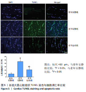
各组大鼠心脏组织TUNEL染色显示,正常细胞核呈蓝色,TUNEL阳性细胞核呈绿色,见图5。与青年安静组比较,老年安静组和老年运动组大鼠心肌细胞凋亡率增加(P < 0.05);与老年安静组比较,老年运动组大鼠心肌细胞凋亡率降低(P < 0.05)。"

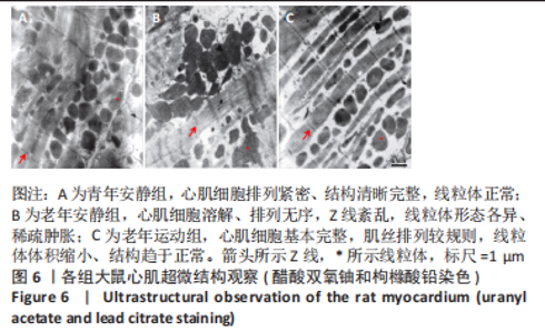
电子显微镜观察心肌超微结构显示,青年安静组大鼠心肌细胞排列紧密、结构清晰完整,线粒体排列整齐、形态呈圆形或卵圆形;老年安静组大鼠心肌细胞溶解、排列无序,Z线紊乱,线粒体形态各异、稀疏肿胀;老年运动组大鼠心肌细胞基本完整,肌丝排列较规则,线粒体体积缩小、结构趋于正常,见图6。"

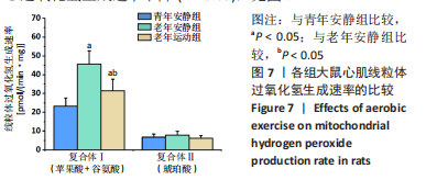
2.5 各组大鼠心肌线粒体功能的比较 线粒体过氧化氢生成速率:与青年安静组比较,老年安静组大鼠心肌线粒体复合体Ⅰ过氧化氢生成速率升高(P < 0.05),复合体Ⅱ过氧化氢生成速率无明显变化(P > 0.05);与老年安静组比较,老年运动组复合体大鼠心肌线粒体复合体Ⅰ过氧化氢生成速率下降(P < 0.05),见图7。"
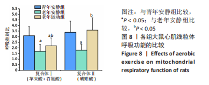
线粒体呼吸功能:与青年安静组比较,老年安静组大鼠心肌线粒体复合体Ⅰ和复合体Ⅱ态3、态4呼吸速率、呼吸控制比均下降(P < 0.05);与老年安静组比较,老年运动组大鼠心肌线粒体复合体Ⅰ和复合体Ⅱ态3、态4呼吸速率、呼吸控制比升高(P < 0.05),见图8。"


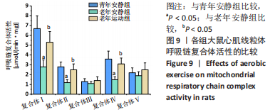
线粒体呼吸链复合体活性:与青年安静组比较,老年安静组大鼠心肌线粒体呼吸链复合体Ⅰ、Ⅱ和Ⅳ活性下降(P < 0.05),复合体Ⅲ和Ⅴ活性无明显变化(P > 0.05);与老年安静组比较,老年运动组大鼠心肌线粒体呼吸链复合体Ⅰ、Ⅱ和Ⅳ活性升高(P < 0.05),见图9。"
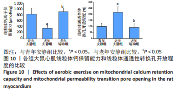
线粒体钙保留能力和线粒体通透性转换孔开放程度:与青年安静组比较,老年安静组大鼠心肌线粒体钙保留能力下降(P < 0.05)、线粒体通透性转换孔开放程度增加(P < 0.05);与老年安静组比较,老年运动组大鼠心肌线粒体钙保留能力升高(P < 0.05),线粒体通透性转换孔开放程度降低(P < 0.05),见图10。"

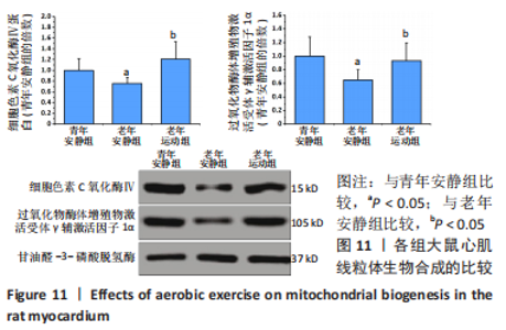
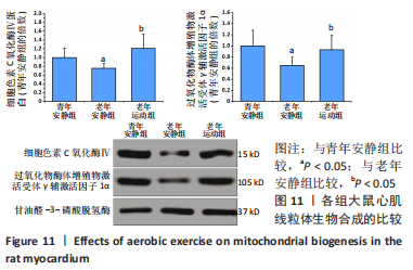
2.6 各组大鼠心肌线粒体质量控制系统的比较 线粒体生物合成:与青年安静组比较,老年安静组大鼠心肌线粒体细胞色素C氧化酶Ⅳ和过氧化物酶体增殖物激活受体γ辅激活因子1α的蛋白表达降低(P < 0.05);与老年安静组比较,老年运动组大鼠心肌线粒体细胞色素C氧化酶Ⅳ和过氧化物酶体增殖物激活受体γ辅激活因子1α的蛋白表达升高(P < 0.05),见图11。"

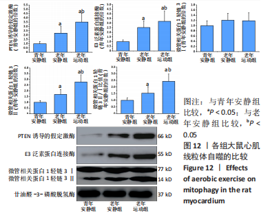
线粒体自噬:与青年安静组比较,老年安静组大鼠心肌线粒体PTEN诱导的假定激酶、E3泛素蛋白连接酶、微管相关蛋白1轻链3Ⅱ和微管相关蛋白1轻链3Ⅱ/Ⅰ比值增加(P < 0.05);与老年安静组比较,老年运动组大鼠心肌线粒体PTEN诱导的假定激酶、E3泛素蛋白连接酶、微管相关蛋白1轻链3Ⅱ和微管相关蛋白1轻链3Ⅱ/Ⅰ比值进一步升高(P < 0.05),见图12。"


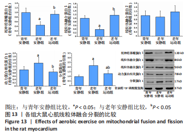
线粒体融合分裂:与青年安静组比较,老年安静组大鼠心肌线粒体视神经萎缩蛋白1和线粒体融合蛋白1表达降低(P < 0.05),动力蛋白相关蛋白1和分裂蛋白1表达升高(P < 0.05);与老年安静组比较,老年运动组大鼠心肌线粒体视神经萎缩蛋白1和线粒体融合蛋白1表达升高(P < 0.05),动力蛋白相关蛋白1和分裂蛋白1蛋白表达下降(P < 0.05)。 见图13。"
| [1] OBAS V, VASAN RS. The aging heart. Clin Sci (Lond). 2018;132(13):1367-1382. [2] TRACY E, ROWE G, LEBLANC AJ. Cardiac tissue remodeling in healthy aging: the road to pathology. Am J Physiol Cell Physiol. 2020;319(1):C166-182. [3] TAHRIR FG, LANGFORD D, AMINI S, et al. Mitochondrial quality control in cardiac cells: mechanisms and role in cardiac cell injury and disease. J Cell Physiol. 2019;234(6):8122-8133. [4] WANG J, LI S, WANG J, et al. Spermidine alleviates cardiac aging by improving mitochondrial biogenesis and function. Aging (Albany NY). 2020;12(1):650-671. [5] MARZETTI E, CALVANI R, CESARI M, et al. Mitochondrial dysfunction and sarcopenia of aging: from signaling pathways to clinical trials. Int J Biochem Cell Biol. 2013;45(10):2288-2301. [6] HONG YX, WU WY, SONG F, et al. Cardiac senescence is alleviated by the natural flavone acacetin via enhancing mitophagy. Aging (Albany NY). 2021;13(12):16381-16403. [7] PICCA A, MANKOWSKI RT, BURMAN JL, et al. Mitochondrial quality control mechanisms as molecular targets in cardiac ageing. Nat Rev Cardiol. 2018;15(9):543-554. [8] JAKOVLJEVIC DG. Physical activity and cardiovascular aging: physiological and molecular insights. Exp Gerontol. 2018;109:67-74. [9] 首健,陈佩杰,肖卫华.线粒体在骨骼肌老化中的作用及运动的改善效应[J].中国康复医学杂志,2021,36(4):499-504. [10] 李德生,陈宁,孟思进.运动对骨骼肌线粒体质量控制的调节作用[J].武汉体育学院学报,2014,48(5):56-59. [11] 陶丽婵,贾方.运动训练对心脏衰老的保护机制[J].上海大学学报(自然科学版), 2017,23(6):828-834. [12] 黄伟.有氧运动对衰老大鼠心肌保护作用的机制研究[J].山东体育科技,2013,35(6):67-71. [13] 张艳,何瑞波,王庆博,等.不同负荷量有氧运动对肥胖大鼠骨骼肌炎症反应和胰岛素信号途径的影响及机制[J].中国组织工程研究,2023,27(8):1237-1244. [14] 何炜,李玉明,周欣,等.短时高强度间歇运动训练心肌梗死模型大鼠心室重构及线粒体变化[J]. 中国组织工程研究,2016,20(40):5986-5993. [15] 马玉珍,彭朋,秦永生,等.NLRP3炎性小体抑制剂VX-765对急性心肌梗死大鼠心功能及线粒体能量代谢的影响[J].武警后勤学院学报(医学版),2017,26(9):742-745. [16] 张桂忠,姜宁,薄海,等.急性运动中线粒体能量转换调节的生物力能学分析:ROS和UCP3的作用[J].中国运动医学杂志,2006,25(2):161-167. [17] 王增喜,李洁,王悦.高强度间歇训练对大鼠心肌线粒体呼吸链复合体活性的影响[J].中国运动医学杂志,2018,37(4):315-322. [18] 彭成欢,向常清,张世忠,等.基于线粒体通透性转换孔调控的脓毒症心肌损伤及普拉克索保护机制的研究[J].中国医院药学杂志,2022,42(20):2115-2120. [19] THAM YK, BERNARDO BC, OOI JY, et al. Pathophysiology of cardiac hypertrophy and heart failure:signaling pathways and novel therapeutic targets. Arch Toxicol. 2015;89(9):1401-1438. [20] MA ZG, YUAN YP, WU HM, et al. Cardiac fibrosis: new insights into the pathogenesis. Int J Biol Sci. 2018;14(12):1645-1657. [21] KYSELOVIČ J, LEDDY JJ. Cardiac fibrosis: the beneficial effects of exercise in cardiac fibrosis. Adv Exp Med Biol. 2017;999:257-268. [22] OLDFIELD CJ, DUHAMEL TA, DHALLA NS. Mechanisms for the transition from physiological to pathological cardiac hypertrophy. Can J Physiol Pharmacol. 2020;98(2):74-84. [23] GEORGE K, WHYTE GP, GREEN DJ, et al. The endurance athletes heart: acute stress and chronic adaptation. Br J Sports Med. 2012;46(Suppl 1):29-36. [24] POMATTO L, DAVIES K. Adaptive homeostasis and the free radical theory of ageing. Free Radic Biol Med. 2018;124:420-430. [25] SHADEL GS, HORVATH TL. Mitochondrial ROS signaling in organismal homeostasis. Cell. 2015;163(3):560-569. [26] ZOROV DB, JUHASZOVA M, SOLLOTT SJ. Mitochondrial reactive oxygen species (ROS) and ROS-induced ROS release. Physiol Rev. 2014;94(3):909-950. [27] PFANNER N, WARSCHEID B, WIEDEMANN N. Mitochondrial proteins: from biogenesis to functional networks. Nat Rev Mol Cell Biol. 2019;20(5):267-284. [28] CAMPOS JC, QUELICONI BB, BOZI L, et al. Exercise reestablishes autophagic flux and mitochondrial quality control in heart failure. Autophagy. 2017;13(8):1304-1317. [29] BAUER TM, MURPHY E. Role of mitochondrial calcium and the permeability transition pore in regulating cell death. Circ Res. 2020;126(2):280-293. [30] BERTERO E, MAACK C. Calcium signaling and reactive oxygen species in mitochondria. Circ Res. 2018;122(10):1460-1478. [31] PELLEGRINO-COPPOLA D. Regulation of the mitochondrial permeability transition pore and its effects on aging. Microb Cell. 2020;7(9):222-233. [32] THOMAS MM, VIGNA C, BETIK AC, et al. Initiating treadmill training in late middle age offers modest adaptations in Ca2+ handling but enhances oxidative damage in senescent rat skeletal muscle. Am J Physiol Regul Integr Comp Physiol. 2010;298(5):R1269-1278. [33] PALIKARAS K, LIONAKI E, TAVERNARAKIS N. Coordination of mitophagy and mitochondrial biogenesis during ageing in C. elegans. Nature. 2015;521(7553):525-528. [34] POPOV LD. Mitochondrial biogenesis: An update. J Cell Mol Med. 2020;24(9):4892-4899. [35] HALLING JF, PILEGAARD H. PGC-1α-mediated regulation of mitochondrial function and physiological implications. Appl Physiol Nutr Metab. 2020;45(9):927-936. [36] PICKLES S, VIGIÉ P, YOULE RJ. Mitophagy and quality control mechanisms in mitochondrial maintenance. Curr Biol. 2018;28(4):R170-185. [37] KIM YA, KIM YS, OH SL, et al. Autophagic response to exercise training in skeletal muscle with age. J Physiol Biochem. 2013;69(4):697-705. [38] 薄海,李玲,段富强,等.低氧联合运动对大鼠骨骼肌线粒体自噬的影响[J].中国康复医学杂志,2014,29(10):908-912. [39] ADEBAYO M, SINGH S, SINGH AP, et al. Mitochondrial fusion and fission: the fine-tune balance for cellular homeostasis. FASEB J. 2021;35(6):e21620. [40] CHAN DC. Mitochondrial dynamics and its involvement in disease. Annu Rev Pathol. 2020;15:235-259. [41] IKEDA Y, SHIRAKABE A, BRADY C, et al. Molecular mechanisms mediating mitochondrial dynamics and mitophagy and their functional roles in the cardiovascular system. J Mol Cell Cardiol. 2015;78:116-122. [42] BHANDARI P, SONG M, DORN GW 2ND. Dissociation of mitochondrial from sarcoplasmic reticular stress in Drosophila cardiomyopathy induced by molecularly distinct mitochondrial fusion defects. J Mol Cell Cardiol. 2015;80:71-80. [43] FANG HY, CHEN CY, CHIOU SH, et al. Overexpression of optic atrophy 1 protein increases cisplatin resistance via inactivation of caspase-dependent apoptosis in lung adenocarcinoma cells. Hum Pathol. 2012;43(1):105-114. [44] WANG K, LIU CY, ZHANG XJ, et al. miR-361-regulated prohibitin inhibits mitochondrial fission and apoptosis and protects heart from ischemia injury. Cell Death Differ. 2015; 22(6):1058-1068. [45] JIANG HK, WANG YH, SUN L, et al. Aerobic interval training attenuates mitochondrial dysfunction in rats post-myocardial infarction: roles of mitochondrial network dynamics. Int J Mol Sci. 2014;15(4):5304-5322. [46] VEERANKI S, GIVVIMANI S, KUNDU S, et al. Moderate intensity exercise prevents diabetic cardiomyopathy associated contractile dysfunction through restoration of mitochondrial function and connexin 43 levels in db/db mice. J Mol Cell Cardiol. 2016;92:163-173. |
| [1] | Wang Ji, Zhang Min, Li Wenbo, Yang Zhongya, Zhang Long. Effect of aerobic exercise on glycolipid metabolism, skeletal muscle inflammation and autophagy in type 2 diabetic rats [J]. Chinese Journal of Tissue Engineering Research, 2024, 28(8): 1200-1205. |
| [2] | Pan Xiaolong, Fan Feiyan, Ying Chunmiao, Liu Feixiang, Zhang Yunke. Effect and mechanism of traditional Chinese medicine on inhibiting the aging of mesenchymal stem cells [J]. Chinese Journal of Tissue Engineering Research, 2024, 28(7): 1091-1098. |
| [3] | Wang Tihui, Wang Xu, Wu Jinqing, Chen Jiliang, Wang Xiaolu, Miao Juan. Application of three-dimensional simulated osteotomy of the distal femur in total knee arthroplasty [J]. Chinese Journal of Tissue Engineering Research, 2024, 28(6): 905-910. |
| [4] | Liu Changzhen, Liu Xin, Li Yuefei, Wang Jianye, Feng Zhimeng, Sun Zhaozhong. Imaging landmarks of one-hole split endoscope in the treatment of upper lumbar intervertebral disc herniation under the guidance of three-dimensional reconstruction [J]. Chinese Journal of Tissue Engineering Research, 2024, 28(6): 939-944. |
| [5] | Wang Shijie, Wen Dengtai, Wang Jingfeng, Gao Yinghui. Mammalian target of rapamycin in relation to exercise, high fat/high salt diet, and aging [J]. Chinese Journal of Tissue Engineering Research, 2024, 28(4): 574-580. |
| [6] | Sun Yuan, Wang Qingbo, Pi Yihua, Lu Chunmin, Xu Chuanyi, Zhang Yan. Effects of early and late aerobic exercise on right heart failure induced by monocrotaline in rats with pulmonary hypertension [J]. Chinese Journal of Tissue Engineering Research, 2024, 28(2): 177-185. |
| [7] | Yin Linwei, Huang Xiarong, Qu Mengjian, Yang Lu, Wang Jinling, Jia Feiyang, Liao Yang, Zhou Jun. Effects of treadmill exercise on osteoporosis and wnt/beta-catenin signal pathway in aged rats [J]. Chinese Journal of Tissue Engineering Research, 2024, 28(2): 231-236. |
| [8] | Xie Peng, Zhang Jiang, Deng Xiaolei, Wei Bo, Hou Decai. A systematic review of mouse model construction for sarcopenia [J]. Chinese Journal of Tissue Engineering Research, 2024, 28(2): 263-266. |
| [9] | Wang Jingfeng, Wen Dengtai, Wang Shijie, Gao Yinghui. Atg-mediated autophagy, exercise and skeletal muscle aging [J]. Chinese Journal of Tissue Engineering Research, 2024, 28(2): 295-301. |
| [10] | Long Yi, Yang Jiaming, Ye Hua, Zhong Yanbiao, Wang Maoyuan. Extracellular vesicles in sarcopenic obesity: roles and mechanisms [J]. Chinese Journal of Tissue Engineering Research, 2024, 28(2): 315-320. |
| [11] | Deng Longfei, Zhang Yeting, Fu Yan. Aerobic exercise inhibits neuroinflammation and alleviates cognitive impairment in Alzheimer’s disease model mice [J]. Chinese Journal of Tissue Engineering Research, 2024, 28(14): 2209-2214. |
| [12] | Wu Yuzhen, Sun Qing, Liu Xia, Zhou Yu, Jin Qiguan. Variation in renal function of type 2 diabetic rats undergoing aerobic exercise [J]. Chinese Journal of Tissue Engineering Research, 2024, 28(14): 2145-2151. |
| [13] | Lin Jianjian, Song Jie. Research progress in mitochondrial dynamic changes regulated by exercise [J]. Chinese Journal of Tissue Engineering Research, 2024, 28(11): 1767-1771. |
| [14] | Xu Kangli, An Lanhua, Zhang Jinsheng, Du Xiaoyan, Yin Lele, Zhang Xixian. Research hotspots and frontiers of functional magnetic resonance imaging in treatment of ischemic stroke by traditional Chinese medicine [J]. Chinese Journal of Tissue Engineering Research, 2024, 28(11): 1789-1796. |
| [15] | Du Xueting, Zhang Xiaodong, Chen Yanjun, Wang Mei, Chen Wubiao, Huang Wenhua. Application of compressed sensing technology in two-dimensional magnetic resonance imaging of the ankle joint [J]. Chinese Journal of Tissue Engineering Research, 2023, 27(9): 1396-1402. |
| Viewed | ||||||
|
Full text |
|
|||||
|
Abstract |
|
|||||