[1] AMBROSINI G, EBERT B, CARVAJAL RD, et al. Use of antibody arrays to probe exosome and extracellular vesicle mediated functional changes in cells. Methods Enzymol. 2020;645:43-53.
[2] BOBRIE A, COLOMBO M, RAPOSO G, et al. Exosome secretion: molecular mechanisms and roles in immune responses. Traffic. 2011;12(12):1659-1668.
[3] KALLURI R, LEBLEU VS. The biology, function, and biomedical applications of exosomes. Science. 2020;367(6478):eaau6977.
[4] HUNG WT, NAVAKANITWORAKUL R, KHAN T, et al. Stage-specific follicular extracellular vesicle uptake and regulation of bovine granulosa cell proliferation. Biol Reprod. 2017;97(4):644-655.
[5] DA SILVEIRA JC, ANDRADE GM, DEL COLLADO M, et al. Supplementation with small-extracellular vesicles from ovarian follicular fluid during in vitro production modulates bovine embryo development. PLoS One. 2017;12(6):e0179451.
[6] RODRIGUES TA, TUNA KM, ALLI AA, et al. Follicular fluid exosomes act on the bovine oocyte to improve oocyte competence to support development and survival to heat shock. Reprod Fertil Dev. 2019;31(5):888-897.
[7] ANDRADE GM, MEIRELLES FV, PERECIN F, et al. Cellular and extracellular vesicular origins of miRNAs within the bovine ovarian follicle. Reprod Domest Anim. 2017; 52(6):1036-1045.
[8] SUTTON-MCDOWALL ML, GILCHRIST RB, THOMPSON JG. The pivotal role of glucose metabolism in determining oocyte developmental competence. Reproduction. 2010;139(4):685-695.
[9] MAALOUF WE, LEE JH, CAMPBELL KH. Effects of caffeine, cumulus cell removal and aging on polyspermy and embryo development on in vitro matured and fertilized ovine oocytes. Theriogenology. 2009;71(7):1083-1092.
[10] FATEHI AN, ZEINSTRA EC, KOOIJ RV, et al. Effect of cumulus cell removal of in vitro matured bovine oocytes prior to in vitro fertilization on subsequent cleavage rate. Theriogenology. 2002;57(4):1347-1355.
[11] KOSSOWSKA-TOMASZCZUK K, DE GEYTER C, DE GEYTER M, et al. The multipotency of luteinizing granulosa cells collected from mature ovarian follicles. Stem Cells. 2009;27(1):210-219.
[12] KOWAL J, TKACH M, THÉRY C. Biogenesis and secretion of exosomes. Curr Opin Cell Biol. 2014;29:116-125.
[13] 龚春梅,徐远飞,周继昌.外泌体分离与鉴定方法的研究进展[J].生命科学, 2018,30(3):319-326.
[14] VAN DER POL E, HOEKSTRA AG, STURK A, et al. Optical and non-optical methods for detection and characterization of microparticles and exosomes. J Thromb Haemost. 2010;8(12):2596-2607.
[15] CAPONNETTO F, MANINI I, SKRAP M, et al. Size-dependent cellular uptake of exosomes. Nanomedicine. 2017;13(3):1011-1020.
[16] PLUCHINO S, SMITH JA. Explicating Exosomes: Reclassifying the Rising Stars of Intercellular Communication. Cell. 2019;177(2):225-227.
[17] XIAO W, DONG W, ZHANG C, et al. Effects of the epigenetic drug MS-275 on the release and function of exosome-related immune molecules in hepatocellular carcinoma cells. Eur J Med Res. 2013;18(1):61.
[18] DOYLE LM, WANG MZ. Overview of Extracellular Vesicles, Their Origin, Composition, Purpose, and Methods for Exosome Isolation and Analysis. Cells. 2019;8(7):727.
[19] POGGIO M, HU T, PAI CC, et al. Suppression of Exosomal PD-L1 Induces Systemic Anti-tumor Immunity and Memory. Cell. 2019;177(2):414-427.e13.
[20] VALADI H, EKSTRÖM K, BOSSIOS A, et al. Exosome-mediated transfer of mRNAs and microRNAs is a novel mechanism of genetic exchange between cells. Nat Cell Biol. 2007;9(6):654-659.
[21] XU R, GREENING DW, ZHU HJ, et al. Extracellular vesicle isolation and characterization: toward clinical application. J Clin Invest. 2016;126(4):1152-1162.
[22] WHITFORD W, GUTERSTAM P. Exosome manufacturing status. Future Med Chem. 2019;11(10):1225-1236.
[23] FAFIÁN-LABORA JA, RODRÍGUEZ-NAVARRO JA, O’LOGHLEN A. Small Extracellular Vesicles Have GST Activity and Ameliorate Senescence-Related Tissue Damage. Cell Metab. 2020;32(1):71-86.e5.
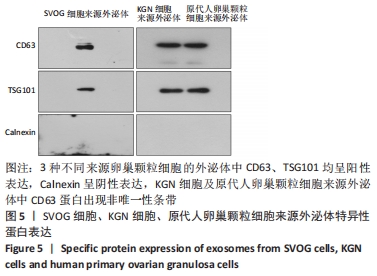
|