Chinese Journal of Tissue Engineering Research ›› 2022, Vol. 26 ›› Issue (24): 3790-3795.doi: 10.12307/2022.555
Previous Articles Next Articles
Bone marrow mesenchymal stem cells may alleviate brain damage caused by the microglial overactivation in the cortex around ischemic site of stroke
Yan Nan1, Wu Yanlong2, Tang Xiaohui2, Zhang Xiaoyan2, Wang Hui2, Yang Tianze2, Zhou Maochun2, Wang Zhengdong3, Yang Xiaoxia1
- 1School of Medical Applied Technology, 2Basic Medical College, 3Department of Anatomy, Shenyang Medical College, Shenyang 110034, Liaoning Province, China
-
Received:2021-02-25Accepted:2021-04-10Online:2022-08-28Published:2022-01-22 -
Contact:Yang Xiaoxia, Master, Professor, School of Medical Applied Technology, Shenyang Medical College, Shenyang 110034, Liaoning Province, China -
About author:Yan Nan, MD, Associate professor, School of Medical Applied Technology, Shenyang Medical College, Shenyang 110034, Liaoning Province, China -
Supported by:the Youth Science Fund Project of National Natural Science Foundation of China, No. 81803200 (to YN); Science and Technology Program Project of Liaoning Province, No. 2017225059 (to YXX); Youth Science and Technology Innovation Talent Support Program of Shenyang in 2020, No. RC200238 (to YN); Scientific Research Fund of Shenyang Medical College, No. 20161009 (to WZD)
CLC Number:
Cite this article
Yan Nan, Wu Yanlong, Tang Xiaohui, Zhang Xiaoyan, Wang Hui, Yang Tianze, Zhou Maochun, Wang Zhengdong, Yang Xiaoxia. Bone marrow mesenchymal stem cells may alleviate brain damage caused by the microglial overactivation in the cortex around ischemic site of stroke[J]. Chinese Journal of Tissue Engineering Research, 2022, 26(24): 3790-3795.
share this article
Add to citation manager EndNote|Reference Manager|ProCite|BibTeX|RefWorks
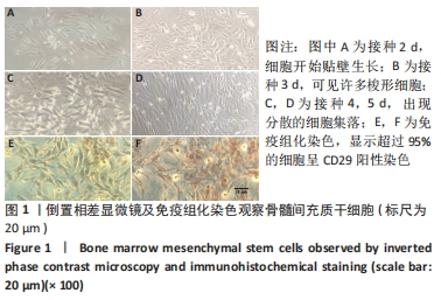
2.1 骨髓间充质干细胞培养与鉴定结果 接种2 d后,细胞开始贴壁生长,见图1A;3 d后镜下可见许多梭形细胞,见图1B;4 d或5 d出现分散的细胞集落,见图1C,D;6 d后梭形细胞充满培养瓶底部,7 d后大多数细胞融合。免疫组化染色显示,超过95%的细胞呈CD29阳性染色,见图1E,F。"
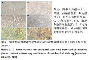
2.2 实验动物数量分析 根据Zea Longa评分评估大鼠瘫痪肢体的运动功能。模型组和移植组大鼠苏醒后对侧肢体瘫痪,无法站立或不稳定地向前移动,抬起尾巴时身体转向一侧。假手术组大鼠均为0级,其余两组8只大鼠为2级,12只大鼠为3级,均进入结果分析。 2.3 各组大鼠BBB评分 术后,模型组和移植组大鼠对侧肢体立即瘫痪,BBB评分为0。术后第1周末,模型组和移植组大鼠关节运动次数、运动负荷、伸展范围以及前后肢的协调程度都有一定程度的改善,BBB评分也逐渐升高,移植组与模型组之间的BBB评分无显著差异(P > 0.05),但是移植组与模型组的BBB评分显著低于假手术组(P < 0.001);术后第2周末,移植组BBB评分明显高于模型组(P < 0.01),移植组和模型组的BBB评分仍低于假手术组(P < 0.001),见图2。"

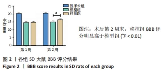
2.4 苏木精-伊红染色结果 假手术组皮质神经元正常,神经纤维未见异常,观察到少量神经胶质。模型组大鼠皮质结构不清楚,液化坏死溶解,结构规则破坏,炎症细胞浸润,可见部分胶质细胞,神经元数量减少,一些神经元的细胞核变小,而胞质肥大,细胞质明显减少,小血管和毛细血管充血、水肿、渗出。移植组皮质神经元的形态和结构正常,神经细胞分布相对均匀,可以看到细胞核和丰富的细胞质,在血管周围仍有一些炎症细胞,见图3。"

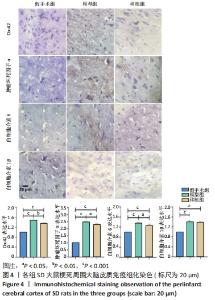
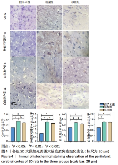
2.5 免疫组化染色结果 与假手术组相比,造模后小胶质细胞活化的特异性标志物Ox-42蛋白表达水平显著增加(P < 0.05),移植组Ox-42蛋白表达水平显著低于模型组(P < 0.01)。模型组肿瘤坏死因子α、白细胞介素6、白细胞介素1β的表达水平明显高于假手术组(P < 0.001),移植组肿瘤坏死因子α、白细胞介素6的表达水平明显低于模型组(P < 0.05),移植组白细胞介素1β的表达水平与模型组比较差异无显著性意义(P > 0.05),见图4。"
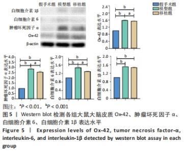
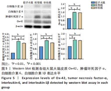
2.6 免疫印迹结果 造模后小胶质细胞被激活,与假手术组相比,模型组Ox-42蛋白表达水平显著增加(P < 0.001),移植组Ox-42蛋白表达水平显著低于模型组(P < 0.01)。模型组大脑皮质中激活的小胶质细胞会影响炎症因子的表达。模型组肿瘤坏死因子α、白细胞介素6、白细胞介素1β的表达水平明显高于假手术组(P < 0.001),移植组肿瘤坏死因子α、白细胞介素6的表达水平明显低于模型组(P < 0.01),移植组白细胞介素1β的表达水平与模型组比较差异无显著性意义(P > 0.05),见图5。"

2.7 生物相容性 各组大鼠均未出现过敏反应、免疫反应及排斥反应,说明骨髓间充质干细胞具有良好的生物相容性。"

| [1] KERR N, DIETRICH DW, BRAMLETT HM, et al. Sexually dimorphic microglia and ischemic stroke. CNS Neurosci Ther. 2019;25(12):1308-1317. [2] ZHANG D, LU Z, MAN J, et al. Wnt-3a alleviates neuroinflammation after ischemic stroke by modulating the responses of microglia/macrophages and astrocytes. Int Immunopharmacol. 2019;75:105760. [3] YAN RY, WANG SJ, YAO GT, et al. The protective effect and its mechanism of 3-n-butylphthalide pretreatment on cerebral ischemia reperfusion injury in rats. Eur Rev Med Pharmacol Sci. 2017;21(22):5275-5282. [4] GOMEZ-NICOLA D, PERRY VH. Microglial dynamics and role in the healthy and diseased brain: a paradigm of functional plasticity. Neuroscientist. 2015; 21(2):169-184. [5] DU L, ZHANG Y, CHEN Y, et al. Role of Microglia in Neurological Disorders and Their Potentials as a Therapeutic Target. Mol Neurobiol. 2017;54(10): 7567-7584. [6] YEH CF, CHUANG TY, HUNG YW, et al. Soluble epoxide hydrolase inhibition enhances anti-inflammatory and antioxidative processes, modulates microglia polarization, and promotes recovery after ischemic stroke. Neuropsychiatr Dis Treat. 2019;15:2927-2941. [7] ABDALLAH BM, HAACK-SØRENSEN M, BURNS JS, et al. Maintenance of differentiation potential of human bone marrow mesenchymal stem cells immortalized by human telomerase reverse transcriptase gene despite [corrected] extensive proliferation. Biochem Biophys Res Commun. 2005; 326(3):527-538. [8] CHAO YH, PENG CT, HARN HJ, et al. Poor potential of proliferation and differentiation in bone marrow mesenchymal stem cells derived from children with severe aplastic anemia. Ann Hematol. 2010;89(7):715-723. [9] SUN LY, HSIEH DK, SYU WS, et al. Cell proliferation of human bone marrow mesenchymal stem cells on biodegradable microcarriers enhances in vitro differentiation potential. Cell Prolif. 2010;43(5):445-456. [10] XIE DM, LI YL, LI J, et al. CD51 distinguishes a subpopulation of bone marrow mesenchymal stem cells with distinct migratory potential: a novel cell-based strategy to treat acute myocardial infarction in mice. Stem Cell Res Ther. 2019;10(1):331. [11] WEISS JN, LEVY S, BENES SC. Stem Cell Ophthalmology Treatment Study (SCOTS): bone marrow-derived stem cells in the treatment of Leber’s hereditary optic neuropathy. Neural Regen Res. 2016;11(10):1685-1694. [12] WANG X, ZHAI W, ZHU J, et al. Treatment of the bone marrow stromal stem cell supernatant by nasal administration-a new approach to EAE therapy. Stem Cell Res Ther. 2019;10(1):325. [13] NAKANO M, NAGAISHI K, KONARI N, et al. Bone marrow-derived mesenchymal stem cells improve diabetes-induced cognitive impairment by exosome transfer into damaged neurons and astrocytes. Sci Rep. 2016; 6:24805. [14] HAO F, LI A, YU H, et al. Enhanced Neuroprotective Effects of Combination Therapy with Bone Marrow-Derived Mesenchymal Stem Cells and Ginkgo biloba Extract (EGb761) in a Rat Model of Experimental Autoimmune Encephalomyelitis. Neuroimmunomodulation. 2016;23(1):41-57. [15] LI J, LIU Z, WANG L, et al. Thousand and one kinase 1 protects MCAO-induced cerebral ischemic stroke in rats by decreasing apoptosis and pro-inflammatory factors. Biosci Rep. 2019;39(10):BSR20190749. [16] NASOUTI R, KHAKSARI M, MIRZAEE M, et al. Trehalose protects against spinal cord injury through regulating heat shock proteins 27 and 70 and caspase-3 genes expression. J Basic Clin Physiol Pharmacol. 2019;31(1):1-8. [17] QIN C, ZHOU LQ, MA XT, et al. Dual Functions of Microglia in Ischemic Stroke. Neurosci Bull. 2019;35(5):921-933. [18] ELDAHSHAN W, FAGAN SC, ERGUL A. Inflammation within the neurovascular unit: Focus on microglia for stroke injury and recovery. Pharmacol Res. 2019; 147:104349. [19] HEINDL S, GESIERICH B, BENAKIS C, et al. Automated Morphological Analysis of Microglia After Stroke. Front Cell Neurosci. 2018;12:106. [20] SANDVIG I, AUGESTAD IL, HÅBERG AK, et al. Neuroplasticity in stroke recovery. The role of microglia in engaging and modifying synapses and networks. Eur J Neurosci. 2018;47(12):1414-1428. [21] XIONG XY, LIU L, YANG QW. Functions and mechanisms of microglia/macrophages in neuroinflammation and neurogenesis after stroke. Prog Neurobiol. 2016;142:23-44. [22] CHEN E, LIU G, ZHOU X, et al. Concentration-dependent, dual roles of IL-10 in the osteogenesis of human BMSCs via P38/MAPK and NF-κB signaling pathways. FASEB J. 2018;32(9):4917-4929. [23] PAN J, JIN JL, GE HM, et al. Malibatol A regulates microglia M1/M2 polarization in experimental stroke in a PPARγ-dependent manner. J Neuroinflammation. 2015;12:51. [24] LI D, WANG C, YAO Y, et al. mTORC1 pathway disruption ameliorates brain inflammation following stroke via a shift in microglia phenotype from M1 type to M2 type. FASEB J. 2016;30(10):3388-3399. [25] WON S, LEE JK, STEIN DG. Recombinant tissue plasminogen activator promotes, and progesterone attenuates, microglia/macrophage M1 polarization and recruitment of microglia after MCAO stroke in rats. Brain Behav Immun. 2015;49:267-279. [26] COLLMANN FM, PIJNENBURG R, HAMZEI-TAJ S, et al. Individual in vivo Profiles of Microglia Polarization After Stroke, Represented by the Genes iNOS and Ym1. Front Immunol. 2019;10:1236. [27] TIAN X, LIU H, XIANG F, et al. β-Caryophyllene protects against ischemic stroke by promoting polarization of microglia toward M2 phenotype via the TLR4 pathway. Life Sci. 2019;237:116915. [28] WANG J, XING H, WAN L, et al. Treatment targets for M2 microglia polarization in ischemic stroke. Biomed Pharmacother. 2018;105:518-525. [29] CHEN AQ, FANG Z, CHEN XL, et al. Microglia-derived TNF-α mediates endothelial necroptosis aggravating blood brain-barrier disruption after ischemic stroke. Cell Death Dis. 2019;10(7):487. [30] LIU Z, RAN Y, HUANG S, et al. Curcumin Protects against Ischemic Stroke by Titrating Microglia/Macrophage Polarization. Front Aging Neurosci. 2017; 9:233. [31] FONFRIA E, MATTEI C, HILL K, et al. TRPM2 is elevated in the tMCAO stroke model, transcriptionally regulated, and functionally expressed in C13 microglia. J Recept Signal Transduct Res. 2006;26(3):179-198. [32] ZHOU W, LIU Q, XU B. Improvement of bone defect healing in rats via mesenchymal stem cell supernatant. Exp Ther Med. 2018;15(2):1500-1504. [33] LIN J, WANG C, WU Z. Preliminary study on effects of human brain-derived neurotrophic factor gene-modified bone marrow mesenchymal stem cells by intravenous transplantation on structure and function of rat injured spinal cord. Zhongguo Xiu Fu Chong Jian Wai Ke Za Zhi. 2010;24(8):982-987. [34] WANG W, HUANG X, LI J, et al. Methane Suppresses Microglial Activation Related to Oxidative, Inflammatory, and Apoptotic Injury during Spinal Cord Injury in Rats. Oxid Med Cell Longev. 2017;2017:2190897. [35] HE Y, MA X, LI D, et al. Thiamet G mediates neuroprotection in experimental stroke by modulating microglia/macrophage polarization and inhibiting NF-κB p65 signaling. J Cereb Blood Flow Metab. 2017;37(8):2938-2951. [36] 朱婷,孙桂波,孟祥宝,等.小胶质细胞/巨噬细胞的极化及在脑卒中修复中的作用[J].中国药理学通报,2019,35(8):1046-1050. [37] VON LEDEN RE, KHAYRULLINA G, MORITZ KE, et al. Age exacerbates microglial activation, oxidative stress, inflammatory and NOX2 gene expression, and delays functional recovery in a middle-aged rodent model of spinal cord injury. J Neuroinflammation. 2017;14(1):161. [38] 樊文香.缺血性脑卒中的机制研究进展[J].中国药科大学学报,2018, 49(6):751-759. |
| [1] | Wang Jing, Xiong Shan, Cao Jin, Feng Linwei, Wang Xin. Role and mechanism of interleukin-3 in bone metabolism [J]. Chinese Journal of Tissue Engineering Research, 2022, 26(8): 1260-1265. |
| [2] | Xiao Hao, Liu Jing, Zhou Jun. Research progress of pulsed electromagnetic field in the treatment of postmenopausal osteoporosis [J]. Chinese Journal of Tissue Engineering Research, 2022, 26(8): 1266-1271. |
| [3] | Tian Chuan, Zhu Xiangqing, Yang Zailing, Yan Donghai, Li Ye, Wang Yanying, Yang Yukun, He Jie, Lü Guanke, Cai Xuemin, Shu Liping, He Zhixu, Pan Xinghua. Bone marrow mesenchymal stem cells regulate ovarian aging in macaques [J]. Chinese Journal of Tissue Engineering Research, 2022, 26(7): 985-991. |
| [4] | Hou Jingying, Guo Tianzhu, Yu Menglei, Long Huibao, Wu Hao. Hypoxia preconditioning targets and downregulates miR-195 and promotes bone marrow mesenchymal stem cell survival and pro-angiogenic potential by activating MALAT1 [J]. Chinese Journal of Tissue Engineering Research, 2022, 26(7): 1005-1011. |
| [5] | Zhou Ying, Zhang Huan, Liao Song, Hu Fanqi, Yi Jing, Liu Yubin, Jin Jide. Immunomodulatory effects of deferoxamine and interferon gamma on human dental pulp stem cells [J]. Chinese Journal of Tissue Engineering Research, 2022, 26(7): 1012-1019. |
| [6] | Liang Xuezhen, Yang Xi, Li Jiacheng, Luo Di, Xu Bo, Li Gang. Bushen Huoxue capsule regulates osteogenic and adipogenic differentiation of rat bone marrow mesenchymal stem cells via Hedgehog signaling pathway [J]. Chinese Journal of Tissue Engineering Research, 2022, 26(7): 1020-1026. |
| [7] | Wang Jifang, Bao Zhen, Qiao Yahong. miR-206 regulates EVI1 gene expression and cell biological behavior in stem cells of small cell lung cancer [J]. Chinese Journal of Tissue Engineering Research, 2022, 26(7): 1027-1031. |
| [8] | Liu Feng, Peng Yuhuan, Luo Liangping, Wu Benqing. Plant-derived basic fibroblast growth factor maintains the growth and differentiation of human embryonic stem cells [J]. Chinese Journal of Tissue Engineering Research, 2022, 26(7): 1032-1037. |
| [9] | Wen Dandan, Li Qiang, Shen Caiqi, Ji Zhe, Jin Peisheng. Nocardia rubra cell wall skeleton for extemal use improves the viability of adipogenic mesenchymal stem cells and promotes diabetes wound repair [J]. Chinese Journal of Tissue Engineering Research, 2022, 26(7): 1038-1044. |
| [10] | Zhu Bingbing, Deng Jianghua, Chen Jingjing, Mu Xiaoling. Interleukin-8 receptor enhances the migration and adhesion of umbilical cord mesenchymal stem cells to injured endothelium [J]. Chinese Journal of Tissue Engineering Research, 2022, 26(7): 1045-1050. |
| [11] | Luo Xiaoling, Zhang Li, Yang Maohua, Xu Jie, Xu Xiaomei. Effect of naringenin on osteogenic differentiation of human periodontal ligament stem cells [J]. Chinese Journal of Tissue Engineering Research, 2022, 26(7): 1051-1056. |
| [12] | Wang Xinmin, Liu Fei, Xu Jie, Bai Yuxi, Lü Jian. Core decompression combined with dental pulp stem cells in the treatment of steroid-associated femoral head necrosis in rabbits [J]. Chinese Journal of Tissue Engineering Research, 2022, 26(7): 1074-1079. |
| [13] | Fang Xiaolei, Leng Jun, Zhang Chen, Liu Huimin, Guo Wen. Systematic evaluation of different therapeutic effects of mesenchymal stem cell transplantation in the treatment of ischemic stroke [J]. Chinese Journal of Tissue Engineering Research, 2022, 26(7): 1085-1092. |
| [14] | Guo Jia, Ding Qionghua, Liu Ze, Lü Siyi, Zhou Quancheng, Gao Yuhua, Bai Chunyu. Biological characteristics and immunoregulation of exosomes derived from mesenchymal stem cells [J]. Chinese Journal of Tissue Engineering Research, 2022, 26(7): 1093-1101. |
| [15] | Huang Chenwei, Fei Yankang, Zhu Mengmei, Li Penghao, Yu Bing. Important role of glutathione in stemness and regulation of stem cells [J]. Chinese Journal of Tissue Engineering Research, 2022, 26(7): 1119-1124. |
| Viewed | ||||||
|
Full text |
|
|||||
|
Abstract |
|
|||||