Chinese Journal of Tissue Engineering Research ›› 2022, Vol. 26 ›› Issue (13): 2006-2011.doi: 10.12307/2022.325
Previous Articles Next Articles
Low-intensity pulsed ultrasound promotes the proliferation of bone marrow mesenchymal stem cells by inducing cyclin D1 up-regulation
Li Zhi1, 2, 3, Hua Yongyong4, Zhang Jianquan4, Fu Zhen4
- 1Ward Three, Department of General Surgery, 2Hubei Key Laboratory of Embryonic Stem Cell Research, Taihe Hospital, Hubei University of Medicine, Shiyan 442000, Hubei Province, China; 3Taihe Hospital Affiliated to Xi’an Jiaotong University Health Science Center, Shiyan 442000, Hubei Province, China; 4Department of Hepatobiliary Surgery, Haikou Hospital Affiliated to Xiangya School of Medicine, Central South University, Haikou 570000, Hainan Province, China
-
Received:2020-12-16Revised:2020-12-19Accepted:2021-01-23Online:2022-05-08Published:2021-12-18 -
Contact:Zhang Jianquan, Chief physician, Professor, Department of Hepatobiliary Surgery, Haikou Hospital Affiliated to Xiangya School of Medicine, Central South University, Haikou 570000, Hainan Province, China -
About author:Li Zhi, Master, Physician, Ward Three, Department of General Surgery, and Hubei Key Laboratory of Embryonic Stem Cell Research, Taihe Hospital, Hubei University of Medicine, Shiyan 442000, Hubei Province, China; Taihe Hospital Affiliated to Xi’an Jiaotong University Health Science Center, Shiyan 442000, Hubei Province, China -
Supported by:Hainan Science and Technology Fund Project, No. ZDXM2015082 (to ZJQ); the Hainan Natural Science Fund Project of China, No. 817382 (to FZ)
CLC Number:
Cite this article
Li Zhi, Hua Yongyong, Zhang Jianquan, Fu Zhen. Low-intensity pulsed ultrasound promotes the proliferation of bone marrow mesenchymal stem cells by inducing cyclin D1 up-regulation[J]. Chinese Journal of Tissue Engineering Research, 2022, 26(13): 2006-2011.
share this article
Add to citation manager EndNote|Reference Manager|ProCite|BibTeX|RefWorks
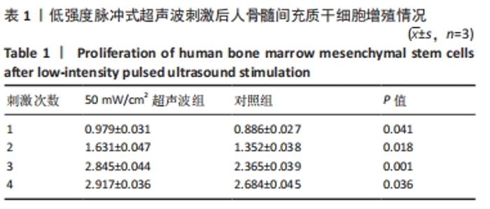
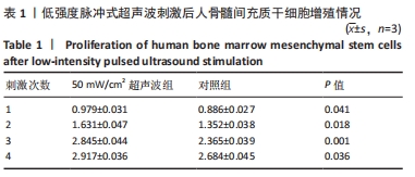
2.1 低强度脉冲式超声波促进人骨髓间充质干细胞增殖 低强度脉冲式超声波刺激1,2,3,4次后,50 mW/cm2超声波组吸光度值均高于对照组(0 mW/cm2超声波),差异有显著性意义(P < 0.05,P < 0.01),其中50 mW/cm2低强度脉冲式超声波刺激3次作用最明显,见表1。后续实验选取50 mW/cm2低强度脉冲式超声波刺激3次。"
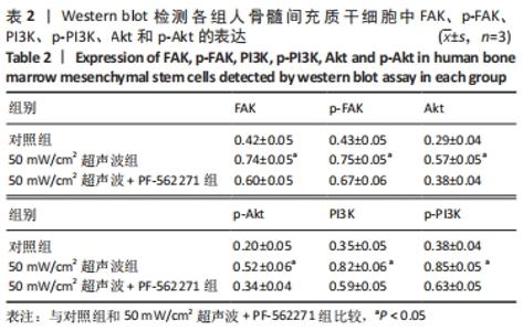
2.2 低强度脉冲式超声波激活FAK/PI3K/Akt信号通路 与对照组相比,50 mW/cm2超声波组FAK、p-FAK、PI3K、p-PI3K、Akt和p-Akt的表达均上调,差异有显著性意义(P < 0.05)。使用PF-562271抑制FAK/PI3K/Akt信号通路后,FAK、p-FAK、PI3K、p-PI3K、Akt和p-Akt的表达与50 mW/cm2超声波组相比明显降低,差异有显著性意义(P < 0.05);抑制FAK/PI3K/Akt信号通路后,FAK、p-FAK、PI3K、p-PI3K、Akt和p-Akt的表达与对照组相比无明显差异(P > 0.05)。因此,作者认为,低强度脉冲式超声波刺激人骨髓间充质干细胞可以上调FAK的表达,引起FAK磷酸化为p-FAK,进而激活下游靶基因PI3K、p-PI3K、Akt和p-Akt,见表2和图1。"
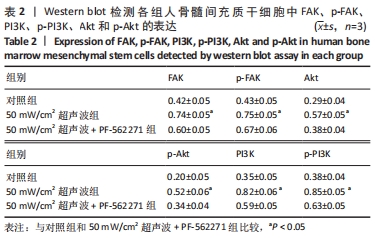
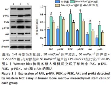
"


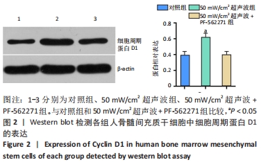
2.3 FAK/PI3K/Akt信号通路与低强度脉冲式超声波促进人骨髓间充质干细胞增殖的相关性 与对照组(2.365±0.039)相比,50 mW/cm2超声波组(2.845±0.044)细胞增殖能力明显增强,差异有显著性意义(P < 0.05);与50 mW/cm2超声波组相比,使用PF-562271抑制FAK/PI3K/Akt信号通路后,细胞增殖能力明显降低(2.135±0.468),差异有显著性意义(P < 0.001)。 2.4 FAK/PI3K/Akt信号通路与低强度脉冲式超声波上调细胞周期蛋白D1的相关性 与对照组(0.39±0.05)相比,50 mW/cm2超声波组细胞周期蛋白D1的表达(0.62±0.05)上调,差异有显著性意义(P=0.002)。使用PF-562271抑制FAK/PI3K/Akt信号通路后,细胞周期蛋白D1的表达(0.40±0.04)降低,差异有显著性意义(P=0.002),说明低强度脉冲式超声波可以通过FAK/PI3K/Akt信号通路上调细胞周期蛋白D1表达,见图2。"
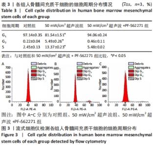
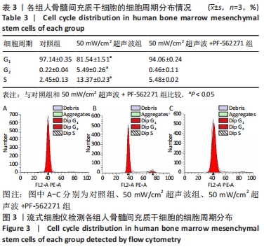
2.5 FAK/PI3K/Akt信号通路与低强度脉冲式超声波调控细胞周期的相关性 50 mW/cm2超声波组G1期细胞比例小于对照组(P=0.026),S期细胞比例大于对照组(P=0.002)。使用 PF-562271抑制FAK/PI3K/Akt信号通路后,50 mW/cm2超声波+ PF-562271组G1期细胞比例升高(P=0.023),S期细胞比例降低(P=0.035),差异有显著性意义。低强度脉冲式超声波可能通过激活FAK/PI3K/Akt信号通路促进人骨髓间充质干细胞从G1期进入S期从而促进其增殖,见表3和图3。"
| [1] 谢树才,张剑权,蒋锡丽,等.骨髓间充质干细胞诱导分化为肝细胞的方法及机制研究与进展[J].中国组织工程研究,2016,20(50): 7586-7593. [2] Fleming MM, Liu F, Zhang Y, et al. Model for End-Stage Liver Disease Underestimates Morbidity and Mortality in Patients with Ascites Undergoing Colectomy. World J Surg. 2018;42(10):3390-3397. [3] Hatzistergos KE, Quevedo H, Oskouei BN, et al. Bone marrow mesenchymal stem cells stimulate cardiac stem cell proliferation and differentiation. Circ Res. 2010;107(7):913-922. [4] Wu Y, Gao Q, Zhu S, et al. Low-intensity pulsed ultrasound regulates proliferation and differentiation of neural stem cells through notch signaling pathway. Biochem Biophys Res Commun. 2020;526(3):793-798. [5] Doan N, Reher P, Meghji S, et al. In vitro effects of therapeutic ultrasound on cell proliferation, protein synthesis, and cytokine production by human fibroblasts, osteoblasts, and monocytes. J Oral Maxillofac Surg. 1999;57(4):409-419. [6] Miyasaka M, Nakata H, Hao J, et al. Low-Intensity Pulsed Ultrasound Stimulation Enhances Heat-Shock Protein 90 and Mineralized Nodule Formation in Mouse Calvaria-Derived Osteoblasts. Tissue Eng Part A. 2015;21(23-24):2829-2839. [7] Huang D, Gao Y, Wang S, et al. Impact of low-intensity pulsed ultrasound on transcription and metabolite compositions in proliferation and functionalization of human adipose-derived mesenchymal stromal cells. Sci Rep. 2020;10(1):13690. [8] Gao Q, Walmsley AD, Cooper PR, et al. Ultrasound Stimulation of Different Dental Stem Cell Populations: Role of Mitogen-activated Protein Kinase Signaling. J Endod. 2016;42(3):425-431. [9] Korstjens CM, van der Rijt RH, Albers GH, et al. Low-intensity pulsed ultrasound affects human articular chondrocytes in vitro. Med Biol Eng Comput. 2008;46(12):1263-1270. [10] Ishihara Y, Ueki K, Sotobori M, et al. Bone regeneration by statin and low-intensity pulsed ultrasound (LIPUS) in rabbit nasal bone. J Craniomaxillofac Surg. 2014;42(3):185-193. [11] Sato M, Motoyoshi M, Shinoda M, et al. Low-intensity pulsed ultrasound accelerates nerve regeneration following inferior alveolar nerve transection in rats. Eur J Oral Sci. 2016;124(3):246-250. [12] 李志,谢树才,张剑权,等.低强度超声波对人骨髓间充质干细胞增殖影响的体外研究[J].中华实验外科杂志,2018,35(11):2006-2009. [13] Ren C, Chen X, Du N, et al. Low-intensity pulsed ultrasound promotes Schwann cell viability and proliferation via the GSK-3β/β-catenin signaling pathway. Int J Biol Sci. 2018;14(5):497-507. [14] Xu T, Gu J, Li C, et al. Low-intensity pulsed ultrasound suppresses proliferation and promotes apoptosis via p38 MAPK signaling in rat visceral preadipocytes. Am J Transl Res. 2018;10(3):948-956. [15] Wu S, Xu X, Sun J, et al. Low-Intensity Pulsed Ultrasound Accelerates Traumatic Vertebral Fracture Healing by Coupling Proliferation of Type H Microvessels. J Ultrasound Med. 2018;37(7):1733-1742. [16] Coskun ME, Coskun KA, Tutar Y. Determination of Optimum Operation Parameters for Low-Intensity Pulsed Ultrasound and Low-Level Laser Based Treatment to Induce Proliferation of Osteoblast and Fibroblast Cells. Photomed Laser Surg. 2018;36(5):246-252. [17] Akca H, Demiray A, Tokgun O, et al. Invasiveness and anchorage independent growth ability augmented by PTEN inactivation through the PI3K/AKT/NFkB pathway in lung cancer cells. Lung Cancer. 2011; 73(3):302-309. [18] Xu R, Chen J, Cong X, et al. Lovastatin protects mesenchymal stem cells against hypoxia- and serum deprivation-induced apoptosis by activation of PI3K/Akt and ERK1/2. J Cell Biochem. 2008;103(1): 256-269. [19] Aliabouzar M, Lee SJ, Zhou X, et al. Effects of scaffold microstructure and low intensity pulsed ultrasound on chondrogenic differentiation of human mesenchymal stem cells. Biotechnol Bioeng. 2018;115(2):495-506. [20] Bayat M, Virdi A, Rezaei F, et al. Comparison of the in vitro effects of low-level laser therapy and low-intensity pulsed ultrasound therapy on bony cells and stem cells. Prog Biophys Mol Biol. 2018;133:36-48. [21] Bruder SP, Jaiswal N, Haynesworth SE. Growth kinetics, self-renewal, and the osteogenic potential of purified human mesenchymal stem cells during extensive subcultivation and following cryopreservation. J Cell Biochem. 1997;64(2):278-294. [22] Baker N, Sohn J, Tuan RS. Promotion of human mesenchymal stem cell osteogenesis by PI3-kinase/Akt signaling, and the influence of caveolin-1/cholesterol homeostasis. Stem Cell Res Ther. 2015;6:238. [23] Liu Y, Zhang Y, Lin L, et al. Effects of bone marrow-derived mesenchymal stem cells on the axonal outgrowth through activation of PI3K/AKT signaling in primary cortical neurons followed oxygen-glucose deprivation injury. PLoS One. 2013;8(11):e78514. [24] Xu P, Gul-Uludag H, Ang WT, et al. Low-intensity pulsed ultrasound-mediated stimulation of hematopoietic stem/progenitor cell viability, proliferation and differentiation in vitro. Biotechnol Lett. 2012;34(10):1965-1973. [25] Zhang J, Guan J, Qi X, et al. Dimethyloxaloylglycine Promotes the Angiogenic Activity of Mesenchymal Stem Cells Derived from iPSCs via Activation of the PI3K/Akt Pathway for Bone Regeneration. Int J Biol Sci. 2016;12(6):639-652. [26] Presneau N, Shalaby A, Idowu B, et al. Potential therapeutic targets for chordoma: PI3K/AKT/TSC1/TSC2/mTOR pathway. Br J Cancer. 2009;100(9):1406-1414. [27] Liu H, Xu J, Zhou L, et al. Hepatitis B virus large surface antigen promotes liver carcinogenesis by activating the Src/PI3K/Akt pathway. Cancer Res. 2011;71(24):7547-7557. [28] Cheng K, Xia P, Lin Q, et al. Effects of low-intensity pulsed ultrasound on integrin-FAK-PI3K/Akt mechanochemical transduction in rabbit osteoarthritis chondrocytes. Ultrasound Med Biol. 2014;40(7):1609-1618. [29] Liu XB, Jiang J, Gui C, et al. Angiopoietin-1 protects mesenchymal stem cells against serum deprivation and hypoxia-induced apoptosis through the PI3K/Akt pathway. Acta Pharmacol Sin. 2008;29(7):815-822. [30] Matsuo T, Sato K, Matsui T, et al. Inhibitory effects of low-intensity pulsed ultrasound sonication on the proliferation of osteosarcoma cells. Oncol Lett. 2017;14(3):3071-3076. [31] Jung YJ, Kim R, Ham HJ, et al. Focused low-intensity pulsed ultrasound enhances bone regeneration in rat calvarial bone defect through enhancement of cell proliferation. Ultrasound Med Biol. 2015; 41(4):999-1007. [32] Uddin SM, Richbourgh B, Ding Y, et al. Chondro-protective effects of low intensity pulsed ultrasound. Osteoarthritis Cartilage. 2016;24(11):1989-1998. [33] Puts R, Rikeit P, Ruschke K, et al. Functional regulation of YAP mechanosensitive transcriptional coactivator by Focused Low-Intensity Pulsed Ultrasound (FLIPUS) enhances proliferation of murine mesenchymal precursors. PLoS One. 2018;13(10):e0206041. [34] Li L, Yang Z, Zhang H, et al. Low-intensity pulsed ultrasound regulates proliferation and differentiation of osteoblasts through osteocytes. Biochem Biophys Res Commun. 2012;418(2):296-300. [35] Go MJ, Takenaka C, Ohgushi H. Effect of forced expression of basic fibroblast growth factor in human bone marrow-derived mesenchymal stromal cells. J Biochem. 2007;142(6):741-748. [36] Jäger M, Feser T, Denck H, et al. Proliferation and osteogenic differentiation of mesenchymal stem cells cultured onto three different polymers in vitro. Ann Biomed Eng. 2005;33(10):1319-1332. [37] Song G, Ju Y, Soyama H, et al. Regulation of cyclic longitudinal mechanical stretch on proliferation of human bone marrow mesenchymal stem cells. Mol Cell Biomech. 2007;4(4):201-210. [38] Zhong C, Zhang X, Xu Z, et al. Effects of low-intensity electromagnetic fields on the proliferation and differentiation of cultured mouse bone marrow stromal cells. Phys Ther. 2012;92(9): 1208-1219. [39] Tsai MT, Li WJ, Tuan RS, et al. Modulation of osteogenesis in human mesenchymal stem cells by specific pulsed electromagnetic field stimulation. J Orthop Res. 2009;27(9):1169-1174. [40] 蒋柳宏,郑有华,张志光,等.bFGF基因转染对兔骨髓间充质干细胞生物学特性的影响[J].中山大学学报(医学科学版), 2009,30(S2): 41-46. [41] 朱磊,沈洪,刘丽,等.健脾补肾、清肠化湿方对骨髓间充质干细胞增殖迁移的影响[J].中国中西医结合杂志,2016,36(2):191-195. |
| [1] | Xiao Hao, Liu Jing, Zhou Jun. Research progress of pulsed electromagnetic field in the treatment of postmenopausal osteoporosis [J]. Chinese Journal of Tissue Engineering Research, 2022, 26(8): 1266-1271. |
| [2] | Wang Baojuan, Zheng Shuguang, Zhang Qi, Li Tianyang. Miao medicine fumigation can delay extracellular matrix destruction in a rabbit model of knee osteoarthritis [J]. Chinese Journal of Tissue Engineering Research, 2022, 26(8): 1180-1186. |
| [3] | Wang Jing, Xiong Shan, Cao Jin, Feng Linwei, Wang Xin. Role and mechanism of interleukin-3 in bone metabolism [J]. Chinese Journal of Tissue Engineering Research, 2022, 26(8): 1260-1265. |
| [4] | Zhang Jinglin, Leng Min, Zhu Boheng, Wang Hong. Mechanism and application of stem cell-derived exosomes in promoting diabetic wound healing [J]. Chinese Journal of Tissue Engineering Research, 2022, 26(7): 1113-1118. |
| [5] | Huang Chenwei, Fei Yankang, Zhu Mengmei, Li Penghao, Yu Bing. Important role of glutathione in stemness and regulation of stem cells [J]. Chinese Journal of Tissue Engineering Research, 2022, 26(7): 1119-1124. |
| [6] | Hui Xiaoshan, Bai Jing, Zhou Siyuan, Wang Jie, Zhang Jinsheng, He Qingyong, Meng Peipei. Theoretical mechanism of traditional Chinese medicine theory on stem cell induced differentiation [J]. Chinese Journal of Tissue Engineering Research, 2022, 26(7): 1125-1129. |
| [7] | An Weizheng, He Xiao, Ren Shuai, Liu Jianyu. Potential of muscle-derived stem cells in peripheral nerve regeneration [J]. Chinese Journal of Tissue Engineering Research, 2022, 26(7): 1130-1136. |
| [8] | Fan Yiming, Liu Fangyu, Zhang Hongyu, Li Shuai, Wang Yansong. Serial questions about endogenous neural stem cell response in the ependymal zone after spinal cord injury [J]. Chinese Journal of Tissue Engineering Research, 2022, 26(7): 1137-1142. |
| [9] | Gao Yujin, Peng Shuanglin, Ma Zhichao, Lu Shi, Cao Huayue, Wang Lang, Xiao Jingang. Osteogenic ability of adipose stem cells in diabetic osteoporosis mice [J]. Chinese Journal of Tissue Engineering Research, 2022, 26(7): 999-1004. |
| [10] | Hou Jingying, Guo Tianzhu, Yu Menglei, Long Huibao, Wu Hao. Hypoxia preconditioning targets and downregulates miR-195 and promotes bone marrow mesenchymal stem cell survival and pro-angiogenic potential by activating MALAT1 [J]. Chinese Journal of Tissue Engineering Research, 2022, 26(7): 1005-1011. |
| [11] | Zhou Ying, Zhang Huan, Liao Song, Hu Fanqi, Yi Jing, Liu Yubin, Jin Jide. Immunomodulatory effects of deferoxamine and interferon gamma on human dental pulp stem cells [J]. Chinese Journal of Tissue Engineering Research, 2022, 26(7): 1012-1019. |
| [12] | Liang Xuezhen, Yang Xi, Li Jiacheng, Luo Di, Xu Bo, Li Gang. Bushen Huoxue capsule regulates osteogenic and adipogenic differentiation of rat bone marrow mesenchymal stem cells via Hedgehog signaling pathway [J]. Chinese Journal of Tissue Engineering Research, 2022, 26(7): 1020-1026. |
| [13] | Wang Jifang, Bao Zhen, Qiao Yahong. miR-206 regulates EVI1 gene expression and cell biological behavior in stem cells of small cell lung cancer [J]. Chinese Journal of Tissue Engineering Research, 2022, 26(7): 1027-1031. |
| [14] | Liu Feng, Peng Yuhuan, Luo Liangping, Wu Benqing. Plant-derived basic fibroblast growth factor maintains the growth and differentiation of human embryonic stem cells [J]. Chinese Journal of Tissue Engineering Research, 2022, 26(7): 1032-1037. |
| [15] | Wen Dandan, Li Qiang, Shen Caiqi, Ji Zhe, Jin Peisheng. Nocardia rubra cell wall skeleton for extemal use improves the viability of adipogenic mesenchymal stem cells and promotes diabetes wound repair [J]. Chinese Journal of Tissue Engineering Research, 2022, 26(7): 1038-1044. |
| Viewed | ||||||
|
Full text |
|
|||||
|
Abstract |
|
|||||