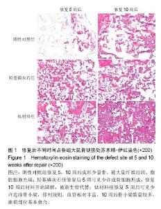
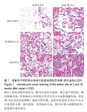
| [1]Yun KJ,Choi BH,Cong Z,et al.Bioengineered mussel glue incorporated with a cell recognition motif as an osteostimulating bone adhesive for titanium implants.J Mater Chem B. 2015;3(41): 8102-8114.[2]冯冬阳,王磊,柴瑜,等.中等强度跑台运动结合改性羟基磷灰石/壳聚糖复合水凝胶对大鼠髌股关节软骨缺损修复影响的研究[J].中华创伤骨科杂志,2016,18(6):518-525.[3]Betekhtin VI,Kolobov YR,Sklenicka V,et al.Effect of a defect structure on the static and long-term strength of submicrocrystalline VT1-0 titanium fabricated by plastic deformation during screw and lengthwise rolling.Tech Phys. 2015;60(1):66-71.[4]庄亚瑟,方志成,刘伯毅,等.基于CT灌注评价早期钛网修补颅骨缺损对脑血流量及神经功能康复的影响:随机对照临床试验[J].中国组织工程研究,2017,21(26):4228-4233.[5]Mosbahi S,Oudadesse H,Wers E,et al.Study of bioactive glass ceramic for use as bone biomaterial in vivo: investigation by Nuclear Magnetic Resonance and Histology. Ceram Int. 2016; 42(4):4827-4836.[6]黄俊红,叶党华,桂志勇,等.钛表面梯度生物活性涂层材料在颅骨修补中的生物性能[J].中国组织工程研究,2016,20(12):1772-1778.[7]van Houdt CI,Preethanath RS,van Oirschot BA,et al.Toward accelerated bone regeneration by altering poly(d, l-lactic-co-glycolic) acid porogen content in calcium phosphate cement. J Biomed Mater Res A.2016;104(2):483-492. [8]于祥茹,韩晓谦,程梁,等.载辛伐他汀PLGA/CPC支架材料复合BMSCs修复大鼠颅骨缺损的实验研究[J].口腔医学研究, 2015, 31(10):1032-1036.[9]杨玉鹏,赵海静,谷建琦,等.钛芯与骨形成蛋白复合材料修复即刻种植牙槽骨缺陷的评价[J].中国组织工程研究, 2017,21(22): 3536-3540.[10]Miron RJ,Bosshardt DD,Buser D,et al.Comparison of the capacity of enamel matrix derivative gel and enamel matrix derivative in liquid formulation to adsorb to bone grafting materials.J Periodontol. 2015;86(4):578.[11]陈彪,屈鹏飞,刘耀强,等.腓骨重建及小钛板固定下颌骨体部缺损的三维有限元分析[J].中国组织工程研究,2015,19(47):7550-7555.[12]殷渠东,顾三军,孙振中,等.钛网包裹打压植骨修复大段骨缺损初步报告[J].中国临床解剖学杂志,2015,33(1):101-104.[13]Hirota M,Ikeda T,Tabuchi M,et al.Effects of Ultraviolet Photofunctionalization on Bone Augmentation and Integration Capabilities of Titanium Mesh and Implants. Int J Oral Maxillofac Implants. 2017;32(1):52-62. [14]高永建,欧云生,邓乾兴,等.钛笼与髂骨块植骨修复椎骨缺损治疗胸腰椎结核的疗效比较[J].中国矫形外科杂志,2017,25(5):404-409.[15]张云绮,贾保军,敖建华,等.3D打印技术在下颌骨缺损修复中应用的初步临床研[J].口腔医学研究,2016,32(5):517-520.[16]刘忠.钛网支架结合前臂游离皮瓣修复上颌部缺损:可更好地维护吞咽和语言功能[J].中国组织工程研究,2015,19(30):4903-4907.[17]Chatterjee M,Hatori K,Duyck J,et al.High-frequency loading positively impacts titanium implant osseointegration in impaired bone.Osteoporosis Int.2015;26(1):281.[18]王晨辰,刘向辉,孙卫革,等.钛网应用于即刻种植骨缺损骨再生的实验研究[J].安徽医科大学学报,2017,52(6):814-818.[19]邓跃飞,刘正豪,郑眉光,等.帽状腱膜下层骨膜瓣和钛板联合修复前颅底沟通肿瘤术后颅底巨大骨缺损的疗效观察[J].中华神经外科杂志,2015,31(2):155-157.[20]罗晟,何永生,陈隆益,等.数字化塑型钛网颅骨修补对颅骨缺损患者颅内压、脑血流动力学及神经功能康复的影响[J].中华神经医学杂志,2015,14(11):1128-1132.[21]Vand SJ,Lozano D,Chai YC,et al.Osteostatin-coated porous titanium can improve early bone regeneration of cortical bone defects in rats.Tissue Eng Part A.2015;21(9-10):1495-1506.[22]鄢荣曾,骆丹媚,秦晓宇,等.3D打印制作个性化下颌骨三维网状修复体支架数字化建模方法的研究[J].中华口腔医学杂志, 2016,51(5): 280-285.[23]刘林,夏德林,孙黎波,等.个性化预成型重建板技术联合血管化髂骨肌瓣在下颌骨修复重建中的应用[J].中华整形外科杂志, 2016, 32(4):258-263.[24]Yan Q,Li Y,Cheng N,et al.Effect of retinoic acid on the function of lipopolysaccharide-stimulated bone marrow stromal cells grown on titanium surfaces.Inflamm Res.2015;64(1):63-70. [25]陈哲峰,范卫民,王青,等.同种异体颗粒骨打压植骨钛网杯重建髋臼骨缺损的疗效[J].中华骨科杂志,2016,36(23):1512-1516.[26]林鸿雷,王月燕,卢阳.不同材料支架式可摘局部义齿修复牙列缺损的生物相容性分析[J].中国组织工程研究, 2016,20(8):1171-1176.[27]Li H,Oppenheimer SM,Stupp SI,et al.Effects of Pore Morphology and Bone Ingrowth on Mechanical Properties of Microporous Titanium as an Orthopaedic Implant Material. Mater Trans. 2015; 45(4):1124-1131.[28]蓝平衡,郭征,吴智钢,等.带陶瓷涂层的钛合金关节表面假体治疗羊膝关节软骨全层缺损的实验研究[J].中华创伤骨科杂志, 2016,18(8): 708-714. |
