Chinese Journal of Tissue Engineering Research ›› 2017, Vol. 21 ›› Issue (22): 3483-3487.doi: 10.3969/j.issn.2095-4344.2017.22.007
Previous Articles Next Articles
Tissue-engineered cartilage construction using microfibrillar collagen
- 1Key Laboratory of Oral Medicine, Guangzhou Institute of Oral Disease, Stomatology Hospital of Guangzhou Medical University, Guangzhou 510140, Guangdong Province, China; 2Department of Otolaryngology Head and Neck Surgery, the 306th Hospital of PLA, Beijing 100101, China
-
Received:2017-02-17Online:2017-08-08Published:2017-09-01 -
Contact:Wu Wei, M.D., Professor, Chief physician, Department of Otolaryngology Head and Neck Surgery, the 306th Hospital of PLA, Beijing 100101, China -
About author:Zhou Li-bin, M.D., Associate chief physician, Key Laboratory of Oral Medicine, Guangzhou Institute of Oral Disease, Stomatology Hospital of Guangzhou Medical University, Guangzhou 510140, Guangdong Province, China -
Supported by:the National Natural Science Foundation of China, No. 81401609
CLC Number:
Cite this article
Zhou Li-bin, Xu Bing-xin, Ding Rui-ying, Han Hao-lun, Wang Gang, Li Bao-wei, Wang Hong-nan, Wu Wei.
share this article
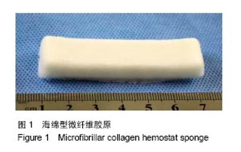
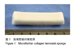
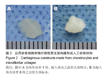
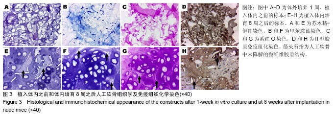
2.1 实验动物数量分析 4只BALB/c裸鼠全部进入结果分析,中途无脱落。 2.2 大体观察结果 微纤维胶原呈稀疏的海绵状结构(图1),具有很强的可压缩性和吸附性。在细胞接种过程中,湿水之后的微纤维胶原支架无明显缩水,仍然能够保持其立体形态。体外培养1周后无明显体积和形态变化(图2A)。 植入裸鼠体内,进行体内培育8周后,形成白色半透明且富有弹性的成熟软骨块(图2B)。取材时,人工软骨被纤维组织包裹,这层组织囊可以通过剥离而被轻易去除。空白对照的微纤维胶原材料未完全吸收,但体积明显减小,呈白色柔软的胶样结构,被周围结缔组织包裹,较难解剖。"

"
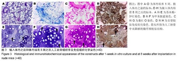
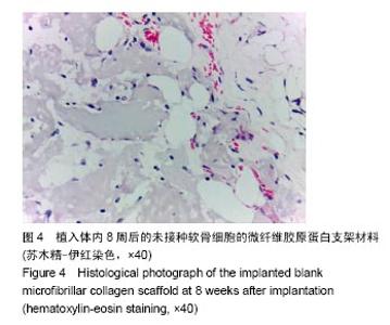
2.3 组织学及免疫组化观察结果 软骨细胞接种于微纤维胶原支架后,细胞与支架材料贴附,微纤维支架材料在甲苯胺蓝染色中不显色,在番红O染色中被染成粉红色,另外,在Ⅱ型胶原免疫组化染色中,观察到支架材料与Ⅱ型胶原抗体反应,说明微纤维胶原支架中包含有Ⅱ型胶原成分(图3)。 体内培养8周后,组织学观察人工软骨结构,可见软骨细胞形态成熟,软骨细胞位于软骨陷窝之中,软骨陷窝在组织中分布均匀,软骨基质丰富,基质中可分辨出尚未降解的微纤维胶原支架材料。甲苯胺蓝染色和番红O染色证明人工软骨基质中糖胺聚糖含量丰富,免疫组化染色观察到软骨基质中Ⅱ型胶原强阳性表达(图3),而在人工软骨的间隙内,可见到未降解的微纤维胶原支架材料中Ⅱ型胶原的表达(图3H)。 空白对照组镜下可见未被降解的微纤维胶原蛋白结构,间隙内裸鼠结缔组织细胞长入,新生血管生成(图4)。"
"

| [1]Bichara DA, O'Sullivan NA, Pomerantseva I, et al. The tissue-engineered auricle: past, present, and future. Tissue Eng Part B Rev. 2012;18(1):51-61.[2]Wu L, Zhang H, Zhang J, et al. Fabrication of three-dimensional porous scaffolds of complicated shape for tissue engineering. I. Compression molding based on flexible-rigid combined mold. Tissue Eng. 2005;11(7-8):1105-1114.[3]Lin X, Wang W, Zhang W, et al. Hyaluronic Acid Coating Enhances Biocompatibility of Nonwoven PGA Scaffold and Cartilage Formation. Tissue Eng Part C Methods. 2017; 23(2):86-97.[4]Toosi S, Naderi-Meshkin H, Kalalinia F, et al. PGA-incorporated collagen: Toward a biodegradable composite scaffold for bone-tissue engineering. J Biomed Mater Res A. 2016;104(8):2020-2028.[5]Jiang L, Li Y, Xiong C, et al. Preparation and Properties of Bamboo Fiber/Nano-hydroxyapatite/Poly(lactic-co-glycolic) Composite Scaffold for Bone Tissue Engineering. ACS Appl Mater Interfaces. 2017;9(5):4890-4897.[6]Cao Y, Xiong D, Wang K, et al. Semi-degradable porous poly (vinyl alcohol) hydrogel scaffold for cartilage repair: Evaluation of the initial and cell-cultured tribological properties. J Mech Behav Biomed Mater. 2017;68:163-172.[7]Frasca S, Norol F, Le Visage C, et al. Calcium-phosphate ceramics and polysaccharide-based hydrogel scaffolds combined with mesenchymal stem cell differently support bone repair in rats. J Mater Sci Mater Med. 2017;28(2):35.[8]Shirakata Y, Nakamura T, Shinohara Y, et al. An exploratory study on the efficacy of rat dedifferentiated fat cells (rDFATs) with a poly lactic-co-glycolic acid/hydroxylapatite (PLGA/HA) composite for bone formation in a rat calvarial defect model. J Mater Sci Mater Med. 2014;25(3):899-908.[9]Moura D, Mano JF, Paiva MC, et al. Chitosan nanocomposites based on distinct inorganic fillers for biomedical applications. Sci Technol Adv Mater. 2016;17(1):626-643.[10]Fujioka-Kobayashi M, Mottini M, Kobayashi E, et al. An in vitro study of fibrin sealant as a carrier system for recombinant human bone morphogenetic protein (rhBMP)-9 for bone tissue engineering. J Craniomaxillofac Surg. 2017;45(1):27-32. [11]Zhou L, Pomerantseva I, Bassett EK, et al. Engineering ear constructs with a composite scaffold to maintain dimensions. Tissue Eng Part A. 2011;17(11-12):1573-1581.[12]Schweiger M, Knirsch W, Cesarovic N, et al. Surgical technique: establishing a pre-clinical large animal model to test aortic valve leaflet substitute. J Thorac Dis. 2016;8(12): 3733-3738.[13]Kesireddy V, Kasper FK. Approaches for building bioactive elements into synthetic scaffolds for bone tissue engineering. J Mater Chem B Mater Biol Med. 2016;4(42): 6773-6786.[14]Lam AT, Li J, Toh JP, et al. Biodegradable poly-ε-caprolactone microcarriers for efficient production of human mesenchymal stromal cells and secreted cytokines in batch and fed-batch bioreactors. Cytotherapy. 2017;19(3):419-432.[15]Radhakrishnan J, Subramanian A, Krishnan UM, et al. Injectable and 3D Bioprinted Polysaccharide Hydrogels: From Cartilage to Osteochondral Tissue Engineering. Biomacromolecules. 2017;18(1):1-26.[16]Kiyotake EA, Beck EC, Detamore MS. Cartilage extracellular matrix as a biomaterial for cartilage regeneration. Ann N Y Acad Sci. 2016;1383(1):139-159.[17]Schneider C, Lehmann J, van Osch GJ, et al. Systematic Comparison of Protocols for the Preparation of Human Articular Cartilage for Use as Scaffold Material in Cartilage Tissue Engineering. Tissue Eng Part C Methods. 2016;22(12): 1095-1107.[18]Eslahi N, Abdorahim M, Simchi A. Smart Polymeric Hydrogels for Cartilage Tissue Engineering: A Review on the Chemistry and Biological Functions. Biomacromolecules. 2016;17(11):3441-3463.[19]Asghari F, Samiei M, Adibkia K, et al. Biodegradable and biocompatible polymers for tissue engineering application: a review. Artif Cells Nanomed Biotechnol. 2017;45(2):185-192.[20]Qi N, Li WJ, Tian H. A systematic review of animal and clinical studies on the use of scaffolds for urethral repair. J Huazhong Univ Sci Technolog Med Sci. 2016;36(1):111-117.[21]Thomas D, Gaspar D, Sorushanova A, et al. Scaffold and scaffold-free self-assembled systems in regenerative medicine. Biotechnol Bioeng. 2016;113(6):1155-1163.[22]Fujimoto Y, Kobayashi T, Komori M, et al. Modified hemostatic technique using microfibrillar collagen hemostat in endoscopic endonasal transsphenoidal surgery: technical note. Neurol Med Chir (Tokyo). 2014;54(8):617-621.[23]Struk D, Rankin RN, Karlik SJ. Stability studies on chemoembolization mixtures. Dialysis studies of doxorubicin and lipiodol with Avitene, Gelfoam, and Angiostat. Invest Radiol. 1993;28(11):1024-1027.[24]Minkin P, Bertetti R, Lindsey S, et al. Management of tooth extraction in a patient with a rare bleeding disorder associated with Hermansky-Pudlak syndrome: a case report. J Oral Maxillofac Surg. 2015;73(2):219-223.[25]Magro-Ernica N, Magro-Filho O, Rangel-Garcia I. Histologic study of use of microfibrillar collagen hemostat in rat dental sockets. Braz Dent J. 2003;14(1):12-15.[26]Sirlak M, Eryilmaz S, Yazicioglu L, et al. Comparative study of microfibrillar collagen hemostat (Colgel) and oxidized cellulose (Surgicel) in high transfusion-risk cardiac surgery.J Thorac Cardiovasc Surg. 2003;126(3):666-670.[27]Horton JW, Garcia NM, Stone KR. Evaluation of a new hemostatic agent in experimental splenic laceration. Arch Surg. 1995;130(2):161-164.[28]Watanabe G, Misaki T, Kotoh K. Microfibrillar collagen (Avitene) and antibiotic-containing fibrin-glue after median sternotomy. J Card Surg. 1997;12(2):110-111.[29]Robicsek F. Microfibrillar collagen hemostat in cardiac surgery. J Thorac Cardiovasc Surg. 2004;127(4):1228.[30]钟江龙,张大明,陈伟良,等.经口内镜辅助下切除咽旁间隙良性肿瘤6例报道[J].中国口腔颌面外科杂志, 2015, 13(3):258-261.[31]罗伟初,谢道远,李翠芳,等.63例人工髋关节置换手术的治疗体会[J].中国当代医药, 2012, 19(10):28-30.[32]陈运,邵大畏,任伟业,等.胶原海绵填充甲状腺手术残腔与止血疗效临床观察[J].中国医药指南, 2012, 10(5):79-80.[33]缪雪华,周勇,任伟业,等.胶原海绵促进肉芽生长与创面愈合实验研究[J].中国当代医药, 2012, 19(10):26-27.[34]周勇,任伟业,邵大畏,等.胶原海绵促进创面止血与血管新生的实验研究[J].湖南中医杂志, 2012, 28(3):140-141.[35]Wagner WR, Pachence JM, Ristich J, et al. Comparative in vitro analysis of topical hemostatic agents. J Surg Res. 1996; 66(2):100-108.[36]Ereth M, Sibonga J, Oliver W, et al. Microporous polysaccharide hemospheres do not inhibit bone healing compared to bone wax or microfibrillar collagen. Orthopedics. 2008;31(3):222.[37]Doita M, Nishida K, Kurosaka M. Radiculopathy due to microfibrillar collagen hemostat mimicking recurrence of disc herniation. Skeletal Radiol. 2006;35(12):953-955.[38]Zhou L, Ding R, Li B, et al. Cartilage engineering using chondrocyte cell sheets and its application in reconstruction of microtia. Int J Clin Exp Pathol. 2015;8(1):73-80.[39]于美丽, 杜智. 可吸收止血材料的研究现状及临床应用[J].北京生物医学工程, 2008,27(2):208-211.[40]O'Shaughnessy BA, Schafernak KT, DiPatri AJ Jr, et al. A granulomatous reaction to Avitene mimicking recurrence of a medulloblastoma. Case report. J Neurosurg. 2006;104 (1 Suppl):33-36. |
| [1] | Tan Xinfang, Guo Yanxing, Qin Xiaofei, Zhang Binqing, Zhao Dongliang, Pan Kunkun, Li Yuzhuo, Chen Haoyu. Effect of uniaxial fatigue exercise on patellofemoral cartilage injury in a rabbit [J]. Chinese Journal of Tissue Engineering Research, 2022, 26(在线): 1-6. |
| [2] | Yao Xiaoling, Peng Jiancheng, Xu Yuerong, Yang Zhidong, Zhang Shuncong. Variable-angle zero-notch anterior interbody fusion system in the treatment of cervical spondylotic myelopathy: 30-month follow-up [J]. Chinese Journal of Tissue Engineering Research, 2022, 26(9): 1377-1382. |
| [3] | Wu Cong, Jia Quanzhong, Liu Lun. Relationship between transforming growth factor beta1 expression and chondrocyte migration in adult articular cartilage after fragmentation [J]. Chinese Journal of Tissue Engineering Research, 2022, 26(8): 1167-1172. |
| [4] | Zhang Jinglin, Leng Min, Zhu Boheng, Wang Hong. Mechanism and application of stem cell-derived exosomes in promoting diabetic wound healing [J]. Chinese Journal of Tissue Engineering Research, 2022, 26(7): 1113-1118. |
| [5] | An Weizheng, He Xiao, Ren Shuai, Liu Jianyu. Potential of muscle-derived stem cells in peripheral nerve regeneration [J]. Chinese Journal of Tissue Engineering Research, 2022, 26(7): 1130-1136. |
| [6] | Xu Lei, Han Xiaoqiang, Zhang Jintao, Sun Haibiao. Hyaluronic acid around articular chondrocytes: production, transformation and function characteristics [J]. Chinese Journal of Tissue Engineering Research, 2022, 26(5): 768-773. |
| [7] | Lin Xuchen, Zhu Hainian, Wang Zengshun, Qi Tengmin, Liu Limin, Suonan Angxiu. Effect of xanthohumol on inflammatory factors and articular cartilage in a mouse mode of osteoarthritis [J]. Chinese Journal of Tissue Engineering Research, 2022, 26(5): 676-681. |
| [8] | Chen Xiaoxu, Luo Yaxin, Bi Haoran, Yang Kun. Preparation and application of acellular scaffold in tissue engineering and regenerative medicine [J]. Chinese Journal of Tissue Engineering Research, 2022, 26(4): 591-596. |
| [9] | Kang Kunlong, Wang Xintao. Research hotspot of biological scaffold materials promoting osteogenic differentiation of bone marrow mesenchymal stem cells [J]. Chinese Journal of Tissue Engineering Research, 2022, 26(4): 597-603. |
| [10] | Shen Jiahua, Fu Yong. Application of graphene-based nanomaterials in stem cells [J]. Chinese Journal of Tissue Engineering Research, 2022, 26(4): 604-609. |
| [11] | Zhang Tong, Cai Jinchi, Yuan Zhifa, Zhao Haiyan, Han Xingwen, Wang Wenji. Hyaluronic acid-based composite hydrogel in cartilage injury caused by osteoarthritis: application and mechanism [J]. Chinese Journal of Tissue Engineering Research, 2022, 26(4): 617-625. |
| [12] | Li Hui, Chen Lianglong. Application and characteristics of bone graft materials in the treatment of spinal tuberculosis [J]. Chinese Journal of Tissue Engineering Research, 2022, 26(4): 626-630. |
| [13] | Gao Cangjian, Yang Zhen, Liu Shuyun, Li Hao, Fu Liwei, Zhao Tianyuan, Chen Wei, Liao Zhiyao, Li Pinxue, Sui Xiang, Guo Quanyi. Electrospinning for rotator cuff repair [J]. Chinese Journal of Tissue Engineering Research, 2022, 26(4): 637-642. |
| [14] | He Yunying, Li Lingjie, Zhang Shuqi, Li Yuzhou, Yang Sheng, Ji Ping. Method of constructing cell spheroids based on agarose and polyacrylic molds [J]. Chinese Journal of Tissue Engineering Research, 2022, 26(4): 553-559. |
| [15] | He Guanyu, Xu Baoshan, Du Lilong, Zhang Tongxing, Huo Zhenxin, Shen Li. Biomimetic orientated microchannel annulus fibrosus scaffold constructed by silk fibroin [J]. Chinese Journal of Tissue Engineering Research, 2022, 26(4): 560-566. |
| Viewed | ||||||
|
Full text |
|
|||||
|
Abstract |
|
|||||