Chinese Journal of Tissue Engineering Research ›› 2017, Vol. 21 ›› Issue (21): 3376-3381.doi: 10.3969/j.issn.2095-4344.2017.21.016
Previous Articles Next Articles
Effect of pulsed magnetic fields on endogenous neural stem cell factors in the brain after craniocerebral injury
Gan Xiao1, Zhang Dong-bo2, Liu Xiang-ye1, Fu Liu-peng3, Bai Xin-xue2, Wu Nan-li4
- 1Department of Traumatic Surgery, 2Department of Neurosurgery, 3Clinical Department of Pharmacy, Nanyang Central Hospital, Zhengzhou University, Nanyang 473009, Henan Province, China; 4First Affiliated Hospital of Zhengzhou University, Zhengzhou 450000, Henan Province, China
-
Revised:2017-03-04Online:2017-07-28Published:2017-08-02 -
About author:Gan Xiao, Attending physician, Department of Traumatic Surgery, Nanyang Central Hospital, Zhengzhou University, Nanyang 473009, Henan Province, China
CLC Number:
Cite this article
Gan Xiao, Zhang Dong-bo, Liu Xiang-ye, Fu Liu-peng, Bai Xin-xue, Wu Nan-li. Effect of pulsed magnetic fields on endogenous neural stem cell factors in the brain after craniocerebral injury[J]. Chinese Journal of Tissue Engineering Research, 2017, 21(21): 3376-3381.
share this article
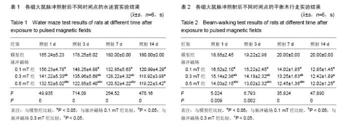
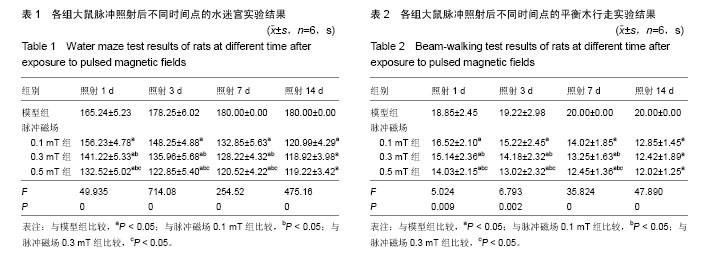
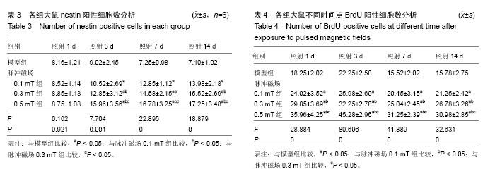
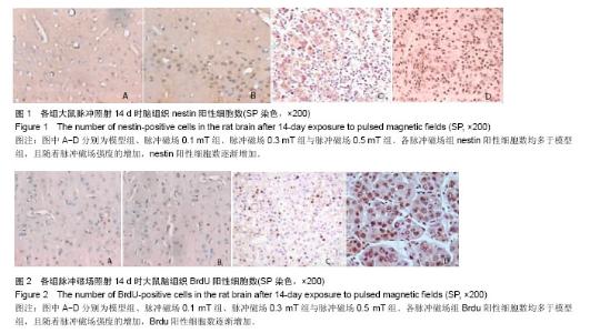
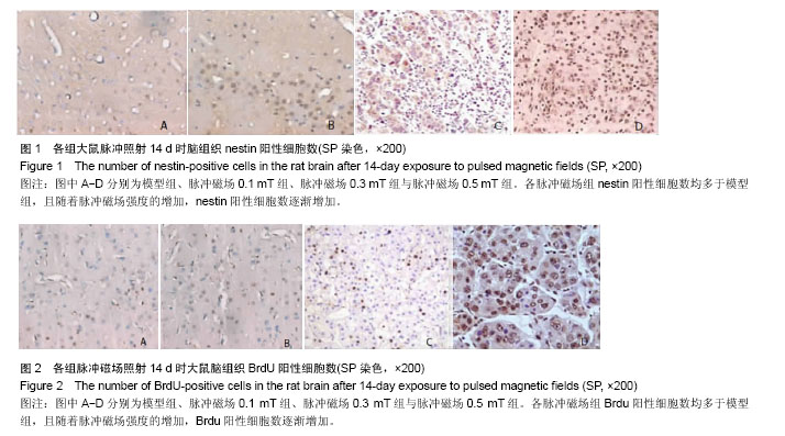
2.1 实验动物数量分析 320只大鼠均进入结果分析。 2.2 各组大鼠水迷宫实验结果 水迷宫实验中,各脉冲磁场组脉冲照射后不同时间点到达高台的时间显著短于模型组(P < 0.05);脉冲照射1,3,7 d,脉冲磁场0.5 mT组达到高台时间明显短于脉冲磁场0.1,0.3 mT组(P < 0.05),脉冲磁场0.3 mT组短于脉冲磁场0.1 mT组(P < 0.05);照射14 d时脉冲磁场各组比较差异无显著性意义(P > 0.05),见表1。 2.3 各组大鼠平衡木行走实验结果 平衡木行走实验中,各脉冲磁场组脉冲照射不同时间点后的通过平衡木时间显著短于模型组(P < 0.05);脉冲照射1,3,7 d,脉冲磁场0.5 mT组通过平衡木的时间明显短于脉冲磁场0.1,0.3 mT组(P < 0.05),脉冲磁场0.3 mT组短于脉冲磁场0.1 mT组 (P < 0.05);照射14 d时脉冲磁场各组通过平衡木的时间比较差异无显著性意义(P > 0.05),见表2。 2.4 各组大鼠不同时间点nestin阳性细胞数分析 脉冲照射3,7,14 d 时,各脉冲磁场组在nestin阳性细胞数多于模型组(P < 0.05),脉冲磁场0.5 mT组nestin阳性细胞数多于脉冲磁场0.1,0.3 mT组,脉冲磁场0.3 mT组多于脉冲磁场0.1 mT组(P < 0.05),见表3,图1。 2.5 各组大鼠不同时间点BrdU阳性细胞数分析 照射3,7,14 d 时,各脉冲磁场组BrdU阳性细胞数多于模型组(P < 0.05),脉冲磁场0.5 mT组BrdU阳性细胞数多于脉冲磁场0.1,0.3 mT组,脉冲磁场0.3 mT组多于脉冲磁场0.1 mT组(P < 0.05),见表4,图2。"
"

"
| [1] 李侠,张磊,陈燕伟,等.852例开放性颅脑损伤的临床救治经验[J].中华神经医学杂志,2014,13(5):451-455.[2] 李春伟,伊志强,李良,等.重型创伤性颅脑损伤的治疗进展[J].中国微创外科杂志,2016,16(7):656-660.[3] 叶玉勤,贺晓生.内源性神经干细胞参与神经再生的分子影像学研究进展[J].中华神经医学杂志,2014,13(6):639-642.[4] Alabovsky VV,Kudryshov YB,Vinokurov AA,et al.Radio Frequency Electromagnetic Field Effect on the State of Na+/Ca2+ Exchange in the Isolated Rat Heart.Radiats Biol Radioecol.2016;56(2):171-176.[5] Zhou Y,Qiu Bland GZ,et al.Effects of electromagnetic pulse exposure on gelatinase of blood-brain barrier in vitro. Electromagn Biol Med.2016;29(2):1-7.[6] Kranjc S,Kranjc M,Scancar J,et al.Electrochemotherapy by pulsed electromagnetic field treatment (PEMF) in mouse melanoma B16F10 in vivo.Radiol Oncol.2016;50(1):39-48.[7] Jiang DP,Li JH,Zhang J,et al.Long-term electromagnetic pulse exposure induces Abeta deposition and cognitive dysfunction through oxidative stress and overexpression of APP and BACE1.Brain Res.2016;1642(2):10-19.[8] Afshari D, Moradian N, Khalili M, et al. Evaluation of pulsing magnetic field effects on paresthesia in multiple sclerosis patients, a randomized, double-blind, parallel-group clinical trial. Clin Neurol Neurosurg. 2016;149(2):171-174.[9] 陈斌,陶静,黄佳,等.从Notch通路探讨电针促进局灶性脑缺血再灌注大鼠海马神经干细胞增殖的作用机制[J].中国康复医学杂志,2014,29(5):399-404.[10] Guerriero F, Ricevuti G.Extremely low frequency electromagnetic felds stimulation modulates autoimmunity and immune responses: a possible immuno-modulatory therapeutic effect in neurodegenerative diseases.Neural Regen Res.2016;11(12): 1888-1895.[11] Zhang F,Duan X,Lu L,et al.In Vivo Targeted MR Imaging of Endogenous Neural Stem Cells in Ischemic Stroke. Molecules. 2016;21(9):143-145.[12] Klein R,Mahlberg N,Ohren M,etal.The Neural Cell Adhesion Molecule-Derived (NCAM)-Peptide FG Loop(FGL)Mobilizes Endogenous Neural Stem Cells and Promotes Endogenous Regenerative Capacity after Stroke.J Neuroimmune Pharmacol. 2016;8(5):85-86.[13] Nakatomi H,Ochi T,Saito N.Endogenous neural stem cell mobilization with intraventricular growth factor administration. Nihon Rinsho.2016;74(4):655-660.[14] Jiang S,Chen W,Zhang Y,etal.Acupuncture Induces the Proliferation and Differentiation of Endogenous Neural Stem Cells in Rats with Traumatic Brain Injury].Evid Based Complement Alternat Med.2016;20(2):89-92.[15] 蒋婷,许涛,向威,等.脉冲强磁场诱导体外新生大鼠神经干细胞向神经元方向分化[J].中华物理医学与康复杂志,2014,36(10): 740-744.[16] Ma F,Zhu T,Xu F,et al.Neural stem/progenitor cells on collagen with anchored basic fibroblast growth factor as potential natural nerve conduits for facial nerve regeneration. Acta Biomater. 2016;8(5):78-80.[17] Ye LJ,Bian H,Fan YD,et al.Rhesus monkey neural stem cell transplantation promotes neural regeneration in rats with hippocampal lesions.Neural Regen Res.2016;11(9):1464- 1470.[18] Li QQ,Qiao GQ,Ma J,et al.Cortical neurogenesis in adult rats after ischemic brain injury: most new neurons fail to mature. Neural Regen Res. 2015;10(2): 277-285.[19] Yao Y,Zheng XR,Zhang SS,et al.Transplantation of vascular endothelial growth factor-modified neural stem/progenitor cells promotes the recovery of neurological function following hypoxic-ischemic brain damage.Neural Regen Res.2016; 11(9):1456-1463.[20] Ye Y,Peng YR,Hu SQ,et al.In Vitro Differentiation of Bone Marrow Mesenchymal Stem Cells into Neuron-Like Cells by Cerebrospinal Fluid Improves Motor Function of Middle Cerebral Artery Occlusion Rats.Front Neurol.2016;7(5): 183-185.[21] Kim JY,Lee JH,Sun W.Isolation and Culture of Adult Neural Stem Cells from the Mouse Subcallosal Zone.J Vis Exp. 2016;15(118):78-80.[22] Gonzalez R,Garitaonandia I,Poustovoitov M,et al.Neural Stem Cells Derived From Human Parthenogenetic Stem Cells Engraft and Promote Recovery in a Nonhuman Primate Model of Parkinsons Disease.Cell Transplant.2016; 25(11): 1945-1966.[23] Alizadeh R,Hassanzadeh G,Joghataei MT,et al.In vitro differentiation of neural stem cells derived from human olfactory bulb into dopaminergic-like neurons.Eur J Neurosci. 2016;17(2):52-58.[24] 杨永凯,张帆,陈春美,等.高压氧对大鼠颅脑损伤后海马区神经干细胞增殖分化的影响[J].中华实验外科杂志,2016,33(11): 2543-2545.[25] 隋立森,余佳彬,姜晓丹,等.大鼠创伤性颅脑损伤后脑室下区内源性神经干细胞的增殖与分化[J].南方医科大学学报,2016,36(8): 1094-1099.[26] Kandalam S,Sindji L,Delcroix GJ,et al.Pharmacologically active microcarriers delivering BDNF within a hydrogel: Novel strategy for human bone marrow-derived stem cells neural/neuronal differentiation guidance and therapeutic secretome enhancement.Acta Biomater.2016;15(2):89-90.[27] Wang X,Seekaew P,Gao X,et al.Traumatic Brain Injury Stimulates Neural Stem Cell Proliferation via Mammalian Target of Rapamycin Signaling Pathway Activation. eNeuro. 2016;3(5):78-80.[28] Malloy KE,Li J,Choudhury GR,et al.Magnetic Resonance Imaging-Guided Delivery of Neural Stem Cells Into the Basal Ganglia of Nonhuman Primates Reveals a Pulsatile Mode of Cell Dispersion.Stem Cells Transl Med.2016;18(4):85-86.[29] 黄佳,郑薏,姚建宁,等.MicroRNA-9调控室管膜下区干细胞增殖在电针治疗局灶性脑缺血中的作用[J].中国康复理论与实践, 2016,22(11):1252-1258.[30] Tomokiyo A,Hynes K,Ng J,et al.Generation of Neural Crest-Like Cells From Human Periodontal Ligament Cell-Derived Induced Pluripotent Stem Cells.J Cell Physiol. 2017;232(2):402-416.[31] Glud AN,Bjarkam CR,Azimi N,et al.Feasibility of Three-Dimensional Placement of Human Therapeutic Stem Cells Using the Intracerebral Microinjection Instrument. Neuromodulation.2016;19(7):708-716. [32] Sui LS,Yu JB,Jiang XD.Proliferation and differentiation of endogenous neural stem cells in subventricular zone in rats after traumatic craniocerebral injury.Nan Fang Yi Ke Da Xue Xue Bao.2016;36(8):1094-1099. [33] Haus DL,López-Velázquez L,Gold EM,et al.Transplantation of human neural stem cells restores cognition in an immunodeficient rodent model of traumatic brain injury.Exp Neurol.2016;281:1-16.[34] Zhou HX,Liu ZG,Liu XJ,et al.Umbilical cord-derived mesenchymal stem cell transplantation combined with hyperbaric oxygen treatment for repair of traumatic brain injury.Neural Regen Res.2016;11(1):107-113. [35] Patel K,Sun D.Strategies targeting endogenous neurogenic cell response to improve recovery following traumatic brain injury.Brain Res.2016;1640(Pt A):104-113. [36] Wei ZZ,Lee JH,Zhang Y,et al.Intracranial Transplantation of Hypoxia-Preconditioned iPSC-Derived Neural Progenitor Cells Alleviates Neuropsychiatric Defects After Traumatic Brain Injury in Juvenile Rats.Cell Transplant. 2016;25(5): 797-809. [37] Koutsoudaki PN,Papastefanaki F,Stamatakis A,et al.Neural stem/progenitor cells differentiate into oligodendrocytes, reduce inflammation, and ameliorate learning deficits after transplantation in a mouse model of traumatic brain injury. Glia.2016;64(5):763-779. [38] Kim JY,Choi K,Shaker MR,et al.Promotion of Cortical Neurogenesis from the Neural Stem Cells in the Adult Mouse Subcallosal Zone.Stem Cells.2016;34(4):888-901.[39] Bohrer C,Pfurr S,Mammadzada K,et al.The balance of Id3 and E47 determines neural stem/precursor cell differentiation into astrocytes.EMBO J. 2015;34(22):2804-2819. [40] Zhao ML,Chen YS,Li XH,et al.Effect of chemical microenvironment after traumatic brain injury on temperature-sensitive umbilical cord mesenchymal stem cells.Zhongguo Ying Yong Sheng Li Xue Za Zhi.2015; 31(3):207-210,215. |
| [1] | Yao Xiaoling, Peng Jiancheng, Xu Yuerong, Yang Zhidong, Zhang Shuncong. Variable-angle zero-notch anterior interbody fusion system in the treatment of cervical spondylotic myelopathy: 30-month follow-up [J]. Chinese Journal of Tissue Engineering Research, 2022, 26(9): 1377-1382. |
| [2] | Wang Jing, Xiong Shan, Cao Jin, Feng Linwei, Wang Xin. Role and mechanism of interleukin-3 in bone metabolism [J]. Chinese Journal of Tissue Engineering Research, 2022, 26(8): 1260-1265. |
| [3] | Xiao Hao, Liu Jing, Zhou Jun. Research progress of pulsed electromagnetic field in the treatment of postmenopausal osteoporosis [J]. Chinese Journal of Tissue Engineering Research, 2022, 26(8): 1266-1271. |
| [4] | Wen Dandan, Li Qiang, Shen Caiqi, Ji Zhe, Jin Peisheng. Nocardia rubra cell wall skeleton for extemal use improves the viability of adipogenic mesenchymal stem cells and promotes diabetes wound repair [J]. Chinese Journal of Tissue Engineering Research, 2022, 26(7): 1038-1044. |
| [5] | Zhu Bingbing, Deng Jianghua, Chen Jingjing, Mu Xiaoling. Interleukin-8 receptor enhances the migration and adhesion of umbilical cord mesenchymal stem cells to injured endothelium [J]. Chinese Journal of Tissue Engineering Research, 2022, 26(7): 1045-1050. |
| [6] | Luo Xiaoling, Zhang Li, Yang Maohua, Xu Jie, Xu Xiaomei. Effect of naringenin on osteogenic differentiation of human periodontal ligament stem cells [J]. Chinese Journal of Tissue Engineering Research, 2022, 26(7): 1051-1056. |
| [7] | Wang Xinmin, Liu Fei, Xu Jie, Bai Yuxi, Lü Jian. Core decompression combined with dental pulp stem cells in the treatment of steroid-associated femoral head necrosis in rabbits [J]. Chinese Journal of Tissue Engineering Research, 2022, 26(7): 1074-1079. |
| [8] | Fang Xiaolei, Leng Jun, Zhang Chen, Liu Huimin, Guo Wen. Systematic evaluation of different therapeutic effects of mesenchymal stem cell transplantation in the treatment of ischemic stroke [J]. Chinese Journal of Tissue Engineering Research, 2022, 26(7): 1085-1092. |
| [9] | Guo Jia, Ding Qionghua, Liu Ze, Lü Siyi, Zhou Quancheng, Gao Yuhua, Bai Chunyu. Biological characteristics and immunoregulation of exosomes derived from mesenchymal stem cells [J]. Chinese Journal of Tissue Engineering Research, 2022, 26(7): 1093-1101. |
| [10] | Zhang Jinglin, Leng Min, Zhu Boheng, Wang Hong. Mechanism and application of stem cell-derived exosomes in promoting diabetic wound healing [J]. Chinese Journal of Tissue Engineering Research, 2022, 26(7): 1113-1118. |
| [11] | Huang Chenwei, Fei Yankang, Zhu Mengmei, Li Penghao, Yu Bing. Important role of glutathione in stemness and regulation of stem cells [J]. Chinese Journal of Tissue Engineering Research, 2022, 26(7): 1119-1124. |
| [12] | Hui Xiaoshan, Bai Jing, Zhou Siyuan, Wang Jie, Zhang Jinsheng, He Qingyong, Meng Peipei. Theoretical mechanism of traditional Chinese medicine theory on stem cell induced differentiation [J]. Chinese Journal of Tissue Engineering Research, 2022, 26(7): 1125-1129. |
| [13] | Tian Chuan, Zhu Xiangqing, Yang Zailing, Yan Donghai, Li Ye, Wang Yanying, Yang Yukun, He Jie, Lü Guanke, Cai Xuemin, Shu Liping, He Zhixu, Pan Xinghua. Bone marrow mesenchymal stem cells regulate ovarian aging in macaques [J]. Chinese Journal of Tissue Engineering Research, 2022, 26(7): 985-991. |
| [14] | Hou Jingying, Guo Tianzhu, Yu Menglei, Long Huibao, Wu Hao. Hypoxia preconditioning targets and downregulates miR-195 and promotes bone marrow mesenchymal stem cell survival and pro-angiogenic potential by activating MALAT1 [J]. Chinese Journal of Tissue Engineering Research, 2022, 26(7): 1005-1011. |
| [15] | Zhou Ying, Zhang Huan, Liao Song, Hu Fanqi, Yi Jing, Liu Yubin, Jin Jide. Immunomodulatory effects of deferoxamine and interferon gamma on human dental pulp stem cells [J]. Chinese Journal of Tissue Engineering Research, 2022, 26(7): 1012-1019. |
| Viewed | ||||||
|
Full text |
|
|||||
|
Abstract |
|
|||||