Chinese Journal of Tissue Engineering Research ›› 2016, Vol. 20 ›› Issue (29): 4269-4276.doi: 10.3969/j.issn.2095-4344.2016.29.002
Previous Articles Next Articles
Insulin effects on fracture healing and cytokines in the osteotylus in experimental diabetic rats
Zhou Qiang1, Lu Hua1, Wang Zhan-chao2, Zhang Hao-jie2, Jiang Lei-sheng1
- 1Department of Orthopedics, Xin Hua Hospital Affiliated to Shanghai Jiao Tong University School of Medicine, Shanghai 200092, China; 2Department of Orthopedics, Chongming Branch, Xin Hua Hospital Affiliated to Shanghai Jiao Tong University School of Medicine, Shanghai 202150, China
-
Received:2016-04-24Online:2016-07-08Published:2016-07-08 -
Contact:iang Lei-sheng, M.D., Chief physician, Doctoral supervisor, Department of Orthopedics, Xin Hua Hospital Affiliated to Shanghai Jiao Tong University School of Medicine, Shanghai 200092, China -
About author:Zhou Qiang, Master, Attending physician, Xin Hua Hospital Affiliated to Shanghai Jiao Tong University School of Medicine, Shanghai 200092, China
CLC Number:
Cite this article
Zhou Qiang, Lu Hua, Wang Zhan-chao, Zhang Hao-jie, Jiang Lei-sheng. Insulin effects on fracture healing and cytokines in the osteotylus in experimental diabetic rats[J]. Chinese Journal of Tissue Engineering Research, 2016, 20(29): 4269-4276.
share this article
"

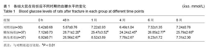
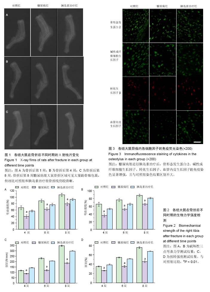
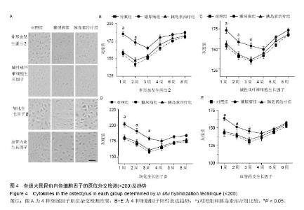
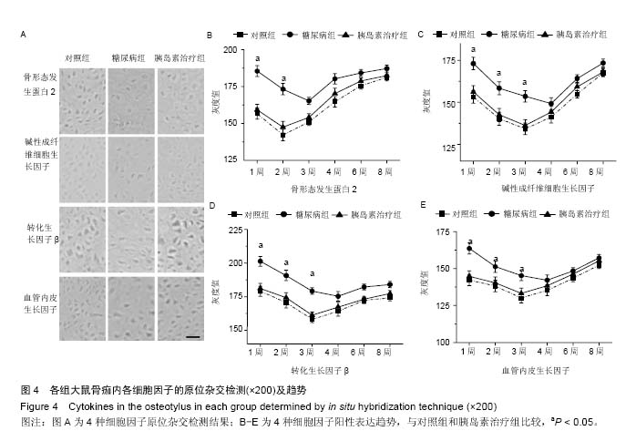
静脉注射5%四氧嘧啶,选取连续3 d血糖> 16.7 mmol/L的糖尿病造模成功大鼠进行实验。成功造模74只,建模成功率82.2%。将建模成功的大鼠分为2组,糖尿病组37只,胰岛素治疗组37只。 2.2 不同时期的血糖水平变化 分别于建模前和建模后的48 h以及第1,4,6,8周测量空腹血糖值,见表1。造模前,对照组、糖尿病组和胰岛素治疗组之间的基础血糖没有差异。大鼠注射四氧嘧啶48 h后,血糖浓度与对照组比较显著升高(P < 0.01),达到(28.71±2.20) mmol/L。骨折造模1周后,胰岛素治疗组血糖值明显低于糖尿病组,且糖尿病组相较于胰岛素治疗组和对照组,血糖值差异有显著性意义(P < 0.01)。结果表明,实验成功建立了糖尿病和胰岛素治疗组的大鼠骨折模型。 2.3 各组大鼠X射线片观察骨折愈合情况 见图1。骨折后第1周(图1A)骨折端骨折线清楚,骨膜增厚,对照组和胰岛素治疗组可见形成少量骨痂,然而糖尿病组中并未观察到明显的骨痂形成现象。骨折后第4周(图1B)对照组和胰岛素治疗组可以看到,骨折区域的X射线片透亮度显著下降,骨量较多,并能观察到骨小梁发生连接,而在糖尿病组观察到骨折区域的X射线片透亮度较高,骨量较少,仍能观察到清晰的骨折线。骨折后第8周(图1C)对照组和胰岛素治疗组可以看到,骨折区域的骨量进一步增加,观察到的骨折线模糊不清,而在糖尿病组的骨折区域可见大量的骨痂包裹,但相比对照组和胰岛素治疗组骨折线仍较清晰。 2.4 生物力学检测结果 将对照组、糖尿病组、胰岛素治疗组造模后的第4,6,8周大鼠取出完整胫骨干置于力学测试机的夹具上固定测定。破坏性三点弯曲力学测试结果见图2A,B,扭转强度测试结果见图2C,D。由计算机软件自动计算弯曲强度、扭转强度和每对标本的参数比率。结果显示,糖尿病组生物力学特征的数值相比对照组差异有显著性意义(P < 0.01),而胰岛素治疗后检测数值明显上升,差异有显著性意义(P < 0.01)。 2.5 免疫荧光检测相关细胞因子的表达 为了检测糖尿病和胰岛素干预治疗对糖尿病大鼠骨折愈合的影响,实验通过免疫荧光技术检测骨痂内多种常见细胞因子的表达情况。在第4周,分别选取对照组、糖尿病组、胰岛素治疗组中骨形态发生蛋白2、碱性成纤维细胞生长因子、转化生长因子、血管内皮生长因子的免疫荧光结果(图3)。骨形态发生蛋白2、碱性成纤维细胞生长因子、血管内皮生长因子呈绿色荧光的阳性表达,可以明显观察到对照组和胰岛素治疗组的荧光强度较高,糖尿病组中则较弱;转化生长因子呈红色荧光的阳性表达,同样可以观察到对照组和胰岛素治疗组的荧光强度较高,糖尿病组中则较弱。结果表明,糖尿病组中各细胞因子骨形态发生蛋白2、碱性成纤维细胞生长因子、转化生长因子、血管内皮生长因子相比于对照组免疫染色结果均下降,而糖尿病组进行胰岛素治疗后,骨形态发生蛋白2、碱性成纤维细胞生长因子、转化生长因子、血管内皮生长因子的免疫染色显著增强,且与对照组染色结果区别不大。 2.6 原位杂交检测相关细胞因子的表达 为了进一步观察3组大鼠骨折愈合过程中细胞因子的动态变化,实验运用原位杂交技术检测骨形态发生蛋白2、碱性成纤维细胞生长因子、转化生长因子、血管内皮生长因子在造模后1,2,3,4,6,8周的表达情况(图4A),并计算平均灰度值,代表各细胞因子的含量。对照组和胰岛素治疗组的骨形态发生蛋白2在第1周和第2周的阳性表达逐步升高,第2周达顶峰,第3,4,6,8周逐渐呈下降趋势,而糖尿病组骨形态发生蛋白2阳性表达第3周达顶峰,且阳性表达明显低于对照组(P < 0.01)(图4B)。对照组和胰岛素治疗组的碱性成纤维细胞生长因子表达自第1周至第3周逐步提高,呈现高阳性的持续表达,第3周达表达顶峰,在第4,6,8周表达逐渐降低,而糖尿病组碱性成纤维细胞生长因子阳性表达在第4周达顶峰,且阳性表达明显低于对照组(P < 0.01)(图4C)。对照组和胰岛素治疗组的转化生长因子阳性表达自第1周至第3周逐步升高,第3周达顶峰,第4,6,8周逐渐下降,而糖尿病组转化生长因子阳性表达在第4周达顶峰,且阳性表达明显低于对照组(P < 0.01)(图4D)。对照组和胰岛素治疗组的血管内皮生长因子表达第1至3周逐步升高,呈持续性的高阳性表达,在第3周时达顶峰,第4,6,8周逐渐下降,而糖尿病组血管内皮生长因子阳性表达在第4周达顶峰,且阳性表达明显低于对照组(P < 0.01)(图4E)。"
"
| [1] Ishikawa K, Fukui T, Nagai T,et al.Type 1 diabetes patients have lower strength in femoral bone determined by quantitative computed tomography: A cross-sectional study. J Diabetes Investig. 2015;6(6): 726-733. [2] 李溪,向盈盈,龚跃昆,等. 糖尿病骨折大鼠骨痂组织中成骨细胞增殖和骨钙素表达与骨形态发生蛋白2干预的关系[J].中国组织工程研究与临床康复, 2009, 13(20): 3857-3861. [3] Zhang Q, Dong H, Li Y,et al. Microgrooved Polymer Substrates Promote Collective Cell Migration To Accelerate Fracture Healing in an in Vitro Model. ACS applied materials & interfaces.2015;7(41):23336-23345. [4] Takeyama K,Chatani M,Takano Y,et al.In-vivo imaging of the fracture healing in medaka revealed two types of osteoclasts before and after the callus formation by osteoblasts.Developmental biology.2014;394(2): 292-304. [5] Murata K,Ito H,Yoshitomi H,et al.Inhibition of miR-92a enhances fracture healing via promoting angiogenesis in a model of stabilized fracture in young mice.J Bone Miner Res.2014;29(2):316-326. [6] Wang CY, Yang HB, Hsu HS,et al. Mesenchymal stem cell-conditioned medium facilitates angiogenesis and fracture healing in diabetic rats. Journal of tissue engineering and regenerative medicine. 2012;6(7): 559-569. [7] Alblowi J, Tian C, Siqueira MF,et al.Chemokine expression is upregulated in chondrocytes in diabetic fracture healing. Bone. 2013;53(1):294-300. [8] Kottstorfer J,Kaiser G,Thomas A,et al.The influence of non-osteogenic factors on the expression of M-CSF and VEGF during fracture healing. Injury.2013;44(7): 930-934. [9] 马骋,高岩,苟三怀,等.神经生长因子对大鼠胫骨骨折愈合的影响[J].中国组织工程研究与临床康复,2008, 12(46): 9032-9035. [10] Krakauer JC, McKenna MJ, Buderer NF,et al.Bone loss and bone turnover in diabetes.Diabetes. 1995; 44(7):775-782. [11] Krakauer JC, McKenna MJ, Rao DS,et al. Bone mineral density in diabetes. Diabetes care. 1997; 20(8):1339-1340. [12] Li R,Nauth A,Gandhi R,et al.BMP-2 mRNA expression after endothelial progenitor cell therapy for fracture healing. J Orthop Trauma.2014;28 Suppl 1:S24-27. [13] Saito W, Uchida K, Matsushita O,et al.Acceleration of callus formation during fracture healing using basic fibroblast growth factor-kidney disease domain- collagen-binding domain fusion protein combined with allogenic demineralized bone powder. Journal of orthopaedic surgery and research. 2015;10:59. [14] Poniatowski LA,Wojdasiewicz P,Gasik R,et al. Transforming growth factor Beta family: insight into the role of growth factors in regulation of fracture healing biology and potential clinical applications. Mediators of inflammation.2015;2015:137823. [15] Wang CJ,Huang KE,Sun YC,et al.VEGF modulates angiogenesis and osteogenesis in shockwave- promoted fracture healing in rabbits. J Surg Res. 2011; 171(1):114-119. [16] Levy JR, Murray E, Manolagas S,et al. Demonstration of insulin receptors and modulation of alkaline phosphatase activity by insulin in rat osteoblastic cells. Endocrinology.1986;119(4):1786-1792. [17] 李溪,向盈盈,龚跃昆,刘劲松.胰岛素样生长因子1在糖尿病骨折大鼠骨痂组织中对成骨细胞增殖和骨钙素表达的影响[J].中国组织工程研究与临床康复, 2009, (15):2865-2868. [18] Feng X, Huang D, Lu X,et al. Insulin-like growth factor 1 can promote proliferation and osteogenic differentiation of human dental pulp stem cells via mTOR pathway.Development, growth & differentiation. 2014;56(9):615-624. [19] Chen Y, Ke J, Long X,et al.Insulin-like growth factor-1 boosts the developing process of condylar hyperplasia by stimulating chondrocytes proliferation. Osteoarthritis and cartilage / OARS, Osteoarthritis Research Society.2012;20(4):279-287. [20] Petrou M, Niemeyer P, Stoddart MJ,et al.Mesenchymal stem cell chondrogenesis: composite growth factor-bioreactor synergism for human stem cell chondrogenesis. Regenerative medicine. 2013;8(2): 157-170. [21] Wong MC, Leung MC, Tsang CS,et al. The rising tide of diabetes mellitus in a Chinese population: a population-based household survey on 121,895 persons. Int J Public Health.2013;58(2):269-276. [22] Driessen JH,van Onzenoort HA,Henry RM, et al.Use of dipeptidyl peptidase-4 inhibitors for type 2 diabetes mellitus and risk of fracture. Bone.2014;68:124-30. [23] Mishima T,Motoyama K,Imanishi Y,et al.Decreased cortical thickness, as estimated by a newly developed ultrasound device, as a risk for vertebral fracture in type 2 diabetes mellitus patients with eGFR of less than 60 mL/min/1.73 m2. Osteoporos Int. 2015;26(1): 229-236. [24] Liao CC,Lin CS,Shih CC,et al.Increased risk of fracture and postfracture adverse events in patients with diabetes: two nationwide population-based retrospective cohort studies. Diabetes care. 2014; 37(8):2246-2252. [25] Roy B.Biomolecular basis of the role of diabetes mellitus in osteoporosis and bone fractures. World J Diabetes. 2013;4(4):101-113. [26] Kayal RA,Siqueira M,Alblowi J, et al.TNF-alpha mediates diabetes-enhanced chondrocyte apoptosis during fracture healing and stimulates chondrocyte apoptosis through FOXO1. J Bone Miner Res. 2010; 25(7):1604-1615. [27] Lee SN,Lee DH,Lee MG,et al.Proprotein convertase 5/6a is associated with bone morphogenetic protein-2-induced squamous cell differentiation. Am J Respir Cell Mol Biol. 2015;52(6):749-761. [28] 徐晓峰,李阳,钱栋,等.骨形态发生蛋白2和血管内皮生长因子mRNA在股骨骨不连大鼠损伤区域的动态表达[J].中国组织工程研究与临床康复,2010, 14(37): 6857- 6860. [29] Hughes-Fulford M, Li CF. The role of FGF-2 and BMP-2 in regulation of gene induction, cell proliferation and mineralization. J Orthop Surg Res. 2011;6:8. [30] Cals FL,Hellingman CA,Koevoet W,et al.Effects of transforming growth factor-beta subtypes on in vitro cartilage production and mineralization of human bone marrow stromal-derived mesenchymal stem cells. J Tissue Eng Regen Med..2012;6(1):68-76. [31] Kazemi-Lomedasht F,Behdani M,Bagheri KP,et al.Inhibition of angiogenesis in human endothelial cell using VEGF specific nanobody.Molecular Immunology. 2015;65(1):58-67. [32] Chamorro-Jorganes A, Lee MY, Araldi E,et al. VEGF-Induced Expression of miR-17~92 Cluster in Endothelial Cells is Mediated by ERK/ELK1 Activation and Regulates Angiogenesis. Circ Res. 2016;118(1): 38-47. [33] Blakytny R, Spraul M, Jude EB. Review: The diabetic bone: a cellular and molecular perspective. Int J Low Extrem Wounds. 2011;10(1):16-32. [34] Courcoulas AP,Belle SH,Neiberg RH,et al.Three-Year Outcomes of Bariatric Surgery vs Lifestyle Intervention for Type 2 Diabetes Mellitus Treatment: A Randomized Clinical Trial. JAMA surgery.2015;150(10):931-940. [35] Pramojanee SN,Phimphilai M,Chattipakorn N,et al. Possible roles of insulin signaling in osteoblasts. Endocrine research.2014;39(4):144-151. |
| [1] | Zhang Tongtong, Wang Zhonghua, Wen Jie, Song Yuxin, Liu Lin. Application of three-dimensional printing model in surgical resection and reconstruction of cervical tumor [J]. Chinese Journal of Tissue Engineering Research, 2021, 25(9): 1335-1339. |
| [2] | Zeng Yanhua, Hao Yanlei. In vitro culture and purification of Schwann cells: a systematic review [J]. Chinese Journal of Tissue Engineering Research, 2021, 25(7): 1135-1141. |
| [3] | Yang Weiqiang, Ding Tong, Yang Weike, Jiang Zhengang. Combined variable stress plate internal fixation affects changes of bone histiocyte function and bone mineral density at the fractured end of goat femur [J]. Chinese Journal of Tissue Engineering Research, 2021, 25(6): 890-894. |
| [4] | Xu Dongzi, Zhang Ting, Ouyang Zhaolian. The global competitive situation of cardiac tissue engineering based on patent analysis [J]. Chinese Journal of Tissue Engineering Research, 2021, 25(5): 807-812. |
| [5] | Wu Zijian, Hu Zhaoduan, Xie Youqiong, Wang Feng, Li Jia, Li Bocun, Cai Guowei, Peng Rui. Three-dimensional printing technology and bone tissue engineering research: literature metrology and visual analysis of research hotspots [J]. Chinese Journal of Tissue Engineering Research, 2021, 25(4): 564-569. |
| [6] | Chang Wenliao, Zhao Jie, Sun Xiaoliang, Wang Kun, Wu Guofeng, Zhou Jian, Li Shuxiang, Sun Han. Material selection, theoretical design and biomimetic function of artificial periosteum [J]. Chinese Journal of Tissue Engineering Research, 2021, 25(4): 600-606. |
| [7] | Liu Fei, Cui Yutao, Liu He. Advantages and problems of local antibiotic delivery system in the treatment of osteomyelitis [J]. Chinese Journal of Tissue Engineering Research, 2021, 25(4): 614-620. |
| [8] | Li Xiaozhuang, Duan Hao, Wang Weizhou, Tang Zhihong, Wang Yanghao, He Fei. Application of bone tissue engineering materials in the treatment of bone defect diseases in vivo [J]. Chinese Journal of Tissue Engineering Research, 2021, 25(4): 626-631. |
| [9] | Zhang Zhenkun, Li Zhe, Li Ya, Wang Yingying, Wang Yaping, Zhou Xinkui, Ma Shanshan, Guan Fangxia. Application of alginate based hydrogels/dressings in wound healing: sustained, dynamic and sequential release [J]. Chinese Journal of Tissue Engineering Research, 2021, 25(4): 638-643. |
| [10] | Chen Jiana, Qiu Yanling, Nie Minhai, Liu Xuqian. Tissue engineering scaffolds in repairing oral and maxillofacial soft tissue defects [J]. Chinese Journal of Tissue Engineering Research, 2021, 25(4): 644-650. |
| [11] | Cheng Shigao, , Wang Wanchun, Jiang Dong, Li Tengfei, Li Xun, Ren Lian. Comparison of the standard and long-stem bone cement prosthesis replacement in the treatment of intertrochanteric fractures in elderly patients [J]. Chinese Journal of Tissue Engineering Research, 2021, 25(3): 362-367. |
| [12] | Xing Hao, Zhang Yonghong, Wang Dong. Advantages and disadvantages of repairing large-segment bone defect [J]. Chinese Journal of Tissue Engineering Research, 2021, 25(3): 426-430. |
| [13] | Chen Ziyang, Pu Rui, Deng Shuang, Yuan Lingyan. Regulatory effect of exosomes on exercise-mediated insulin resistance diseases [J]. Chinese Journal of Tissue Engineering Research, 2021, 25(25): 4089-4094. |
| [14] | Gao Shan, Huang Dongjing, Hong Haiman, Jia Jingqiao, Meng Fei. Comparison on the curative effect of human placenta-derived mesenchymal stem cells and induced islet-like cells in gestational diabetes mellitus rats [J]. Chinese Journal of Tissue Engineering Research, 2021, 25(25): 3981-3987. |
| [15] | Liu Liyong, Zhou Lei. Research and development status and development trend of hydrogel in tissue engineering based on patent information [J]. Chinese Journal of Tissue Engineering Research, 2021, 25(22): 3527-3533. |
| Viewed | ||||||
|
Full text |
|
|||||
|
Abstract |
|
|||||