Chinese Journal of Tissue Engineering Research ›› 2021, Vol. 25 ›› Issue (11): 1664-1669.doi: 10.3969/j.issn.2095-4344.3108
Previous Articles Next Articles
Salidroside inhibits apoptosis of retinal Müller cells induced by high glucose in rats
Zhao Ning1, Yu Hongdan2, Feng Zhen2, Ding Jiayuan2, Liu Xuezheng1
- 1College of Integrative Chinese and Western Medicine, Liaoning University of Traditional Chinese Medicine, Shenyang 110847, Liaoning Province, China; 2Department of Anatomy, School of Basic Medicine, Jinzhou Medical University, Jinzhou 121001, Liaoning Province, China
-
Received:2020-04-03Revised:2020-04-28Accepted:2020-05-27Online:2021-04-18Published:2020-12-21 -
Contact:Liu Xuezheng, MD, Professor, College of Integrative Chinese and Western Medicine, Liaoning University of Traditional Chinese Medicine, Shenyang 110847, Liaoning Province, China -
About author:Zhao Ning, Master, Lecturer, College of Integrative Chinese and Western Medicine, Liaoning University of Traditional Chinese Medicine, Shenyang 110847, Liaoning Province, China
CLC Number:
Cite this article
Zhao Ning, Yu Hongdan, Feng Zhen, Ding Jiayuan, Liu Xuezheng. Salidroside inhibits apoptosis of retinal Müller cells induced by high glucose in rats[J]. Chinese Journal of Tissue Engineering Research, 2021, 25(11): 1664-1669.
share this article

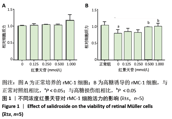
2.1 红景天苷对高糖作用后rMC-1细胞活力的影响 通过CCK8检测不同条件下红景天苷对rMC-1细胞活力的影响。在正常培养基中加入不同浓度红景天苷孵育48 h,对rMC-1细胞活力没有显著影响(P > 0.05),见图1A;在高糖培养基中孵育48 h,与正常对照组比较,高糖损伤组rMC-1细胞活力明显降低(P < 0.05),见图1B;与高糖损伤组比较,红景天苷组(0.5,1 mmol/L)细胞活力显著上升(P < 0.05)。实验筛选红景天苷(0.5 mmol/L)为最佳使用浓度用于后续细胞实验。"

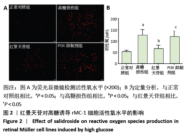
2.2 红景天苷对高糖作用后rMC-1细胞活性氧水平的影响 荧光显微镜检测活性氧水平,见图2。与正常对照组相比,高糖损伤组荧光强度增加(P < 0.05);与高糖损伤组相比,红景天苷组荧光强度减弱(P < 0.05);而PI3K抑制剂组荧光强度与红景天苷组结果相反(P < 0.05)。以上结果提示红景天苷能减少高糖作用后rMC-1细胞活性氧水平,而LY294002 能阻止这种效果。 "
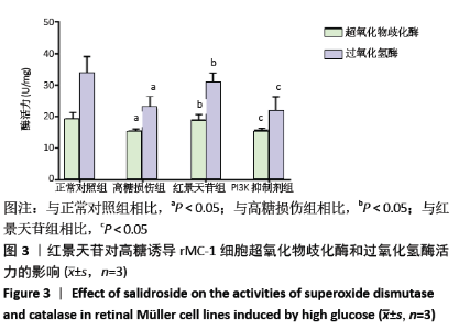
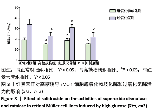
2.3 红景天苷对高糖作用后rMC-1细胞超氧化物歧化酶、过氧化氢酶活力的影响 酶标仪检测rMC-1细胞超氧化物歧化酶、过氧化氢酶的活力,见图3。与正常对照组相比,高糖损伤组rMC-1细胞超氧化物歧化酶、过氧化氢酶活力显著降低(P < 0.05);与高糖损伤组相比,红景天苷组rMC-1细胞超氧化物歧化酶、过氧化氢酶活力显著升高(P < 0.05);而PI3K抑制剂组结果与红景天苷组结果相反(P < 0.05)。以上结果提示红景天苷能增加高糖作用后rMC-1细胞超氧化物歧化酶、过氧化氢酶活力,而LY294002能阻止这种效果。 "

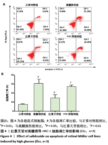
2.4 红景天苷对高糖作用后rMC-1细胞凋亡的影响 流式细胞仪检测各组细胞凋亡,见图4。与正常对照组相比,高糖损伤组rMC-1细胞凋亡率著增加(P < 0.01);与高糖损伤组相比,红景天苷组rMC-1细胞凋亡率显著降低(P < 0.05);而PI3K抑制剂组结果与红景天苷组结果相反(P < 0.01)。以上结果提示红景天苷能减少高糖作用后rMC-1细胞凋亡率,而LY294002 能阻止这种效果。 "
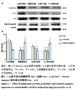
2.5 红景天苷对高糖作用后rMC-1细胞Cleaved caspase-3、PI3K、Akt 、P-Akt蛋白表达的影响 Western blot检测各组细胞Cleaved caspase-3、PI3K、Akt 、P-Akt蛋白的表达情况,见图5。与正常对照组相比,高糖损伤组Cleaved caspase-3蛋白表达明显升高(P < 0.05), PI3K 蛋白表达明显减少(P < 0.01)。以总Akt为参照,P-Akt表达明显减少(P < 0.01);与高糖损伤组相比,红景天苷组Cleaved caspase-3蛋白表达明显减少,PI3K 蛋白表达显著升高,以总Akt为参照,P-Akt表达明显增多(P < 0.05);而PI3K 抑制剂组结果与红景天苷组结果相反(P < 0.05)。以上结果提示红景天苷可能通过激活AKT信号通路减少高糖作用后rMC-1细胞Cleaved caspase-3 的表达,而LY294002 能阻止这种效果。 "

| [1] SALEH I, MARITSKA Z, PARISA N, et al. Inhibition of Receptor for Advanced Glycation End Products as New Promising Strategy Treatment in Diabetic Retinopathy.Open Access Maced J Med Sci.2019;7(23): 3921-3924. [2] OSTRI C, LA COUR M, LUND-ANDERSEN H. Diabetic vitrectomy in a large type 1 diabetes patient population: long-term incidence and risk factors.Acta Ophthalmol. 2014;92(5):439-443. [3] CHEN H, JI Y, YAN X, et al. Berberine attenuates apoptosis in rat retinal Muller cells stimulated with high glucose via enhancing autophagy and the AMPK/mTOR signaling. Biomed Pharmacother. 2018;108: 1201-1207. [4] WANG JJ, ZHU M, LE YZ. Functions of Muller cell-derived vascular endothelial growth factor in diabetic retinopathy. World J Diabetes. 2015;6(5):726-733. [5] ZHOU J, SHEN X, LU Q, et al. Thioredoxin-Interacting Protein (TXNIP) Suppresses Expression of Glutamine Synthetase by Inducing Oxidative Stress in Retinal Muller Glia Under Diabetic Conditions.Med Sci Monit. 2016;22:1460-1466. [6] TIEN T, ZHANG J, MUTO T, et al. High Glucose Induces Mitochondrial Dysfunction in Retinal Muller Cells: Implications for Diabetic Retinopathy. Invest Ophthalmol Vis Sci. 2017;58(7):2915-2921. [7] OLA MS, AL-DOSARI D, ALHOMIDA AS. Role of Oxidative Stress in Diabetic Retinopathy and the Beneficial Effects of Flavonoids. Curr Pharm Des. 2018;24(19):2180-2187. [8] CHEN S, CAI F, WANG J, et al. Salidroside protects SHSY5Y from pathogenic alphasynuclein by promoting cell autophagy via mediation of mTOR/p70S6K signaling.Mol Med Rep. 2019;20(1):529-538. [9] ZHU L, CHEN T, CHANG X, et al. Salidroside ameliorates arthritis-induced brain cognition deficits by regulating Rho/ROCK/NF-kappaB pathway.Neuropharmacology. 2016;103:134-142. [10] TANG H, GAO L, MAO J, et al. Salidroside protects against bleomycin-induced pulmonary fibrosis: activation of Nrf2-antioxidant signaling, and inhibition of NF-kappaB and TGF-beta1/Smad-2/-3 pathways.Cell Stress Chaperones. 2016;21(2):239-249. [11] XING SS, YANG XY, ZHENG T, et al. Salidroside improves endothelial function and alleviates atherosclerosis by activating a mitochondria-related AMPK/PI3K/Akt/eNOS pathway.Vascul Pharmacol. 2015;72: 141-152. [12] SHI K, WANG X, ZHU J, et al. Salidroside protects retinal endothelial cells against hydrogen peroxide-induced injury via modulating oxidative status and apoptosis.Biosci Biotechnol Biochem. 2015;79(9):1406-1413. [13] QIAN C, LIANG S, WAN G, et al. Salidroside alleviates high-glucose-induced injury in retinal pigment epithelial cell line ARPE-19 by down-regulation of miR-138.RNA Biol. 2019;16(10):1461-1470. [14] BERGERHOFF K, CLAR C, RICHTER B. Aspirin in diabetic retinopathy.A systematic review. Endocrinol Metab Clin North Am. 2002;31(3): 779-793. [15] EGAN A, BYRNE M. Effects of medical therapies on retinopathy progression in type 2 diabetes. Ir Med J. 2011;104(2):37. [16] WANG L, WANG N, TAN HY, et al. Protective effect of a Chinese Medicine formula He-Ying-Qing-Re Formula on diabetic retinopathy.J Ethnopharmacol. 2015;169:295-304. [17] ZHAO YQ, LI QS, XIANG MH, et al. Distribution of traditional Chinese medicine syndromes of diabetic retinopathy and correlation between symptoms . Zhongguo Zhong Yao Za Zhi. 2017;42(14):2796-2801. [18] LU H,LI Y,ZHANG T,et al.Salidroside Reduces High-Glucose-Induced Podocyte Apoptosis and Oxidative Stress via Upregulating Heme Oxygenase-1 (HO-1) Expression.Med Sci Monit.2017;23:4067-4076. [19] MA YG, WANG JW, ZHANG YB, et al. Salidroside improved cerebrovascular vasodilation in streptozotocin-induced diabetic rats through restoring the function of BKCa channel in smooth muscle cells.Cell Tissue Res. 2017;370(3):365-377. [20] 韩雪娇.郭娜.朱美宣.等.红景天苷药理作用及其作用机理研究进展[J].中国生化药物杂志,2015,35(1):171-175. [21] NEWMAN E, REICHENBACH A. The Muller cell:a functional element of the retina.Trends Neurosci. 1996;19(8):307-312. [22] OLA MS. Effect of hyperglycemia on insulin receptor signaling in the cultured retinal Muller glial cells.Biochem Biophys Res Commun. 2014; 444(2):264-269. [23] BRINGMANN A, PANNICKE T, GROSCHE J, et al. Muller cells in the healthy and diseased retina. Prog Retin Eye Res. 2006;25(4):397-424. [24] FU QL, WU W, WANG H, et al. Up-regulated endogenous erythropoietin/erythropoietin receptor system and exogenous erythropoietin rescue retinal ganglion cells after chronic ocular hypertension. Cell Mol Neurobiol. 2008;28(2):317-329. [25] 颜赛梅,常青.糖尿病视网膜病变中Müller 细胞氧化还原状态研究[J].中国眼耳鼻喉科杂志,2009,9(4):261-263. [26] TRUDEAU K, MOLINA AJ, GUO W, et al. High glucose disrupts mitochondrial morphology in retinal endothelial cells: implications for diabetic retinopathy.Am J Pathol. 2010;177(1):447-455. [27] KOWLURU RA, KOWLURU A, MISHRA M, et al. Oxidative stress and epigenetic modifications in the pathogenesis of diabetic retinopathy.Prog Retin Eye Res. 2015;48:40-61. [28] LIU L, ZUO Z, LU S, et al. Naringin attenuates diabetic retinopathy by inhibiting inflammation, oxidative stress and NF-kappaB activation in vivo and in vitro. Iran J Basic Med Sci. 2017;20(7):813-821. [29] BRINGMANN A, PANNICKE T, GROSCHE J, et al. Muller cells in the healthy and diseased retina. Prog Retin Eye Res. 2006;25(4):397-424. [30] HAN N, YU L, SONG Z, et al. Agmatine protects Muller cells from high-concentration glucose-induced cell damage via N-methyl-D-aspartic acid receptor inhibition. Mol Med Rep. 2015;12(1):1098-1106. [31] REN X, SUN L, WEI L, et al. Liraglutide Up-regulation Thioredoxin Attenuated Muller Cells Apoptosis in High Glucose by Regulating Oxidative Stress and Endoplasmic Reticulum Stress.Curr Eye Res.2020; 17:1-9. [32] ZHOU X, AI S, CHEN Z, et al. Probucol promotes high glucose-induced proliferation and inhibits apoptosis by reducing reactive oxygen species generation in Muller cells.Int Ophthalmol. 2019;39(12):2833-2842. [33] DEVI TS, LEE I, HUTTEMANN M, et al. TXNIP links innate host defense mechanisms to oxidative stress and inflammation in retinal Muller glia under chronic hyperglycemia: implications for diabetic retinopathy.Exp Diabetes Res. 2012;2012:438238. [34] 庞若宇,关美萍,郑宗基,等.二甲双胍对糖基化终末产物诱导的成纤维细胞凋亡及相关蛋白Caspase3、Bax及Bcl-2 表达的影响[J].南方医科大学学报,2015,35(6):898-902. [35] ZUO T, ZHU M, XU W, et al. Iridoids with Genipin Stem Nucleus Inhibit Lipopolysaccharide-Induced Inflammation and Oxidative Stress by Blocking the NF-kappaB Pathway in Polycystic Ovary Syndrome.Cell Physiol Biochem. 2017;43(5):1855-1865. [36] CUI W, LENG B, WANG G. Klotho protein inhibits H2O2-induced oxidative injury in endothelial cells via regulation of PI3K/AKT/Nrf2/HO-1 pathways. Can J Physiol Pharmacol. 2019;97(5):370-376. [37] 师岩,徐晶,程昊,等.京尼平抑制高糖诱导的大鼠心肌H9c2细胞氧化应激及凋亡损伤[J].中国病理生理杂志,2019,35(2):224-230. [38] 崔伟,王高频,王洪新,等.黄芪甲苷通过激活PI3K/Akt通路抑制内皮细胞内质网应激介导的细胞凋亡的研究[J].中药药理与临床, 2018,34(5):39-44. [39] YANG X, HUO F, LIU B, et al. Crocin Inhibits Oxidative Stress and Pro-inflammatory Response of Microglial Cells Associated with Diabetic Retinopathy Through the Activation of PI3K/Akt Signaling Pathway.J Mol Neurosci. 2017;61(4):581-589. [40] YIN Y, LIU D, TIAN D. Salidroside prevents hydroperoxide-induced oxidative stress and apoptosis in retinal pigment epithelium cells.Exp Ther Med. 2018;16(3):2363-2368. [41] 陶蕊,张怀国,付庆喜,等.红景天苷对肌萎缩侧索硬化症的保护机制[J].中药药理与临床,2019,10(3):2471-2473. |
| [1] | Zhu Xuefen, Huang Cheng, Ding Jian, Dai Yongping, Liu Yuanbing, Le Lixiang, Wang Liangliang, Yang Jiandong. Mechanism of bone marrow mesenchymal stem cells differentiation into functional neurons induced by glial cell line derived neurotrophic factor [J]. Chinese Journal of Tissue Engineering Research, 2021, 25(7): 1019-1025. |
| [2] | Wang Zhengdong, Huang Na, Chen Jingxian, Zheng Zuobing, Hu Xinyu, Li Mei, Su Xiao, Su Xuesen, Yan Nan. Inhibitory effects of sodium butyrate on microglial activation and expression of inflammatory factors induced by fluorosis [J]. Chinese Journal of Tissue Engineering Research, 2021, 25(7): 1075-1080. |
| [3] | Jiao Hui, Zhang Yining, Song Yuqing, Lin Yu, Wang Xiuli. Advances in research and application of breast cancer organoids [J]. Chinese Journal of Tissue Engineering Research, 2021, 25(7): 1122-1128. |
| [4] | Huang Dengcheng, Wang Zhike, Cao Xuewei. Intravenous, topical tranexamic acid alone or their combination in total knee arthroplasty: a meta-analysis of randomized controlled trials [J]. Chinese Journal of Tissue Engineering Research, 2021, 25(6): 948-956. |
| [5] | Zhan Fangbiao, Cheng Jun, Zou Xinsen, Long Jie, Xie Lizhong, Deng Qianrong. Intraoperative intravenous application of tranexamic acid reduces perioperative bleeding in multilevel posterior spinal surgery: a meta-analysis [J]. Chinese Journal of Tissue Engineering Research, 2021, 25(6): 977-984. |
| [6] | Yang Xin, Jin Zhe, Feng Xu, Lu Bing. The current situation of knowledge and attitudes towards organ, eye tissue, body donation of residents in Shenyang [J]. Chinese Journal of Tissue Engineering Research, 2021, 25(5): 779-784. |
| [7] | Zhang Guomei, Zhu Jun, Hu Yang, Jiao Hongwei. Stress of three-dimensional finite element models of E-MAX porcelain inlay [J]. Chinese Journal of Tissue Engineering Research, 2021, 25(4): 537-541. |
| [8] | Liu Fei, Cui Yutao, Liu He. Advantages and problems of local antibiotic delivery system in the treatment of osteomyelitis [J]. Chinese Journal of Tissue Engineering Research, 2021, 25(4): 614-620. |
| [9] | Ye Haimin, Ding Linghua, Kong Weihao, Huang Zutai, Xiong Long. Role and mechanism of hierarchical microchanneled bone scaffolds in promoting osteogenesis and angiogenesis [J]. Chinese Journal of Tissue Engineering Research, 2021, 25(4): 621-625. |
| [10] | Yu Langbo, Qing Mingsong, Zhao Chuntao, Peng Jiachen. Hot issues in clinical application of dynamic contrast-enhanced magnetic resonance imaging in orthopedics [J]. Chinese Journal of Tissue Engineering Research, 2021, 25(3): 449-455. |
| [11] | Huang Maomao, Hu Yue, Wang Binchuan, Zhang Chi, Xie Yujie, Wang Jianxiong, Wang Li, Xu Fangyuan. Bibliometric and visual analysis of international literature addressing ischemic stroke rehabilitation in recent 10 years [J]. Chinese Journal of Tissue Engineering Research, 2021, 25(23): 3725-3733. |
| [12] | Zhu Rui, Zeng Qing, Huang Guozhi. Ferroptosis and stroke [J]. Chinese Journal of Tissue Engineering Research, 2021, 25(23): 3734-3739. |
| [13] | Li Shanshan, You Ran, Guo Xiaoxiao, Zhao Lu, Wang Yanling, Chen Xi. Advances in the mechanisms of optic nerve regeneration [J]. Chinese Journal of Tissue Engineering Research, 2021, 25(23): 3740-3745. |
| [14] | Wang Zhen, Lin Haiqi, He Fei, Lin Wentao. Exercise activates skeletal muscle satellite cells: exercise prevention and treatment for age-related sarcopenia and muscle injury [J]. Chinese Journal of Tissue Engineering Research, 2021, 25(23): 3752-3759. |
| [15] | Song Shilei, Chen Yueping, Zhang Xiaoyun, Li Shibin, Lai Yu, Zhou Yi. Potential molecular mechanism of Wuling powder in treating osteoarthritis based on network pharmacology and molecular docking [J]. Chinese Journal of Tissue Engineering Research, 2021, 25(20): 3185-3193. |
| Viewed | ||||||
|
Full text |
|
|||||
|
Abstract |
|
|||||