Chinese Journal of Tissue Engineering Research ›› 2019, Vol. 23 ›› Issue (7): 1046-1051.doi: 10.3969/j.issn.2095-4344.1073
Previous Articles Next Articles
Human amniotic membrane repairs acute sciatic nerve injury in rat models
Cao Peng, Wang Haonan, Tian Weifeng, Sun Naichao, Bai Jiangbo, Yu Kunlun, Tian Dehu
- (the Third Hospital of Hebei Medical University, Shijiazhuang 050000, Hebei Province, China)
-
Received:2018-10-25Online:2019-03-08Published:2019-03-08 -
Contact:Tian Dehu, MD, Chief physician, Professor, the Third Hospital of Hebei Medical University, Shijiazhuang 050000, Hebei Province, China -
About author:Cao Peng, Master, Attending physician, the Third Hospital of Hebei Medical University, Shijiazhuang 050000, Hebei Province, China
CLC Number:
Cite this article
Cao Peng, Wang Haonan, Tian Weifeng, Sun Naichao, Bai Jiangbo, Yu Kunlun, Tian Dehu. Human amniotic membrane repairs acute sciatic nerve injury in rat models[J]. Chinese Journal of Tissue Engineering Research, 2019, 23(7): 1046-1051.
share this article
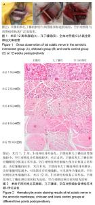
2.1 实验动物数量分析 SD大鼠90只,随机分为3组,每组30只,均进入结果分析。 2.2 大体观察结果 术后3组大鼠伤口均未见感染,无足底溃疡,未见自噬现像,Ⅰ期愈合。 术后1周大鼠左下肢均出现肿胀,拖地现象,肌力差,刺激无收缩,未出现明显缩足及舔足动作。精神状态较差,饮食及尿便较前减少明显。下肢肿胀逐渐减轻,至术后8周羊膜组和几丁糖组跛足现象消失,对外界刺激出现明显缩足及舔足,空白对照组仍有跛足现象,对外界刺激感觉差。术后12周,大鼠对外界刺激敏感,空白对照组稍差。 各组大鼠神经周围恢复情况,见图1。 术后1,2周,3组大鼠损伤段神经压迹清晰可见,损伤神经菲薄,神经外膜充血,羊膜组和几丁糖组神经与周围组织粘连不明显,空白对照组神经与周围组织有薄膜性的粘连。术后4,8周,神经及周围组织充血水肿减轻,损伤段神经较前增粗,羊膜组及几丁糖组外膜充血基本消退,与周围组织粘连较疏松,空白对照组神经外膜充血较明显,质地较硬,与周围组织粘连较致密。羊膜大部分吸收,几丁糖完全吸收。术后12周,神经及周围组织未见明显充血水肿,损伤段神经增粗,接近正常,神经外膜充血消失,羊膜组和几丁糖组神经与周围组织粘连较疏松,羊膜完全吸收,空白对照组与周围组织粘连广泛而致密,钝性分离困难。 2.3 光镜观察结果 见图2。 术后1,2周,3组神经排列紊乱,失去正常形态,髓鞘肿胀,可见大量脱髓鞘的神经,视野内出现大量炎性细胞,可见巨噬细胞,其中羊膜组和几丁糖组炎性细胞较少,空白对照组炎性细胞较多。术后4周,羊膜组和几丁糖组神经髓鞘部分恢复正常形态,可见大量许旺细胞。3组神经炎症细胞较前均有所减少,羊膜组和几丁糖组减少更加明显,空白对照组神经细胞大部分未恢复正常形态,炎症细胞仍较多。术后8周,羊膜组和几丁糖组神经排列整齐,炎症细胞明显减少,可见散在许旺细胞核,可见新生毛细血管。空白对照组炎性细胞较多。术后12周,3组神经均恢复正常形态,羊膜组和几丁糖组神经束间较为疏松,空白对照组神经束间较为致密。 2.4 透射电镜观察结果 见图3。 术后2周,羊膜组和几丁糖组可见髓鞘融合、肿胀、空化,髓鞘不整齐,许旺细胞肿胀,核周系扩张,胞质肿胀,线粒体空泡化,未见明显成纤维细胞。空白对照组可见明显脱髓鞘反应,髓鞘板层结构剥离,髓鞘结构不规则,向内增生,许旺细胞内线粒体空泡化,嵴和膜溶解消失,内质网脱颗粒,核周系扩张,线粒体致密化。偶可见成纤维细胞包裹轴突。术后4周,羊膜组和几丁糖组髓鞘较规则,脱髓鞘反应减少,成纤维细胞及胶原纤维较少,细胞器较空白对照组多。空白对照组髓鞘欠规则,仍可见较多脱髓鞘反应,可见大量成纤维细胞和胶原纤维增生,粗面内质网较多,游离核糖体较发达,线粒体损伤较前减轻。术后8周,羊膜组及几丁糖组成纤维细胞少,轴突排列整齐,粗细较一致,轴突层板较清晰。空白对照组轴突排列较整齐,成纤维细胞和胶原纤维减少。偶可见损伤髓鞘。"
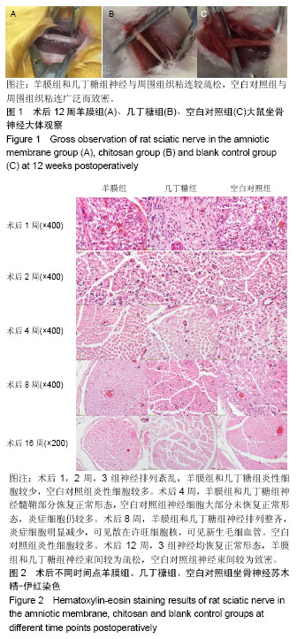
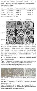
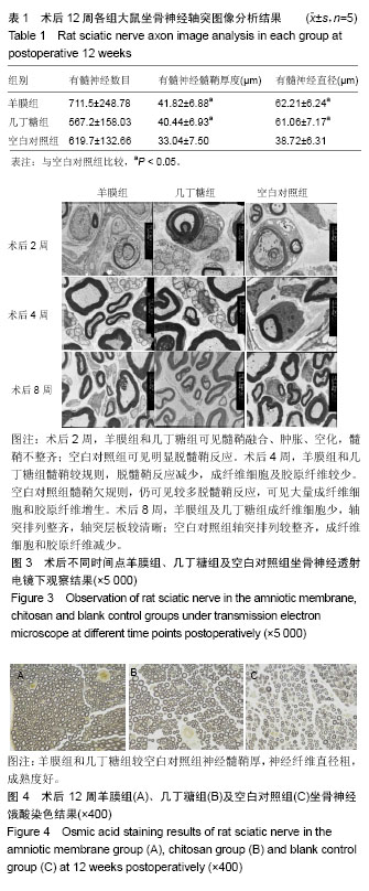
上述结果显示,羊膜组和几丁糖组神经修复过程早于空白对照组,羊膜组与几丁糖组无明显差异。 2.5 轴突图像分析 见图4和表1。术后12周对3组大鼠坐骨神经进行锇酸染色,并在高倍视野下进行有髓神经计数,测量有髓神经髓鞘厚度及直径,羊膜组和几丁糖组较空白对照组神经髓鞘厚,神经纤维直径粗,成熟度好,有髓神经计数并无明显差异。 "
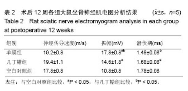
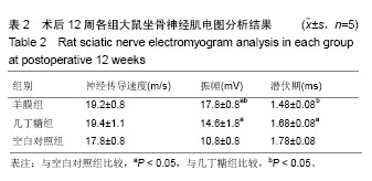
2.6 神经电生理检测结果 见表2。术后12周,对3组大鼠进行肌电图检查,测定神经传导速度、波幅及潜伏期,结果显示:3组间传导速度差异无显著性意义;羊膜组波幅高于几丁糖组,几丁糖组波幅高于空白对照组;羊膜组潜伏期要短于几丁糖组和空白对照组,几丁糖组和空白对照组间差异无显著性意义。"
| [1] 陈琳,张玉琪,左焕琮. 神经生长因子和碱性成纤维细胞生长因子在周围神经修复应用中的研究进展[J]. 中华神经外科杂志,2016, 32(2): 207-209.[2] 刘颜芬,张雪晶,段晓琴,等. 富血小板血浆对周围神经损伤修复的研究现状[J].中国实验诊断学 2018,22(1):145-148.[3] 杜怀栋,周梁. 神经导管在神经修复中的作用[J].复旦学报(医学版), 2006,33(5):709-710.[4] 门永芝, 於子卫. 生物材料构建神经导管修复周围神经损伤的研究进展[J].听力学及言语疾病杂志, 2014,22(6):654-658.[5] Aplin JD, Campbell S, Allen TD. The extracellular matrix of human amniotic epithelium: ultrastructure, composition and deposition. J Cell Sci. 1985;79:119-136.[6] Malak TM, Ockleford CD, Bell SC, et al. Confocal immunofluorescence localization of collagen types I, III, IV, V and VI and their ultrastructural organization in term human fetal membranes. Placenta. 1993;14(4):385-406.[7] Linnala A, Balza E, Zardi L, et al. Human amnion epithelial cells assemble tenascins and three fibronectin isoforms in the extracellular matrix. FEBS Lett. 1993;317(1-2):74-78.[8] Kubo M, Sonoda Y, Muramatsu R, et al. Immunogenicity of human amniotic membrane in experimental xenotransplantation. Invest Ophthalmol Vis Sci. 2001;42(7):1539-1546.[9] 路欣,袁洁,郭昕悦,等. 去细胞人羊膜基质的制备及免疫原性[J]. 解剖学报,2016,47(4):557-562.[10] Grzeti?-Lenac R, Merlak M, Balog T, et al. Transplantation of amniotic membrane in corneal ulcers and persistant epithelial defects. Coll Antropol. 2011;35 Suppl 2:167-169.[11] Grzeti?-Lenac R, Merlak M, Balog T, et al. The expression of interleukin-1 alpha, TNF and VEGF in corneal cells of patients with bullous keratopathy. Coll Antropol. 2011;35 Suppl 2:171-173.[12] Miki T, Strom SC. Amnion-derived pluripotent/multipotent stem cells. Stem Cell Rev. 2006;2(2):133-142.[13] Dluzen DE, McDermott JL, Anderson LI, et al. Age-related changes in nigrostriatal dopaminergic function are accentuated in +/- brain-derived neurotrophic factor mice. Neuroscience. 2004; 128(1):201-208.[14] Ilancheran S, Michalska A, Peh G, et al. Stem cells derived from human fetal membranes display multilineage differentiation potential. Biol Reprod. 2007;77(3):577-588.[15] Solomon A, Wajngarten M, Alviano F, et al. Suppression of inflammatory and fibrotic responses in allergic inflammation by the amniotic membrane stromal matrix. Clin Exp Allergy. 2005; 35(7): 941-948.[16] Trelford-Sauder M, Dawe EJ, Trelford JD. Use of allograft amniotic membrane for control of intra-abdominal adhesions. J Med. 1978;9(4):273-284.[17] Tancer ML, Katz M, Veridiano NP. Vaginal epithelialization with human amnion. Obstet Gynecol. 1979;54(3):345-349.[18] Iakimenko SA, Buznyk OI, Rymgayllo-Jankowska B. Amniotic membrane transplantation in treatment of persistent corneal ulceration after severe chemical and thermal eye injuries. Eur J Ophthalmol. 2013;23(4):496-503.[19] Prabhasawat P, Kosrirukvongs P, Booranapong W, et al. Application of Preserved Human Amniotic Membrane for Corneal Surface Reconstruction. Cell Tissue Bank. 2000;1(3):213-222.[20] Robson MC, Krizek TJ. The effect of human amniotic membranes on the bacteria population of infected rat burns. Ann Surg. 1973;177(2): 144-149.[21] Bennett JP, Matthews R, Faulk WP. Treatment of chronic ulceration of the legs with human amnion. Lancet. 1980;1(8179):1153-1156.[22] 郝守成. 几丁糖预防腹部手术后肠粘连的疗效观察[J]. 山西医药杂志(下半月版),2012, 41(18):921-922.[23] Hu J, Fan D, Lin X, et al. Safety and Efficacy of Sodium Hyaluronate Gel and Chitosan in Preventing Postoperative Peristomal Adhesions After Defunctioning Enterostomy: A Prospective Randomized Controlled Trials. Medicine (Baltimore). 2015;94(51):e2354.[24] McLean J, Batt J, Doering LC, et al. Enhanced rate of nerve regeneration and directional errors after sciatic nerve injury in receptor protein tyrosine phosphatase sigma knock-out mice. J Neurosci. 2002;22(13):5481-5491.[25] Lindborg JA, Mack M, Zigmond RE. Neutrophils Are Critical for Myelin Removal in a Peripheral Nerve Injury Model of Wallerian Degeneration. J Neurosci. 2017;37(43):10258-10277.[26] Boivin A, Pineau I, Barrette B, et al. Toll-like receptor signaling is critical for Wallerian degeneration and functional recovery after peripheral nerve injury. J Neurosci. 2007;27(46):12565-12576.[27] Chen P, Piao X, Bonaldo P. Role of macrophages in Wallerian degeneration and axonal regeneration after peripheral nerve injury. Acta Neuropathol. 2015;130(5):605-618.[28] Fregnan F, Muratori L, Simões AR, et al. Role of inflammatory cytokines in peripheral nerve injury. Neural Regen Res. 2012; 7(29):2259-2266.[29] 叶华隆,张少成,刘芳. 周围神经损伤后的再生微环境以及瘢痕形成[J]. 组织工程与重建外科杂志, 2017,13(2):109-112.[30] 范红石,王艳,陈国平. 周围神经损伤后轴突再生微环境的研究进展[J].中国康复理论与实践, 2015,21(3):288-291.[31] Mackinnon SE, Hudson AR, Hunter DA. Histologic assessment of nerve regeneration in the rat. Plast Reconstr Surg. 1985;75(3): 384-388.[32] Terzis JK, Smith KJ. Repair of severed peripheral nerves: comparison of the "de Medinaceli" and standard microsuture methods. Exp Neurol. 1987;96(3):672-680.[33] Hare GM, Evans PJ, Mackinnon SE, et al. Walking track analysis: a long-term assessment of peripheral nerve recovery. Plast Reconstr Surg. 1992;89(2):251-258.[34] Kokkalis ZT, Pu C, Small GA, et al. Assessment of processed porcine extracellular matrix as a protective barrier in a rabbit nerve wrap model. J Reconstr Microsurg. 2011;27(1):19-28.[35] Atkins S, Loescher AR, Boissonade FM, et al. Interleukin-10 reduces scarring and enhances regeneration at a site of sciatic nerve repair. J Peripher Nerv Syst. 2007;12(4):269-276.[36] Isaacs J, Mallu S, Batchelor M. Modification of commercially available image analysis software for semi-automated qualitative analysis of axon regeneration and myelination in the rat sciatic nerve. J Neurosci Methods. 2014;233:45-49.[37] Johnson EO, Zoubos AB, Soucacos PN. Regeneration and repair of peripheral nerves. Injury. 2005;36 Suppl 4:S24-29.[38] Lee SK, Wolfe SW. Peripheral nerve injury and repair. J Am Acad Orthop Surg. 2000;8(4):243-252. |
| [1] | Zhang Tongtong, Wang Zhonghua, Wen Jie, Song Yuxin, Liu Lin. Application of three-dimensional printing model in surgical resection and reconstruction of cervical tumor [J]. Chinese Journal of Tissue Engineering Research, 2021, 25(9): 1335-1339. |
| [2] | Wan Ran, Shi Xu, Liu Jingsong, Wang Yansong. Research progress in the treatment of spinal cord injury with mesenchymal stem cell secretome [J]. Chinese Journal of Tissue Engineering Research, 2021, 25(7): 1088-1095. |
| [3] | Zeng Yanhua, Hao Yanlei. In vitro culture and purification of Schwann cells: a systematic review [J]. Chinese Journal of Tissue Engineering Research, 2021, 25(7): 1135-1141. |
| [4] | Xu Dongzi, Zhang Ting, Ouyang Zhaolian. The global competitive situation of cardiac tissue engineering based on patent analysis [J]. Chinese Journal of Tissue Engineering Research, 2021, 25(5): 807-812. |
| [5] | Wu Zijian, Hu Zhaoduan, Xie Youqiong, Wang Feng, Li Jia, Li Bocun, Cai Guowei, Peng Rui. Three-dimensional printing technology and bone tissue engineering research: literature metrology and visual analysis of research hotspots [J]. Chinese Journal of Tissue Engineering Research, 2021, 25(4): 564-569. |
| [6] | Chang Wenliao, Zhao Jie, Sun Xiaoliang, Wang Kun, Wu Guofeng, Zhou Jian, Li Shuxiang, Sun Han. Material selection, theoretical design and biomimetic function of artificial periosteum [J]. Chinese Journal of Tissue Engineering Research, 2021, 25(4): 600-606. |
| [7] | Liu Fei, Cui Yutao, Liu He. Advantages and problems of local antibiotic delivery system in the treatment of osteomyelitis [J]. Chinese Journal of Tissue Engineering Research, 2021, 25(4): 614-620. |
| [8] | Li Xiaozhuang, Duan Hao, Wang Weizhou, Tang Zhihong, Wang Yanghao, He Fei. Application of bone tissue engineering materials in the treatment of bone defect diseases in vivo [J]. Chinese Journal of Tissue Engineering Research, 2021, 25(4): 626-631. |
| [9] | Zhang Zhenkun, Li Zhe, Li Ya, Wang Yingying, Wang Yaping, Zhou Xinkui, Ma Shanshan, Guan Fangxia. Application of alginate based hydrogels/dressings in wound healing: sustained, dynamic and sequential release [J]. Chinese Journal of Tissue Engineering Research, 2021, 25(4): 638-643. |
| [10] | Chen Jiana, Qiu Yanling, Nie Minhai, Liu Xuqian. Tissue engineering scaffolds in repairing oral and maxillofacial soft tissue defects [J]. Chinese Journal of Tissue Engineering Research, 2021, 25(4): 644-650. |
| [11] | Xing Hao, Zhang Yonghong, Wang Dong. Advantages and disadvantages of repairing large-segment bone defect [J]. Chinese Journal of Tissue Engineering Research, 2021, 25(3): 426-430. |
| [12] | Chen Siqi, Xian Debin, Xu Rongsheng, Qin Zhongjie, Zhang Lei, Xia Delin. Effects of bone marrow mesenchymal stem cells and human umbilical vein endothelial cells combined with hydroxyapatite-tricalcium phosphate scaffolds on early angiogenesis in skull defect repair in rats [J]. Chinese Journal of Tissue Engineering Research, 2021, 25(22): 3458-3465. |
| [13] | Wang Hao, Chen Mingxue, Li Junkang, Luo Xujiang, Peng Liqing, Li Huo, Huang Bo, Tian Guangzhao, Liu Shuyun, Sui Xiang, Huang Jingxiang, Guo Quanyi, Lu Xiaobo. Decellularized porcine skin matrix for tissue-engineered meniscus scaffold [J]. Chinese Journal of Tissue Engineering Research, 2021, 25(22): 3473-3478. |
| [14] | Mo Jianling, He Shaoru, Feng Bowen, Jian Minqiao, Zhang Xiaohui, Liu Caisheng, Liang Yijing, Liu Yumei, Chen Liang, Zhou Haiyu, Liu Yanhui. Forming prevascularized cell sheets and the expression of angiogenesis-related factors [J]. Chinese Journal of Tissue Engineering Research, 2021, 25(22): 3479-3486. |
| [15] | Liu Chang, Li Datong, Liu Yuan, Kong Lingbo, Guo Rui, Yang Lixue, Hao Dingjun, He Baorong. Poor efficacy after vertebral augmentation surgery of acute symptomatic thoracolumbar osteoporotic compression fracture: relationship with bone cement, bone mineral density, and adjacent fractures [J]. Chinese Journal of Tissue Engineering Research, 2021, 25(22): 3510-3516. |
| Viewed | ||||||
|
Full text |
|
|||||
|
Abstract |
|
|||||