Chinese Journal of Tissue Engineering Research ›› 2017, Vol. 21 ›› Issue (21): 3370-3375.doi: 10.3969/j.issn.2095-4344.2017.21.015
Previous Articles Next Articles
The value of GFAP promoter driven fluorescent reporter system in the neural differentiation tracing of neural stem cells
Chen Jing1, 2, Yu Wei-hua3, Li Fu-gui1
- 1Cancer Research Institution, 2Centre for Molecular Diagnosis, Zhongshan Hospital of Sun Yat-sen University, Zhongshan 528403, Guangdong Province, China; 3Centre of Stem Cells and Tissue Engineering, Sun Yat-sen University, Guangzhou 510080, Guangdong Province, China
-
Revised:2017-06-07Online:2017-07-28Published:2017-08-02 -
Contact:Li Fu-gui, M.D., Associate chief analyst, Cancer Research Institution, Zhongshan Hospital of Sun Yat-sen University, Zhongshan 528403, Guangdong Province, China -
About author:Chen Jing, Master, Technician-in-charge, Cancer Research Institution, Zhongshan Hospital of Sun Yat-sen University, Zhongshan 528403, Guangdong Province, China; Centre for Molecular Diagnosis, Zhongshan Hospital of Sun Yat-sen University, Zhongshan 528403, Guangdong Province, China -
Supported by:the Natural Science Foundation of Guangdong Province, No. 2014A030310013; China Postdoctoral Science Foundation, No. 2014M562244
CLC Number:
Cite this article
Chen Jing, Yu Wei-hua, Li Fu-gui. The value of GFAP promoter driven fluorescent reporter system in the neural differentiation tracing of neural stem cells[J]. Chinese Journal of Tissue Engineering Research, 2017, 21(21): 3370-3375.
share this article
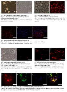
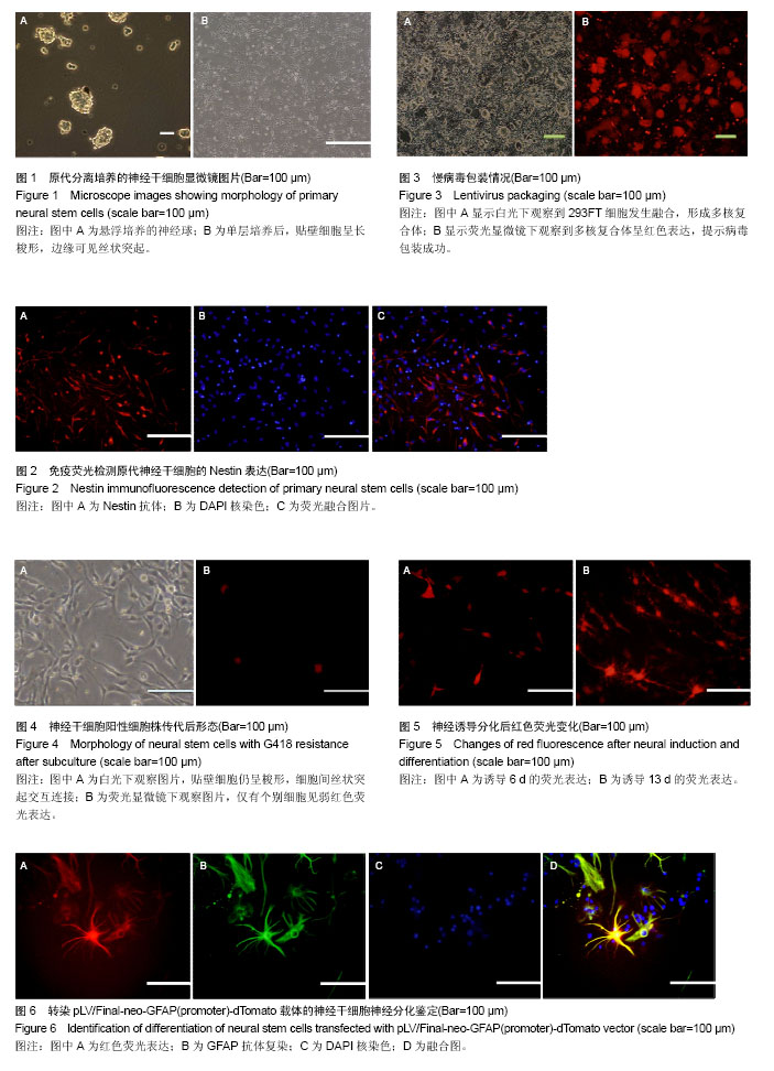
2.1 神经干细胞的形态学观察与鉴定结果 原代分离的小鼠胎脑皮质细胞在神经干细胞悬浮培养液中培养24 h,细胞悬浮生长,部分聚集成团块状,团块周边细胞折光性好。48 h后形成立体感强、折光性好且形态规则的细胞球,即神经球(Neurosphere)(图1A)[21-22]。 用0.25%胰酶消化神经球,辅以机械吹打制成单细胞悬液,接种于包被0.1%多聚赖氨酸的6孔板。采用神经干细胞单层培养液培养,24 h后可观察到贴壁细胞边缘向外伸出发丝状突起,与邻近细胞发出的突起相互连接,交织成网状(图1B)。 细胞免疫化学法鉴定可见贴壁细胞呈Nestin染色强阳性,胞浆部位着色而胞核不着色,证实贴壁培养的细胞为神经干细胞(图2)[23]。 2.2 慢病毒的成功包装 贴壁培养的293FT细胞密度达到90%左右时,更换无抗生素培养基,再以载体质粒和包装质粒共转染8 h,之后用新鲜培养基换液。72 h后,镜下观察到大量293FT细胞发生融合,形成多核复合体并表达强红色荧光(图3),提示慢病毒包装成功。 2.3 慢病毒转导神经干细胞及其纯化 浓缩后的慢病毒以1×10¹⁰ L-1的滴度连续感染神经干细胞3次,3 d后观察到细胞生长状态稍差,部分细胞出现死亡,6-8 d后可见细胞生长状态逐渐转好并开始大量增殖。随后采用G418连续筛选14 d,获得慢病毒载体稳定表达的神经干细胞阳性细胞株。该细胞株经传代后,显微镜下仅见个别细胞有弱红色荧光表达(图4)。 2.4 慢病毒感染神经干细胞后纯化细胞的神经分化及对荧光细胞的鉴定结果 转导pLV/Final-neo-GFAP (promoter)-dTomato载体后的神经干细胞向星形胶质细胞分化过程中,镜下观察可见细胞发生一系列的变化。采用去除生长因子(表皮生长因子、碱性成纤维细胞生长因子)的神经干细胞培养液进行培养后,随着时间延长观察到表达红色荧光的细胞数目开始增多,荧光强度大幅增强(图5 A);荧光表达的高峰期在神经分化的第9-13天(图5B)。 在分化的第13天,利用免疫荧光染色法对表达红色荧光的分化细胞进行GFAP抗体复染,结果发现红色荧光与GFAP的绿色荧光染色的位置保持高度的一致(图6)。此结果证明,慢病毒载体转导后的神经干细胞,经过神经分化后获得的表达红色荧光的细胞就是星形胶质细胞。这一体系的成功建立,为后续的神经干细胞定向分化及GFAP的功能研究等提供了非常好的细胞工具。"
| [1] Galli R, Gritti A, Bonfanti L, et al. Neural stem cells: an overview. Circ Res. 2003;92(6):598-608.[2] Butti E, Cusimano M, Bacigaluppi M, et al. Neurogenic and non-neurogenic functions of endogenous neural stem cells. Front Neurosci. 2014;8:92.[3] Takagi Y. History of Neural Stem Cell Research and Its Clinical Application. Neurol Med Chir (Tokyo). 2016;56(3): 110-124.[4] Akkermann R, Beyer F, Küry P. Heterogeneous populations of neural stem cells contribute to myelin repair. Neural Regen Res. 2017;12(4):509-517.[5] Xing YL, Röth PT, Stratton JA, et al. Adult neural precursor cells from the subventricular zone contribute significantly to oligodendrocyte regeneration and remyelination. J Neurosci. 2014;34(42):14128-14146.[6] Ross HH, Ambrosio F, Trumbower RD, et al. Neural Stem Cell Therapy and Rehabilitation in the Central Nervous System: Emerging Partnerships. Phys Ther. 2016;96(5): 734-742.[7] Skardelly M, Hempel E, Hirrlinger J, et al. Fluorescent protein-expressing neural progenitor cells as a tool for transplantation studies. PLoS One. 2014;9(6):e99819.[8] Yousefifard M, Rahimi-Movaghar V, Nasirinezhad F, et al. Neural stem/progenitor cell transplantation for spinal cord injury treatment; A systematic review and meta-analysis. Neuroscience. 2016;322:377-397.[9] Thomaidou D. Neural stem cell transplantation in an animal model of traumatic brain injury. Methods Mol Biol. 2014;1210: 9-21.[10] Ahmed AI, Shtaya AB, Zaben MJ, et al. Endogenous GFAP-positive neural stem/progenitor cells in the postnatal mouse cortex are activated following traumatic brain injury. J Neurotrauma. 2012;29(5):828-842.[11] Kalyani A, Hobson K, Rao MS. Neuroepithelial stem cells from the embryonic spinal cord: isolation, characterization, and clonal analysis. Dev Biol. 1997;186(2):202-223.[12] Daynac M, Morizur L, Kortulewski T, et al. Cell Sorting of Neural Stem and Progenitor Cells from the Adult Mouse Subventricular Zone and Live-imaging of their Cell Cycle Dynamics. J Vis Exp. 2015;(103): e53247.[13] Kim JY, Lee JH, Sun W. Isolation and Culture of Adult Neural Stem Cells from the Mouse Subcallosal Zone. J Vis Exp. 2016; (118):e54929.[14] Wongpaiboonwattana W, Stavridis MP. Neural differentiation of mouse embryonic stem cells in serum-free monolayer culture. J Vis Exp. 2015;(99):e52823.[15] Yang X, Boehm JS, Yang X, et al. A public genome-scale lentiviral expression library of human ORFs. Nat Methods. 2011;8(8):659-661.[16] Li W, Liu C, Qin J, et al. Efficient genetic modification of cynomolgus monkey embryonic stem cells with lentiviral vectors. Cell Transplant. 2010;19(9):1181-1193.[17] Quan Z, Yang H, Yang Y, et al. Construction and functional analysis of a lentiviral expression vector containing a scavenger receptor (SR-PSOX) that binds uniquely phosphatidylserine and oxidized lipoprotein. Acta Biochim Biophys Sin (Shanghai). 2007;39(3):208-216.[18] De Groote P, Grootjans S, Lippens S, et al. Generation of a new Gateway-compatible inducible lentiviral vector platform allowing easy derivation of co-transduced cells. Biotechniques. 2016;60(5):252-259.[19] Albrecht C, Hosiner S, Tichy B, et al. Comparison of Lentiviral Packaging Mixes and Producer Cell Lines for RNAi Applications. Mol Biotechnol. 2015;57(6):499-505.[20] Zhang X, Wang X, Zhao D, et al. Design and immunogenicity assessment of HIV-1 virus-like particles as a candidate vaccine. Sci China Life Sci. 2011;54(11):1042-1047.[21] Ivanov DP, Al-Rubai AJ, Grabowska AM, et al. Separating chemotherapy-related developmental neurotoxicity from cytotoxicity in monolayer and neurosphere cultures of human fetal brain cells. Toxicol In Vitro. 2016;37:88-96.[22] Shimada IS, Badgandi H, Somatilaka BN, et al. Using Primary Neurosphere Cultures to Study Primary Cilia. J Vis Exp. 2017;(122):e55315.[23] Kawaguchi A, Miyata T, Sawamoto K, et al. Nestin-EGFP transgenic mice: visualization of the self-renewal and multipotency of CNS stem cells. Mol Cell Neurosci. 2001; 17(2):259-273.[24] Molofsky AV, Krencik R, Ullian EM, et al. Astrocytes and disease: a neurodevelopmental perspective. Genes Dev. 2012;26(9):891-907.[25] Segura-Aguilar J.A new mechanism for protection of dopaminergic neurons mediated by astrocytes. Neural Regen Res. 2015;10(8): 1225-1227.[26] Ito K, Sanosaka T, Igarashi K, et al. Identification of genes associated with the astrocyte-specific gene Gfap during astrocyte differentiation. Sci Rep. 2016;6:23903.[27] Ahmed AI, Shtaya AB, Zaben MJ, et al. Endogenous GFAP-positive neural stem/progenitor cells in the postnatal mouse cortex are activated following traumatic brain injury. J Neurotrauma. 2012;29(5):828-842.[28] Gengatharan A, Bammann RR, Saghatelyan A. The Role of Astrocytes in the Generation, Migration, and Integration of New Neurons in the Adult Olfactory Bulb. Front Neurosci. 2016;10:149.[29] Ramos AJ.Astroglial heterogeneity: merely a neurobiological question? Or an opportunity for neuroprotection and regeneration afer brain injury?.Neural Regen Res.2016; 11(11): 1739-1741.[30] Burda JE, Bernstein AM, Sofroniew MV. Astrocyte roles in traumatic brain injury. Exp Neurol. 2016;275 Pt 3:305-315.[31] Lorenzo PI, Ménard C, Miller FD, et al. Thyroid hormone-dependent regulation of Talpha1 alpha-tubulin during brain development. Mol Cell Neurosci. 2002;19(3): 333-343.[32] Suzuki H, Sakabe T, Hirose Y, et al. Development and evaluation of yeast-based GFP and luciferase reporter assays for chemical-induced genotoxicity and oxidative damage. Appl Microbiol Biotechnol. 2017;101(2):659-671.[33] Chen-Roetling J, Lu X, Regan KA, et al. A rapid fluorescent method to quantify neuronal loss after experimental intracerebral hemorrhage. J Neurosci Methods. 2013;216(2): 128-136.[34] Jung HS, Uenishi G, Kumar A, et al. A human VE-cadherin-tdTomato and CD43-green fluorescent protein dual reporter cell line for study endothelial to hematopoietic transition. Stem Cell Res. 2016;17(2):401-405.[35] Lee M, Chea K, Pyda R, et al. Comparative Analysis of Non-viral Transfection Methods in Mouse Embryonic Fibroblast Cells. J Biomol Tech. 2017 Apr 29. [Epub ahead of print][36] Chen C, Akerstrom V, Baus J, et al. Comparative analysis of the transduction efficiency of five adeno associated virus serotypes and VSV-G pseudotype lentiviral vector in lung cancer cells. Virol J. 2013;10:86.[37] Miyazaki M, Sugiyama O, Zou J, et al. Comparison of lentiviral and adenoviral gene therapy for spinal fusion in rats. Spine (Phila Pa 1976). 2008;33(13):1410-1417.[38] Sakoda T, Kasahara N, Kedes L, et al. Lentiviral vector-mediated gene transfer to endotherial cells compared with adenoviral and retroviral vectors. Prep Biochem Biotechnol. 2007;37(1):1-11.[39] 熊玮,董少红,张键,等.慢病毒载体构建小鼠CMKLR1基因缺陷性血管平滑肌细胞系[J].中国组织工程研究, 2015,19(20): 3195-3199.[40] Delzor A, Escartin C, Déglon N. Lentiviral vectors: a powerful tool to target astrocytes in vivo. Curr Drug Targets. 2013; 14(11):1336-1346. |
| [1] | Yao Xiaoling, Peng Jiancheng, Xu Yuerong, Yang Zhidong, Zhang Shuncong. Variable-angle zero-notch anterior interbody fusion system in the treatment of cervical spondylotic myelopathy: 30-month follow-up [J]. Chinese Journal of Tissue Engineering Research, 2022, 26(9): 1377-1382. |
| [2] | An Weizheng, He Xiao, Ren Shuai, Liu Jianyu. Potential of muscle-derived stem cells in peripheral nerve regeneration [J]. Chinese Journal of Tissue Engineering Research, 2022, 26(7): 1130-1136. |
| [3] | Fan Yiming, Liu Fangyu, Zhang Hongyu, Li Shuai, Wang Yansong. Serial questions about endogenous neural stem cell response in the ependymal zone after spinal cord injury [J]. Chinese Journal of Tissue Engineering Research, 2022, 26(7): 1137-1142. |
| [4] | Zhang Jinglin, Leng Min, Zhu Boheng, Wang Hong. Mechanism and application of stem cell-derived exosomes in promoting diabetic wound healing [J]. Chinese Journal of Tissue Engineering Research, 2022, 26(7): 1113-1118. |
| [5] | He Yunying, Li Lingjie, Zhang Shuqi, Li Yuzhou, Yang Sheng, Ji Ping. Method of constructing cell spheroids based on agarose and polyacrylic molds [J]. Chinese Journal of Tissue Engineering Research, 2022, 26(4): 553-559. |
| [6] | He Guanyu, Xu Baoshan, Du Lilong, Zhang Tongxing, Huo Zhenxin, Shen Li. Biomimetic orientated microchannel annulus fibrosus scaffold constructed by silk fibroin [J]. Chinese Journal of Tissue Engineering Research, 2022, 26(4): 560-566. |
| [7] | Chen Xiaoxu, Luo Yaxin, Bi Haoran, Yang Kun. Preparation and application of acellular scaffold in tissue engineering and regenerative medicine [J]. Chinese Journal of Tissue Engineering Research, 2022, 26(4): 591-596. |
| [8] | Kang Kunlong, Wang Xintao. Research hotspot of biological scaffold materials promoting osteogenic differentiation of bone marrow mesenchymal stem cells [J]. Chinese Journal of Tissue Engineering Research, 2022, 26(4): 597-603. |
| [9] | Shen Jiahua, Fu Yong. Application of graphene-based nanomaterials in stem cells [J]. Chinese Journal of Tissue Engineering Research, 2022, 26(4): 604-609. |
| [10] | Zhang Tong, Cai Jinchi, Yuan Zhifa, Zhao Haiyan, Han Xingwen, Wang Wenji. Hyaluronic acid-based composite hydrogel in cartilage injury caused by osteoarthritis: application and mechanism [J]. Chinese Journal of Tissue Engineering Research, 2022, 26(4): 617-625. |
| [11] | Li Hui, Chen Lianglong. Application and characteristics of bone graft materials in the treatment of spinal tuberculosis [J]. Chinese Journal of Tissue Engineering Research, 2022, 26(4): 626-630. |
| [12] | Gao Cangjian, Yang Zhen, Liu Shuyun, Li Hao, Fu Liwei, Zhao Tianyuan, Chen Wei, Liao Zhiyao, Li Pinxue, Sui Xiang, Guo Quanyi. Electrospinning for rotator cuff repair [J]. Chinese Journal of Tissue Engineering Research, 2022, 26(4): 637-642. |
| [13] | Guan Jian, Jia Yanfei, Zhang Baoxin , Zhao Guozhong. Application of 4D bioprinting in tissue engineering [J]. Chinese Journal of Tissue Engineering Research, 2022, 26(3): 446-455. |
| [14] | Huang Bo, Chen Mingxue, Peng Liqing, Luo Xujiang, Li Huo, Wang Hao, Tian Qinyu, Lu Xiaobo, Liu Shuyun, Guo Quanyi . Fabrication and biocompatibility of injectable gelatin-methacryloyl/cartilage-derived matrix particles composite hydrogel scaffold [J]. Chinese Journal of Tissue Engineering Research, 2022, 10(16): 2600-2606. |
| [15] | Liu Jiali, Suo Hairui, Yang Han, Wang Ling, Xu Mingen. Influence of lay-down angles on mechanical properties of three-dimensional printed polycaprolactone scaffolds [J]. Chinese Journal of Tissue Engineering Research, 2022, 10(16): 2612-2617. |
| Viewed | ||||||
|
Full text |
|
|||||
|
Abstract |
|
|||||