Chinese Journal of Tissue Engineering Research ›› 2017, Vol. 21 ›› Issue (21): 3364-3369.doi: 10.3969/j.issn.2095-4344.2017.21.014
Previous Articles Next Articles
Proportion and difference of neural stem cells and neurons from different embryonic mouse brain tissues
Zhang Feng-lan1, Yang Lu-jun2, Xiao Zhi-cheng1, 3
- 1Institute of Molecular and Clinical Medicine, Kunming Medical University, Kunming 650500, Yunnan Province, China; 2Baoan People's Hospital of Shenzhen, Shenzhen 518101, Guangdong Province, China; 3Department of Anatomy and Developmental Biology, Monash University, Victoria 3800, Australia
-
Revised:2017-06-14Online:2017-07-28Published:2017-08-02 -
Contact:Xiao Zhi-cheng, M.D., Professor, Institute of Molecular and Clinical Medicine, Kunming Medical University, Kunming 650500, Yunnan Province, China; Department of Anatomy and Developmental Biology, Monash University, Victoria 3800, Australia -
About author:Zhang Feng-lan, Studying for doctorate, Institute of Molecular and Clinical Medicine, Kunming Medical University, Kunming 650500, Yunnan Province, China -
Supported by:the Talent Program of Yunnan Province, No. 20080A004; the Key Laboratory of Stem Cell and Regenerative Medicine of Yunnan Province, No. 2015DG027
CLC Number:
Cite this article
Zhang Feng-lan, Yang Lu-jun, Xiao Zhi-cheng. Proportion and difference of neural stem cells and neurons from different embryonic mouse brain tissues [J]. Chinese Journal of Tissue Engineering Research, 2017, 21(21): 3364-3369.
share this article
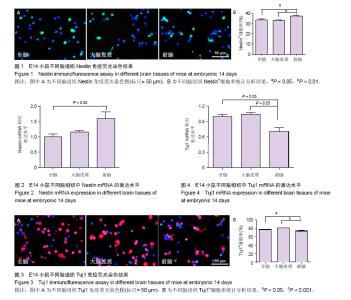
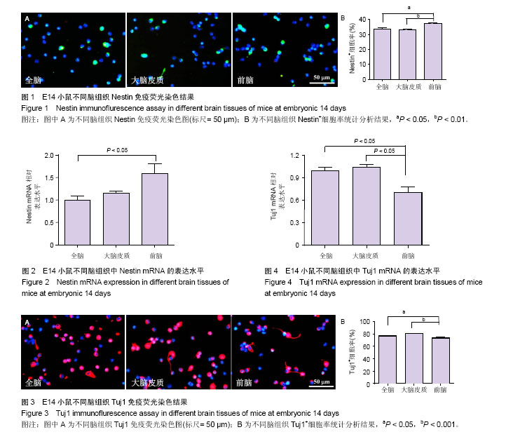
2.1 E14小鼠不同脑组织Nestin免疫荧光染色和分析 观察E14小鼠全脑、大脑皮质和前脑组织分离培养细胞的Nestin免疫荧光染色结果,发现:Nestin阳性细胞的细胞质为绿色荧光,胞核不着色,因培养时间较短(3.0-4.0 h),细胞形态多数为圆形或椭圆形,边缘清晰,只有极少数细胞伸出细长分枝,DAPI阳性细胞核呈蓝色;3种脑组织经消化分离后,几乎全为单个散在细胞,未见明显细胞团块,且几乎没有碎裂细胞残片,整体染色背景较干净;前脑组织中Nestin阳性细胞比例略高于全脑和大脑皮质。见图1A。 将细胞免疫荧光数据进行统计分析,发现:在分离培养的E14小鼠前脑组织中Nestin阳性神经干细胞所占比例最高,为(37.48±0.52)%;大脑皮质组织中比例最低,为(33.07±0.62)%;全脑组织中比例介于二者之间,为(33.92±0.81)%。前脑组织中Nestin阳性神经干细胞的比例显著高于全脑(P < 0.05)和大脑皮质(P < 0.01)。见图1B。该结果表明前脑组织中Nestin阳性神经干细胞比例明显高于全脑和大脑皮质。 2.2 E14小鼠不同脑组织中Nestin mRNA的表达水平 通过Real time PCR实验检测E14小鼠全脑、大脑皮质和前脑组织中Nestin mRNA的表达水平,结果显示:E14小鼠前脑组织中Nestin mRNA的表达水平(1.58±0.23)明显高于全脑组织(设定为1,P < 0.05),略高于大脑皮质(1.15±0.04,P > 0.05),见图2。该结果表明前脑组织中Nestin mRNA的表达水平较高。 2.3 E14小鼠不同脑组织Tuj1免疫荧光染色及分析 观察E14小鼠全脑、大脑皮质和前脑组织中分离培养细胞的Tuj1免疫荧光染色结果,发现:Tuj1阳性神经元的细胞质为红色荧光,胞核不着色,也因培养时间较短(3.0-4.0 h),大部分阳性细胞为圆形或椭圆形,只有少数细胞具有细长的突起,DAPI阳性细胞核呈蓝色;Tuj1免疫荧光染色整体背景较干净,未见非特异性着色;前脑组织中Tuj1阳性细胞比例略低于全脑和大脑皮质,见图3A。 将细胞免疫荧光数据进行统计分析,发现:在分离培养的E14小鼠前脑组织中Tuj1阳性细胞所占比例最低(74.03±0.70)%;全脑组织中比例略低(76.97±0.81)%;大脑皮质组织中比例最高(80.81±0.29)%;前脑组织中Tuj1阳性细胞比例明显低于全脑(P < 0.05)和大脑皮质(P < 0.001);另外,全脑组织中的Tuj1阳性细胞比例低于大脑皮质,且差异有显著性意义(P < 0.01,未标注),见图3B。该结果表明前脑组织中Tuj1阳性神经元比例明显低于全脑和大脑皮质。 2.4 E14小鼠不同脑组织中Tuj1 mRNA的表达水平 通过Real time PCR实验检测E14小鼠全脑、大脑皮质和前脑组织中Tuj1 mRNA的表达水平,结果显示:E14小鼠前脑组织中Tuj1 mRNA的表达水平(0.71±0.08)明显低于全脑组织中的表达水平(设定为1,P < 0.05),也低于大脑皮质组织(1.04±0.04,P < 0.05),但皮质组织与全脑组织之间差异无显著性意义(P > 0.05),见图4。该结果表明前脑组织中Tuj1 mRNA的表达水平较低。"
| [1] Yamaguchi M, Seki T, Imayoshi I, et al. Neural stem cells and neuro/gliogenesis in the central nervous system: understanding the structural and functional plasticity of the developing, mature, and diseased brain. J Physiol Sci. 2016;66(3):197-206.[2] Conti L, Cattaneo E. Neural stem cell systems: physiological players or in vitro entities. Nat Rev Neurosci. 2010;11(3): 176-187.[3] Akerblom M, Sachdeva R, Jakobsson J. Functional Studies of microRNAs in Neural Stem Cells: Problems and Perspectives. Front Neurosci. 2012;6:14.[4] Reynolds BA, Tetzlaff W, Weiss S. A multipotent EGF-responsive striatal embryonic progenitor cell produces neurons and astrocytes. J Neurosci. 1992;12(11):4565- 4574.[5] English D, Sharma NK, Sharma K, et al. Neural stem cells- trends and advances. J Cell Biochem. 2013;114(4): 764-772.[6] Gaspard N, Vanderhaeghen P. From stem cells to neural networks: recent advances and perspectives for neurodevelopmental disorders. Dev Med Child Neurol. 2011;53(1):13-17.[7] Walker T, Huang J, Young K. Neural Stem and Progenitor Cells in Nervous System Function and Therapy. Stem Cells Int. 2016;2016:1890568.[8] Duncan T, Valenzuela M. Alzheimer's disease, dementia, and stem cell therapy. Stem Cell Res Ther. 2017;8(1):111.[9] Ottoboni L, Merlini A, Martino G. Neural Stem Cell Plasticity: Advantages in Therapy for the Injured Central Nervous System. Front Cell Dev Biol. 2017;5:52.[10] Azari H, Sharififar S, Rahman M, et al. Establishing embryonic mouse neural stem cell culture using the neurosphere assay. J Vis Exp. 2011;(47): 2457. [11] Torrado EF, Gomes C, Santos G, et al. Directing mouse embryonic neurosphere differentiation toward an enriched neuronal population. Int J Dev Neurosci. 2014;37:94-99.[12] Ma QH, Futagawa T, Yang WL, et al. A TAG1-APP signalling pathway through Fe65 negatively modulates neurogenesis. Nat Cell Biol. 2008;10(3):283-294.[13] 张涛,李文杰,王峰,等. 小鼠室管膜下区神经干细胞增殖及分化研究[J]. 中国修复重建外科杂志, 2015,29(6):766-771.[14] 许汉鹏,苟琳,杨浩. 中枢神经系统不同部位来源的神经干细胞在体外生长特性的比较[J].解剖学报,2004,35(4): 358-362.[15] 白连琴,刘娜,李腾腾,等. 新生鼠和胎鼠海马神经干细胞分离和培养方法的比较[J].神经解剖学杂志,2015,31(4):481-486.[16] 张蓬勃,李卫松,高明,等. 小鼠胚胎神经干细胞的体外培养与鉴定[J].中国当代儿科杂志,2011, 13(3):244-247.[17] Cao F, Hata R, Zhu P, et al. Conditional deletion of Stat3 promotes neurogenesis and inhibits astrogliogenesis in neural stem cells. Biochem Biophys Res Commun. 2010;394(3): 843-847.[18] Ao X, Liu Y, Qin M, et al. Expression of Dbn1 during mouse brain development and neural stem cell differentiation. Biochem Biophys Res Commun. 2014;449(1):81-87.[19] 杨蓬勃,张军峰,张建水,等. 人胎脑额叶和海马中星形胶质细胞的发育性变化[J].西安交通大学学报,2014,35(5):576-580.[20] Vishwakarma SK, Bardia A, Tiwari SK, et al. Current concept in neural regeneration research: NSCs isolation, characterization and transplantation in various neurodegenerative diseases and stroke: A review. J Adv Res. 2014;5(3):277-294.[21] 闵娜,胡强夫,李晓培,等. 异氟醚对新生大鼠海马神经干细胞增殖及分化的影响[J].中国组织工程研究,2016,20(1):118-122.[22] Telias M, Ben-Yosef D.Neural stem cell replacement: a possible therapy for neurodevelopmental disorders?Neural Regen Res. 2015;10(2): 180-182.[23] 石海杉,吴文,徐建兰.内源性神经干细胞的研究进展[J].实用医学杂志,2013,29(19): 3252-3254.[24] 孙鼐,李春伟,赵伟新,等. 异氟醚抑制海马神经干细胞增殖并可促其向神经元分化[J].中国组织工程研究,2016,20(10): 1488-1493.[25] Jiang XF,Yng K,Yang XQ,et al.Elastic modulus affects the growth and differentiation of neural stem cells. Neural Regen Res. 2015;10(9): 1523-1527.[26] Louis SA, Mak CK, Reynolds BA. Methods to culture, differentiate, and characterize neural stem cells from the adult and embryonic mouse central nervous system. Methods Mol Biol. 2013;946:479-506.[27] De Waele J, Reekmans K, Daans J, et al. 3D culture of murine neural stem cells on decellularized mouse brain sections. Biomaterials. 2015;41:122-131.[28] Lee DY, Yeh TH, Emnett RJ, et al. Neurofibromatosis-1 regulates neuroglial progenitor proliferation and glial differentiation in a brain region-specific manner. Genes Dev. 2010;24(20):2317-2329.[29] 张在金,董艳,卢亦成. 大鼠胎脑不同部位神经干细胞比较性培养的研究[J]. 中华神经外科疾病研究杂志, 2008,7(4):325-327.[30] 马浚宁,高俊玮,候博儒,等, 新生小鼠海马、嗅球及皮质神经干细胞的分离培养及鉴定[J]. 中国组织工程研究,2014,18 (45): 7266-7272.[31] Seaberg RM, Smukler SR, van der Kooy D. Intrinsic differences distinguish transiently neurogenic progenitors from neural stem cells in the early postnatal brain. Dev Biol.2005;278(1):71-85.[32] Maslov AY, Barone TA, Plunkett RJ, et al. Neural stem cell detection, characterization, and age-related changes in the subventricular zone of mice. J Neurosci. 2004;24(7): 1726-1733.[33] Guo W, Patzlaff NE, Jobe EM, et al. Isolation of multipotent neural stem or progenitor cells from both the dentate gyrus and subventricular zone of a single adult mouse. Nat Protoc. 2012;7(11):2005-2012.[34] Bonaventura G, Chamayou S, Liprino A, et al. Different Tissue-Derived Stem Cells: A Comparison of Neural Differentiation Capability. PLoS One. 2015;10(10):e0140790.[35] Xu R, Wu C, Tao Y, et al. Description of distributed features of the nestin-containing cells in brains of adult mice: a potential source of neural precursor cells. J Neurosci Res. 2010;88(5): 945-956.[36] Lee R, Kim IS, Han N, et al. Real-time discrimination between proliferation and neuronal and astroglial differentiation of human neural stem cells. Sci Rep. 2014;4:6319.[37] Zhang J, Jiao J. Molecular Biomarkers for Embryonic and Adult Neural Stem Cell and Neurogenesis. Biomed Res Int. 2015;2015:727542.[38] Farzanehfar P, Lu SS, Dey A, et al. Evidence of functional duplicity of Nestin expression in the adult mouse midbrain. Stem Cell Res. 2017;19:82-93.[39] 刘吉星,候博儒,杨文桢,等.神经干细胞体外培养鉴定及诱导分化的表型特征[J].中国组织工程研究,2015,19(19):3054-3060.[40] Mine Y, Tatarishvili J, Oki K, et al. Grafted human neural stem cells enhance several steps of endogenous neurogenesis and improve behavioral recovery after middle cerebral artery occlusion in rats. Neurobiol Dis. 2013;52:191-203.[41] Lucchinetti E, Zeisberger SM, Baruscotti I, et al. Stem cell-like human endothelial progenitors show enhanced colony-forming capacity after brief sevoflurane exposure: preconditioning of angiogenic cells by volatile anesthetics. Anesth Analg. 2009;109(4):1117-1126.[42] 刘罡,吴毅,贾杰,等.神经干细胞体外分化抗原表达的研究[J].中国运动医学杂志,2009,28(1):55-59. |
| [1] | Yao Xiaoling, Peng Jiancheng, Xu Yuerong, Yang Zhidong, Zhang Shuncong. Variable-angle zero-notch anterior interbody fusion system in the treatment of cervical spondylotic myelopathy: 30-month follow-up [J]. Chinese Journal of Tissue Engineering Research, 2022, 26(9): 1377-1382. |
| [2] | Zhang Jinglin, Leng Min, Zhu Boheng, Wang Hong. Mechanism and application of stem cell-derived exosomes in promoting diabetic wound healing [J]. Chinese Journal of Tissue Engineering Research, 2022, 26(7): 1113-1118. |
| [3] | An Weizheng, He Xiao, Ren Shuai, Liu Jianyu. Potential of muscle-derived stem cells in peripheral nerve regeneration [J]. Chinese Journal of Tissue Engineering Research, 2022, 26(7): 1130-1136. |
| [4] | Fan Yiming, Liu Fangyu, Zhang Hongyu, Li Shuai, Wang Yansong. Serial questions about endogenous neural stem cell response in the ependymal zone after spinal cord injury [J]. Chinese Journal of Tissue Engineering Research, 2022, 26(7): 1137-1142. |
| [5] | Shui Xiaoping, Li Chunying, Li Shunchang, Sun Junzhi, Su Quansheng . Effects of aerobic and resistance exercises on brain-derived neurotrophic factor, nuclear factor-kappa B and inflammatory cytokines in skeletal muscle of type II diabetic rats [J]. Chinese Journal of Tissue Engineering Research, 2022, 26(5): 669-675. |
| [6] | Mo Weibin, Huang Tianchang, Zeng Zhiwei, Yan Linbo. Effects of Pueraria lobata flavonoids on expressions of beta-catenin and glycogen synthase kinase 3beta in the brain of rats undergoing exhaustive exercise after long endurance exercise [J]. Chinese Journal of Tissue Engineering Research, 2022, 26(5): 736-741. |
| [7] | Huang Chuanjun, Zou Yu, Zhou Xiaoting, Zhu Yangqing, Qian Wei, Zhang Wei, Liu Xing. Transplantation of umbilical cord mesenchymal stem cells encapsulated in RADA16-BDNF hydrogel promotes neurological recovery in an intracerebral hemorrhage rat model [J]. Chinese Journal of Tissue Engineering Research, 2022, 26(4): 510-515. |
| [8] | He Yunying, Li Lingjie, Zhang Shuqi, Li Yuzhou, Yang Sheng, Ji Ping. Method of constructing cell spheroids based on agarose and polyacrylic molds [J]. Chinese Journal of Tissue Engineering Research, 2022, 26(4): 553-559. |
| [9] | He Guanyu, Xu Baoshan, Du Lilong, Zhang Tongxing, Huo Zhenxin, Shen Li. Biomimetic orientated microchannel annulus fibrosus scaffold constructed by silk fibroin [J]. Chinese Journal of Tissue Engineering Research, 2022, 26(4): 560-566. |
| [10] | Chen Xiaoxu, Luo Yaxin, Bi Haoran, Yang Kun. Preparation and application of acellular scaffold in tissue engineering and regenerative medicine [J]. Chinese Journal of Tissue Engineering Research, 2022, 26(4): 591-596. |
| [11] | Kang Kunlong, Wang Xintao. Research hotspot of biological scaffold materials promoting osteogenic differentiation of bone marrow mesenchymal stem cells [J]. Chinese Journal of Tissue Engineering Research, 2022, 26(4): 597-603. |
| [12] | Shen Jiahua, Fu Yong. Application of graphene-based nanomaterials in stem cells [J]. Chinese Journal of Tissue Engineering Research, 2022, 26(4): 604-609. |
| [13] | Zhang Tong, Cai Jinchi, Yuan Zhifa, Zhao Haiyan, Han Xingwen, Wang Wenji. Hyaluronic acid-based composite hydrogel in cartilage injury caused by osteoarthritis: application and mechanism [J]. Chinese Journal of Tissue Engineering Research, 2022, 26(4): 617-625. |
| [14] | Li Hui, Chen Lianglong. Application and characteristics of bone graft materials in the treatment of spinal tuberculosis [J]. Chinese Journal of Tissue Engineering Research, 2022, 26(4): 626-630. |
| [15] | Gao Cangjian, Yang Zhen, Liu Shuyun, Li Hao, Fu Liwei, Zhao Tianyuan, Chen Wei, Liao Zhiyao, Li Pinxue, Sui Xiang, Guo Quanyi. Electrospinning for rotator cuff repair [J]. Chinese Journal of Tissue Engineering Research, 2022, 26(4): 637-642. |
| Viewed | ||||||
|
Full text |
|
|||||
|
Abstract |
|
|||||