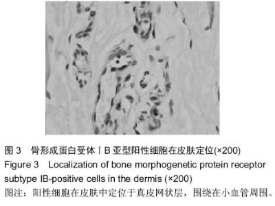
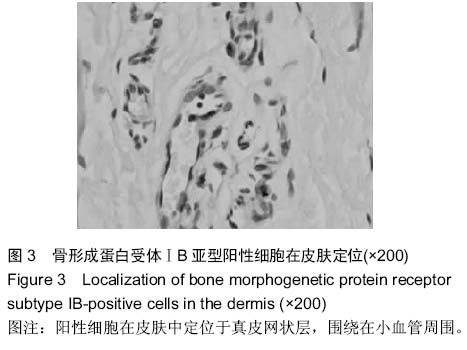
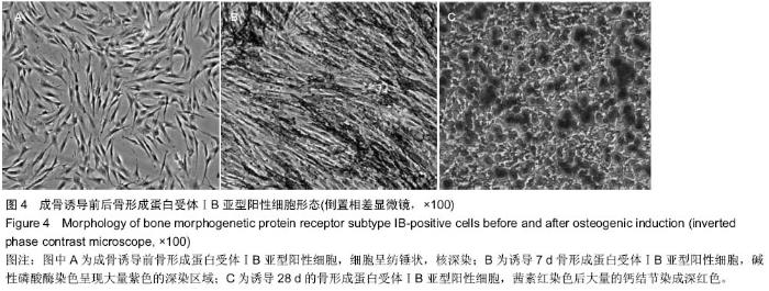
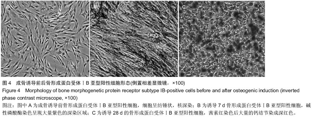
| [1] Toma JG, McKenzie IA, Bagli D, et al. Isolation and characterization of multipotent skin-derived precursors from human skin. Stem Cells. 2005;23(6):727-737.
[2] Singhatanadgit W, Salih V, Olsen I. Shedding of a soluble form of BMP receptor-IB controls bone cell responses to BMP. Bone. 2006;39(5):1008-1017.
[3] Singhatanadgit W, Salih V, Olsen I. RNA interference of the BMPR-IB gene blocks BMP-2-induced osteogenic gene expression in human bone cells. Cell Biol Int. 2008;32(11): 1362-1370.
[4] He J, Dong J, Wang T, et al. Bone morphogenetic protein receptor IB as a marker for enrichment of osteogenic precursor-like cells in human dermis. Arch Dermatol Res. 2011;303(8):581-590.
[5] Manini I, Gulino L, Gava B, et al. Multi-potent progenitors in freshly isolated and cultured human mesenchymal stem cells: a comparison between adipose and dermal tissue. Cell Tissue Res. 2011;344(1):85-95.
[6] Singhatanadgit W, Olsen I. Endogenous BMPR-IB signaling is required for early osteoblast differentiation of human bone cells. In Vitro Cell Dev Biol Anim. 2011;47(3):251-259.
[7] Sahni V, Mukhopadhyay A, Tysseling V, et al. BMPR1a and BMPR1b signaling exert opposing effects on gliosis after spinal cord injury. J Neurosci. 2010;30(5):1839-1855.
[8] Crigler L, Kazhanie A, Yoon TJ, et al. Isolation of a mesenchymal cell population from murine dermis that contains progenitors of multiple cell lineages. FASEB J. 2007;21(9):2050-2063.
[9] Bouacida A, Rosset P, Trichet V, et al. Pericyte-like progenitors show high immaturity and engraftment potential as compared with mesenchymal stem cells. PLoS One. 2012; 7(11):e48648.
[10] Caplan AI. New era of cell-based orthopedic therapies. Tissue Eng Part B Rev. 2009;15(2):195-200.
[11] Driskell RR, Lichtenberger BM, Hoste E, et al. Distinct fibroblast lineages determine dermal architecture in skin development and repair. Nature. 2013;504(7479):277-281.
[12] Chen FG, Zhang WJ, Bi D, et al. Clonal analysis of nestin(-) vimentin(+) multipotent fibroblasts isolated from human dermis. J Cell Sci. 2007;120(Pt 16):2875-2883.
[13] Pittenger MF, Mackay AM, Beck SC, et al. Multilineage potential of adult human mesenchymal stem cells. Science. 1999;284(5411):143-147.
[14] Vaculik C, Schuster C, Bauer W, et al. Human dermis harbors distinct mesenchymal stromal cell subsets. J Invest Dermatol. 2012;132(3 Pt 1):563-574.
[15] Foschi F, Conserva E, Pera P, et al. Graft materials and bone marrow stromal cells in bone tissue engineering. J Biomater Appl. 2012;26(8):1035-1049.
[16] Ma J, Both SK, Yang F, et al. Concise review: cell-based strategies in bone tissue engineering and regenerative medicine. Stem Cells Transl Med. 2014;3(1):98-107. |