Chinese Journal of Tissue Engineering Research ›› 2017, Vol. 21 ›› Issue (18): 2852-2857.doi: 10.3969/j.issn.2095-4344.2017.18.011
Previous Articles Next Articles
Bone regeneration via a novel honeycomb-like polycaprolactone-calcium silicate crystal compound scaffold in extreme-sized cranial defects
- Department of Orthopedic Surgery, Nanfang Hospital, Southern Medical University, Guangzhou 510515, Guangdong Province, China
-
Received:2017-04-15Online:2017-06-28Published:2017-07-07 -
Contact:Zhao Liang, Chief physician, Department of Orthopedic Surgery, Nanfang Hospital, Southern Medical University, Guangzhou 510515, Guangdong Province, China -
About author:Song Bing, Studying for master’s degree, Department of Orthopedic Surgery, Nanfang Hospital, Southern Medical University, Guangzhou 510515, Guangdong Province, China -
Supported by:the Natural Science Foundation of Guangdong Province, No. 2014A030313275, s2013010014253; the National Outstanding Youth Culture Plan of Southern Medical University
CLC Number:
Cite this article
Song Bing, Liao Zhe-ting, Chen Yu-fan, Zhao Liang.
share this article
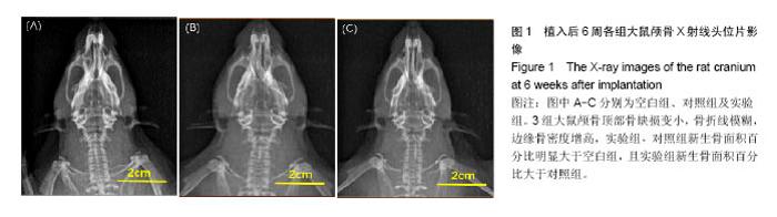
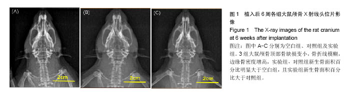
2.1 造模成功动物数量及过程 实验共纳入SD大鼠18只,术后恢复情况均良好,术后1周伤口完全恢复,无伤口裂开、填充材料移位等并发症。 2.2 X射线观察结果 3组大鼠颅骨顶部骨缺损变小,骨折线模糊,边缘骨密度增高,实验组、对照组新生骨面积百分比明显大于空白组,且实验组新生骨面积百分比大于对照组,见图1。"
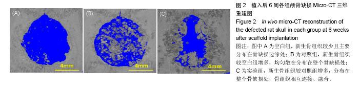
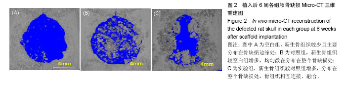
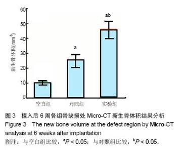
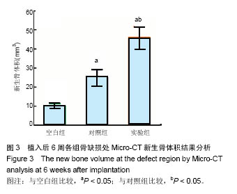
2.3 Micro-CT重建结果 空白组新生骨主要分布在缺损两侧,对照组、实验组新生骨分布于整个颅骨骨缺损处,与空白组比较,实验组与对照组均表现出更强的再生骨能力,与对照组相比,实验组的成骨能力更强一些,见图2。对新生骨体积进行定量分析,结果显示实验组新生骨体积为(46.25±5.71)mm3,明显大于对照组的(23.79±4.82)mm3及空白组的(9.79±1.53)mm3(P < 0.05),见图3。"
"

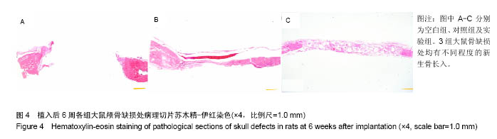
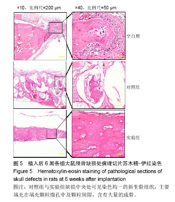
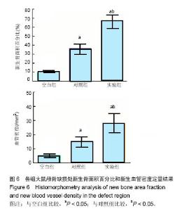
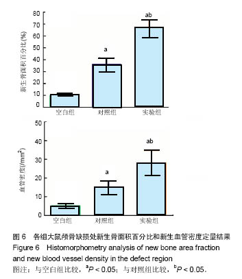
2.4 组织学观察结果 低倍镜下苏木精-伊红染色可见各组大鼠骨缺损处均有不同程度的新生骨长入,见图4;高倍镜下可见对照组与实验组缺损中央处可见染色均一的新生骨组织,主要填充在填充颗粒微孔中及颗粒间隙,含有大量的成骨细胞与骨基质,见图5。对照组与实验组内新生骨部分可见部分散在的新生血管,新生血管内可见红细胞,与对照组相比,实验组新生骨明显增多,两组边缘与周围骨组织无急慢性炎症细胞浸润,无炎性瘢痕组织产生。 新生骨组织面积百分比及新生血管密度以IPP 6.0软件进行定量分析,结果显示:实验组新生骨面积百分比为(66.58±7.28)%,明显高于对照组的(35.41±5.15)%及空白组的(10.14±1.37)%(P < 0.05),对照组新生骨面积明显高于空白组(P < 0.05);实验组血管密度为(27.93± 6.83)个/mm2,明显多于对照组的(15.12±2.93)个/mm2及空白组的(4.89±1.32)个/mm2(P < 0.05),对照组血管密度高于空白组(P < 0.05),见图6。"

"
"
| [1]Finkemeier CG.Bone-grafting and bone-graft substitutes.J Bone Joint Surg Am.2002;84-A(3): 454-464.[2]Dorozhkin SV.Bioceramics of calcium orthophosphates. Biomaterials.2010;31(7):1465-1485.[3]Doblaré M,Garc?a JM,Gómez MJ.Modelling bone tissue fracture and healing: a review.Eng Fract Mech. 2004;71 (13-14):1809-1840.[4]Tatara AM, Mikos AG.Tissue Engineering in Orthopaedics.J Bone Joint Surg.2016;98(13):1132-1139.[5]Jeon OH,Elisseeff J.Orthopedic tissue regeneration: cells, scaffolds, and small molecules.Drug Deliv Transl Res. 2016; 6(2):105-120.[6]Bastami F,Nazeman P,Moslemi H,et al.Induced pluripotent stem cells as a new getaway for bone tissue engineering: A systematic review.Cell Prolif.2017;50(2).doi: 10.1111/cpr.12321. Epub 2016 Dec 1.[7]Kimelman N,Pelled G,Helm GA,et al.Review: Gene- and Stem Cell–Based Therapeutics for Bone Regeneration and Repair.Tissue Eng.2007;13(6):1135-1150.[8]Discher DE, Mooney DJ,Zandstra PW.Growth factors, matrices, and forces combine and control stem cells. Science. 2009;324(5935):1673-1677.[9]Burg KJ,Porter S,Kellam JF.Biomaterial developments for bone tissue engineering.Biomaterials. 2000;21(23): 2347-2359.[10]Bose S,Roy M,Bandyopadhyay A.Recent advances in bone tissue engineering scaffolds.Trends Biotechnol. 2012;30(10): 546-554.[11]Ma PX.Biomimetic materials for tissue engineering.Adv Drug Deliv Rev.2008;60(2):184-198.[12]Fernandez-Yague MA,Abbah SA,McNamara L,et al.Biomimetic approaches in bone tissue engineering: Integrating biological and physicomechanical strategies.Adv Drug Deliv Rev.2015;84:1-29. [13]Henstock JR,Canham LT,Anderson SI.Silicon: The evolution of its use in biomaterials.Acta Biomaterialia. 2015; 11:17-26.[14]Liu W,Zhang J,Rethore G,et al.A novel injectable, cohesive and toughened Si-HPMC (silanized-hydroxypropyl methylcellulose) composite calcium phosphate cement for bone substitution.Acta Biomaterialia.2014; 10(7):3335-3345.[15]Lin K,Liu Y,Huang H,et al.Degradation and silicon excretion of the calcium silicate bioactive ceramics during bone regeneration using rabbit femur defect model. J Mater Sci Mater Med. 2015;26(6):197.[16]Khan AF,Saleem M, Afzal A,et al.Bioactive behavior of silicon substituted calcium phosphate based bioceramics for bone regeneration.Mater Sci Eng C Mater Biol Appl.2014;35: 245-252.[17]Shadjou N,Hasanzadeh M.Silica-based mesoporous nanobiomaterials as promoter of bone regeneration process.J Biomed Mater ResA.2015;103(11):3703.[18]Alfotawei R,Naudi KB,Lappin D,et al.The use of TriCalcium Phosphate (TCP) and stem cells for the regeneration of osteoperiosteal critical-size mandibular bony defects, an in vitro and preclinical study.J Craniomaxillofac Surg. 2014; 42(6):863-869.[19]Samavedi S,Whittington AR,Goldstein AS.Calcium phosphate ceramics in bone tissue engineering: A review of properties and their influence on cell behavior.Acta Biomaterialia. 2013; 9(9):8037-8045.[20]Choo T, Marino V,Bartold PM.Effect of PDGF-BB and beta-tricalcium phosphate (β-TCP) on bone formation around dental implants: a pilot study in sheep.Clin Oral Implants Res. 2013;24(2):158-166.[21]Fujihara K,Kotaki M,Ramakrishna S.Guided bone regeneration membrane made of polycaprolactone/calcium carbonate composite nano-fibers.Biomaterials. 2005;26(19): 4139-4147.[22]Causa F,Netti PA,Ambrosio L,et al.Poly-epsilon-caprolactone/ hydroxyapatite composites for bone regeneration: in vitro characterization and human osteoblast response.J Biomed Mater Res A.2006;76(1):151-162.[23]Yeo MG,Kim GH.Preparation and Characterization of 3D Composite Scaffolds Based on Rapid-Prototyped PCL/β-TCP Struts and Electrospun PCL Coated with Collagen and HA for Bone Regeneration.Chem Mater.2012;24(5):903-913.[24]Wu C.Methods of improving mechanical and biomedical properties of Ca-Si-based ceramics and scaffolds.Expert Rev Med Devices.2009;6(3):237-241. [25]Ni S,Chang J,Chou L.A novel bioactive porous CaSiO3 scaffold for bone tissue engineering.J Biomed Mater Res A. 2006;76(1):196-205.[26]Bohner M,Gbureck U,Barralet JE.Technological issues for the development of more efficient calcium phosphate bone cements: A critical assessment.Biomaterials.2005; 26(33): 6423-6429.[27]Chow LC.Next generation calcium phosphate-based biomaterials.Dent MaterJ.2009;28(1):1-10. [28]Ginebra MP,Espanol M,Montufar EB,et al.New processing approaches in calcium phosphate cements and their applications in regenerative medicine.Acta Biomaterialia. 2010;6(8):2863-2873.[29]Dash TK, Konkimalla VB.Poly-?-caprolactone based formulations for drug delivery and tissue engineering: A review.J Control Release.2012;158(1):15-33.[30]Labet M,Thielemans W.Synthesis of polycaprolactone: a review.Chem Soc Revi.2009;38(12):3484.[31]Kim J,McBride S,Donovan A,et al.Tyrosine-derived polycarbonate scaffolds for bone regeneration in a rabbit radius critical-size defect model.Biomed Mater. 2015;10(3): 035001.[32]Sears NA,Seshadri DR,Dhavalikar PS,et al.A Review of Three-Dimensional Printing in Tissue Engineering.Tissue Eng Part B Rev.2016;22(4):298-310. [33]Black CM,Goriainov V,Gibbs D,et al.Bone Tissue Engineering. CurrMol Biol Rep.2015;1(3):132-140.[34]Amini AR,Laurencin CT,Nukavarapu SP.Bone tissue engineering: recent advances and challenges. Crit Rev Biomed Eng.2012;40(5):363-408.[35]Zhao L,Burguera EF,Xu HH,et al.Fatigue and human umbilical cord stem cell seeding characteristics of calcium phosphate–chitosan–biodegradable fiber scaffolds. Biomaterials. 2010;31(5):840-847. [36]Khan AF,Saleem M,Afzal A,et al.Bioactive behavior of silicon substituted calcium phosphate based bioceramics for bone regeneration.Mater Sci Eng C Mater Biol Appl.2014;35: 245-252. [37]Vila M,García A,Girotti A,et al.3D silicon doped hydroxyapatite scaffolds decorated with Elastin-like Recombinamers for bone regenerative medicine.Acta Biomater.2016;45:349-356. [38]Avolio E,Alvino VV,Ghorbel MT,et al.Perivascular cells and tissue engineering: Current applications and untapped potential. Pharmacol Ther.2017;171:83-92. |
| [1] | Yao Xiaoling, Peng Jiancheng, Xu Yuerong, Yang Zhidong, Zhang Shuncong. Variable-angle zero-notch anterior interbody fusion system in the treatment of cervical spondylotic myelopathy: 30-month follow-up [J]. Chinese Journal of Tissue Engineering Research, 2022, 26(9): 1377-1382. |
| [2] | An Weizheng, He Xiao, Ren Shuai, Liu Jianyu. Potential of muscle-derived stem cells in peripheral nerve regeneration [J]. Chinese Journal of Tissue Engineering Research, 2022, 26(7): 1130-1136. |
| [3] | Wu Weiyue, Guo Xiaodong, Bao Chongyun. Application of engineered exosomes in bone repair and regeneration [J]. Chinese Journal of Tissue Engineering Research, 2022, 26(7): 1102-1106. |
| [4] | Zhou Hongqin, Wu Dandan, Yang Kun, Liu Qi. Exosomes that deliver specific miRNAs can regulate osteogenesis and promote angiogenesis [J]. Chinese Journal of Tissue Engineering Research, 2022, 26(7): 1107-1112. |
| [5] | Zhang Jinglin, Leng Min, Zhu Boheng, Wang Hong. Mechanism and application of stem cell-derived exosomes in promoting diabetic wound healing [J]. Chinese Journal of Tissue Engineering Research, 2022, 26(7): 1113-1118. |
| [6] | He Yunying, Li Lingjie, Zhang Shuqi, Li Yuzhou, Yang Sheng, Ji Ping. Method of constructing cell spheroids based on agarose and polyacrylic molds [J]. Chinese Journal of Tissue Engineering Research, 2022, 26(4): 553-559. |
| [7] | He Guanyu, Xu Baoshan, Du Lilong, Zhang Tongxing, Huo Zhenxin, Shen Li. Biomimetic orientated microchannel annulus fibrosus scaffold constructed by silk fibroin [J]. Chinese Journal of Tissue Engineering Research, 2022, 26(4): 560-566. |
| [8] | Chen Xiaoxu, Luo Yaxin, Bi Haoran, Yang Kun. Preparation and application of acellular scaffold in tissue engineering and regenerative medicine [J]. Chinese Journal of Tissue Engineering Research, 2022, 26(4): 591-596. |
| [9] | Kang Kunlong, Wang Xintao. Research hotspot of biological scaffold materials promoting osteogenic differentiation of bone marrow mesenchymal stem cells [J]. Chinese Journal of Tissue Engineering Research, 2022, 26(4): 597-603. |
| [10] | Shen Jiahua, Fu Yong. Application of graphene-based nanomaterials in stem cells [J]. Chinese Journal of Tissue Engineering Research, 2022, 26(4): 604-609. |
| [11] | Zhang Tong, Cai Jinchi, Yuan Zhifa, Zhao Haiyan, Han Xingwen, Wang Wenji. Hyaluronic acid-based composite hydrogel in cartilage injury caused by osteoarthritis: application and mechanism [J]. Chinese Journal of Tissue Engineering Research, 2022, 26(4): 617-625. |
| [12] | Li Hui, Chen Lianglong. Application and characteristics of bone graft materials in the treatment of spinal tuberculosis [J]. Chinese Journal of Tissue Engineering Research, 2022, 26(4): 626-630. |
| [13] | Gao Cangjian, Yang Zhen, Liu Shuyun, Li Hao, Fu Liwei, Zhao Tianyuan, Chen Wei, Liao Zhiyao, Li Pinxue, Sui Xiang, Guo Quanyi. Electrospinning for rotator cuff repair [J]. Chinese Journal of Tissue Engineering Research, 2022, 26(4): 637-642. |
| [14] | Guan Jian, Jia Yanfei, Zhang Baoxin , Zhao Guozhong. Application of 4D bioprinting in tissue engineering [J]. Chinese Journal of Tissue Engineering Research, 2022, 26(3): 446-455. |
| [15] | Liu Jiali, Suo Hairui, Yang Han, Wang Ling, Xu Mingen. Influence of lay-down angles on mechanical properties of three-dimensional printed polycaprolactone scaffolds [J]. Chinese Journal of Tissue Engineering Research, 2022, 10(16): 2612-2617. |
| Viewed | ||||||
|
Full text |
|
|||||
|
Abstract |
|
|||||