Chinese Journal of Tissue Engineering Research ›› 2022, Vol. 26 ›› Issue (7): 1102-1106.doi: 10.12307/2022.151
Previous Articles Next Articles
Application of engineered exosomes in bone repair and regeneration
Wu Weiyue, Guo Xiaodong, Bao Chongyun
- State Key Laboratory of Oral Diseases, Sichuan University, Department of Alveolar and Maxillofacial Implant Surgery, West China Hospital of Stomatology, Sichuan University, Chengdu 610041, Sichuan Province, China
-
Received:2020-11-02Revised:2020-11-05Accepted:2020-12-07Online:2022-03-08Published:2021-10-29 -
Contact:Bao Chongyun, MD, Professor, State Key Laboratory of Oral Diseases, Sichuan University, Department of Alveolar and Maxillofacial Implant Surgery, West China Hospital of Stomatology, Sichuan University, Chengdu 610041, Sichuan Province, China -
About author:Wu Weiyue, Master candidate, State Key Laboratory of Oral Diseases, Sichuan University, Department of Alveolar and Maxillofacial Implant Surgery, West China Hospital of Stomatology, Sichuan University, Chengdu 610041, Sichuan Province, China -
Supported by:the National Natural Science Foundation of China, No. 81870814 (to BCY)
CLC Number:
Cite this article
Wu Weiyue, Guo Xiaodong, Bao Chongyun. Application of engineered exosomes in bone repair and regeneration[J]. Chinese Journal of Tissue Engineering Research, 2022, 26(7): 1102-1106.
share this article
Add to citation manager EndNote|Reference Manager|ProCite|BibTeX|RefWorks

2.1 骨缺损修复的研究现状 肿瘤、创伤、感染及先天畸形导致的各种骨缺损在临床上十分常见,小范围骨缺损或创伤通常可自我修复;节段性或较大而复杂的骨缺损,不能实现自我形态修复及功能重建,因此需要利用特殊的治疗手段(如牵张成骨)及骨替代物(如自体骨、人工骨替代材料)完成。牵张成骨技术对长骨的缺损修复或部分骨增量的治疗效果较好,但对形态复杂的骨缺损,该技术应用有限,而且治疗周期长、流程繁琐也限制了其临床应用[1]。骨替代物中,自体骨移植来源于受者本身,骨移植后与受植区相容性较好,一直被认为是骨缺损修复的“金标准”,目前在临床应用中仍占主要地位[2]。然而自体骨移植由于需在身体其他部位取骨,常需开辟第二术区,术后会在取骨区域遗留瘢痕,还可能继发取骨区域的感染及功能障碍;另外,自体骨取骨量有限且受供区骨形态影响,常不能准确地恢复缺损区域的形态与功能[3]。异体及异种骨移植虽然可以避免开辟第二术区的问题,但存在潜在的免疫排斥反应及传播疾病的风险,未被广泛应用[4]。骨替代材料如磷酸三钙陶瓷等,因缺乏良好的骨诱导性和力学性能,目前仍无法满足临床需求,而且其骨整合和体内代谢等问题仍处于优化表面结构、材料性质和表面性能的实验阶段[5-7],需要进一步探究。以细胞为基础的骨组织工程具有优秀的促成骨能力[8-9],但其缺点(细胞无限繁殖癌变、免疫抵抗等)也十分明显。外泌体作为细胞的衍生物,不会产生免疫抵抗,同时负载了细胞活性物质,这使其成为目前骨缺损修复研究的热点。 2.2 外泌体与骨修复再生 2.2.1 外泌体的定义及功能 外泌体是天然存在的直径介于30-150 nm的细胞外囊泡,几乎所有细胞都可分泌[10-12],其中包含多种蛋白质、脂质和核酸(mRNA、miRNA等)等生物活性物质[13-14],具有介导细胞间通讯、促进抗原提呈等功能[15]。外泌体膜具有脂质双分子层结构,由丰富的胆固醇、鞘磷脂、磷脂酰丝氨酸组成,与其亲代细胞的细胞膜脂质组成高度相似[16],为选取治疗使用的供体细胞提供依据。现阶段研究发现Rab蛋白、膜联蛋白、整联蛋白、四跨膜蛋白、MHCⅡ类、细胞黏附分子与外泌体运输蛋白质、mRNA/miRNA功能有关[17-20]。除此之外,相对于细胞移植、人工合成材料,外泌体还具有相对无毒性、高稳定性、可进入血脑屏障和具有靶向识别能力等优点[21-25],这些特点使外泌体成为目前组织再生领域的明星物质,在疾病治疗、组织再生领域展现出优秀的潜力[26]。 2.2.2 外泌体在骨修复再生中的应用 由于外泌体的天然优势,近年来许多研究开始探究其促进骨缺损修复再生的可能。QI等[27]将人诱导多功能干细胞诱导分化形成间充质干细胞并提取其分泌的外泌体,然后对患有骨质疏松症的颅骨缺损小鼠模型进行体内实验,结果表明:人诱导多功能干细胞衍生的间充质干细胞来源外泌体(hiPSC-MSC-Exos)可以刺激骨形成,进一步研究表明hiPSC-MSC-Exos通过激活PI3K/Akt信号通路,上调Runx2、Ⅰ型胶原和碱性磷酸酶的表达,从而促进成骨细胞的骨向分化。ZHANG等[28]在研究大鼠颅骨缺损修复时,使用了人骨髓间充质干细胞来源外泌体与β-磷酸三钙组成的复合支架(实验组)和纯β-磷酸三钙支架(对照组),结果发现实验组的外泌体可以通过激活PI3K/Akt途径,诱导人骨髓间充质干细胞的增殖、迁移和分化,促进骨再生。除此之外,天然外泌体还可以通过促进血管内皮细胞增殖、迁移及管样分化[29-30],为新骨提供养分从而促进骨再生。 2.3 工程化外泌体的优势及一般制备方法 2.3.1 工程化外泌体的优势 随着研究的深入,天然外泌体作为药物递送系统修复骨缺损的缺陷也逐渐暴露。①天然外泌体靶向性差:成熟外泌体被供体细胞排出进入体液循环,循环过程中部分外泌体被体内其他细胞摄入而无法全部精准达到需要治疗的区域;②有效的天然外泌体数量过少:细胞产生的外泌体全部进入体液循环导致细胞摄入的外泌体不全都具有治疗作用;③负载分子含量无法达到治疗浓度:外泌体在供体细胞中加工组装,继承了供体细胞的生物活性物质,导致外泌体负载的治疗所需物质含量占比低。因此,研究者开始人工制备特殊的外泌体——工程化外泌体来达到治疗目的[31]。工程化外泌体是为达成目的人工改性制备的外泌体,相比于天然外泌体,它可以在外泌体膜上添加特定的配体从而提高其靶向性,也可以改变供体细胞的培养环境提高某种基因的表达从而得到所需的高表达外泌体,也可以直接改变供体细胞的DNA、RNA得到富含目的成分的外泌体。 2.3.2 工程化外泌体的一般制备方法 目前,可通过电穿孔、化学共轭法、孵育法等方法制备得到含有选定siRNA、miRNA、药物和酶等物质的工程化外泌体[32]。除此之外,也可以通过改变供体细胞生长环境、遗传物质等方式,得到特殊的工程化外泌体。常见的工程化外泌体制备方法和各自的优缺点见表1[33-42]。 "
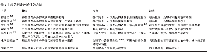

使用上述方法,工程化外泌体不仅可以装载亲水/不亲水小分子,还可以运输蛋白质、RNA、DNA等大分子物质,还可以通过脂质膜上的共价键、配体特异性结合抗体等,见图2[43]。 2.4 工程化外泌体在骨修复再生中的应用 研究发现改变供体细胞培养环境得到的工程化外泌体可以显著促进骨再生。LI等[44]发现缺氧环境下培养的骨髓间充质干细胞来源外泌体可以促进成骨,体外实验发现该外泌体可以增强血清骨钙素和碱性磷酸酶的基因表达,从而促进骨髓间充质干细胞的成骨分化;该外泌体还可以促进人脐静脉内皮细胞的增殖、迁移和管状结构的形成;对类固醇诱导的股骨头缺血性坏死模型进行体内实验,工程化外泌体组检测到了更高的血管密度和更密集的骨小梁组织生成。SAKAGUCHI等[45]也发现低氧环境刺激骨髓间充质干细胞产生特殊的外泌体,与正常环境培养得到的骨髓间充质干细胞来源外泌体相比,不仅能增强ALP、Runx2、COL1和COL2的基因表达,还能增强人脐带内皮细胞的管结构形成,体内实验结果也显示工程化外泌体组的新骨形成率高于天然外泌体组。 使用孵育法也可得到促成骨的工程化外泌体。LIANG 等[46]使用低剂量的二甲基草酰甘氨酸处理骨髓间充质干细胞来源外泌体用于大鼠临界颅骨缺损修复,实验发现该工程化外泌体通过激活AKT/mTOR途径,促进人脐静脉内皮细胞生成血管和骨再生。 使用化学共轭法可以使具有促成骨作用的外泌体靶向到指定细胞。LUO等[47]为解决骨髓基质细胞来源外泌体不能与骨髓间充质干细胞靶向结合的问题,使用化学共轭法制备了可与骨髓间充质干细胞特异性结合的工程化外泌体;对切除卵巢的骨质疏松小鼠模型进行体内实验,结果显示工程化外泌体组骨量恢复明显高于天然外泌体组,且骨折愈合速度也有所加快,此实验证实了该工程化外泌体具有靶向骨髓间充质干细胞并促进骨再生的作用。 使用转染法可得到精确表达促进成骨基因的工程化外泌体。罗月等[48]利用miR-764慢病毒载体转染大鼠骨髓间充质干细胞,得到过表达促成骨miR-764的外泌体;体内实验结果显示,脱钙骨基质与工程化外泌体组成的混合支架植入大鼠颅骨缺损部位后,显著促进骨缺损部位的细胞浸润、胶原沉积、骨钙素和骨桥蛋白表达量增加以及微血管结构生成。另一项研究显示Akt修饰的人脐带间充质干细胞来源外泌体显著促进血管内皮细胞的增殖和迁移、管状结构的形成和体内血管的形成[49]。"

| [1] 韩志琪,蒋伟东,周诺.干细胞组织工程技术辅助下颌牵张成骨的研究与进展[J].中国组织工程研究,2019,23(25):4037-4043. [2] VELASCO MA, NARVÁEZ-TOVAR CA, GARZÓN-ALVARADO DA. Design, materials, and mechanobiology of biodegradable scaffolds for bone tissue engineering. Biomed Res Int. 2015;2015:729076. [3] 王良,李军,沈毅,等.虚拟手术辅助的血管化腓骨肌(皮)瓣行下颌骨精确重建[J].中国耳鼻咽喉颅底外科杂志,2016,22(3):220-224. [4] 张赛,张龙城.下颌骨缺损修复的研究进展[J].中国耳鼻咽喉颅底外科杂志,2017,23(2):177-181. [5] RUSTOM LE, BOUDOU T, NEMKE BW, et al. Multiscale Porosity Directs Bone Regeneration in Biphasic Calcium Phosphate Scaffolds. ACS Biomater Sci Eng. 2017;3(11):2768-2778. [6] BABAIE E, BHADURI SB. Fabrication Aspects of Porous Biomaterials in Orthopedic Applications: A Review. ACS Biomater Sci Eng. 2017;4(1):1-39. [7] GARCÍA-GARETA E, HUA J, ORERA A, et al. Biomimetic surface functionalization of clinically relevant metals used as orthopaedic and dental implants. Biomed Mater. 2017;13(1):015008. [8] ROSETI L, PARISI V, PETRETTA M, et al. Scaffolds for Bone Tissue Engineering: State of the art and new perspectives. Mater Sci Eng C Mater Biol Appl. 2017;78:1246-1262. [9] EL-BADAWY A, AHMED SM, EL-BADRI N. Adipose-Derived Stem Cell-Based Therapies in Regenerative Medicine. Advances in Stem Cell Therapy. 2017;10:117-138. [10] GUAY C, KRUIT JK, ROME S, et al. Lymphocyte-Derived Exosomal MicroRNAs Promote Pancreatic β Cell Death and May Contribute to Type 1 Diabetes Development. Cell Metab. 2019;29(2):348-361. [11] CHEUNG KL, JARRETT R, SUBRAMANIAM S, et al. Psoriatic T cells recognize neolipid antigens generated by mast cell phospholipase delivered by exosomes and presented by CD1a. J Exp Med. 2016; 213(11):2399-2412. [12] BOURDONNAY E, ZASŁONA Z, PENKE LR, et al. Transcellular delivery of vesicular SOCS proteins from macrophages to epithelial cells blunts inflammatory signaling. J Exp Med. 2015;212(5):729-742. [13] 贺娇,许泼实.外泌体提取方法及鉴定分析研究进展[J].中华实用诊断与治疗杂志,2018,32(7):718-721. [14] 宋赛赛,贺娇,程琳.外泌体的研究进展[J].智慧健康,2019,5(22): 57-59. [15] RODRIGUES M, FAN J, LYON C, et al. Role of Extracellular Vesicles in Viral and Bacterial Infections: Pathogenesis, Diagnostics, and Therapeutics. Theranostics. 2018;8(10):2709-2721. [16] SKOTLAND T, HESSVIK NP, SANDVIG K, et al. Exosomal lipid composition and the role of ether lipids and phosphoinositides in exosome biology. J Lipid Res. 2019;60(1):9-18. [17] SCHILLER LT, LEMUS-DIAZ N, RINALDI FERREIRA R, et al. Enhanced Production of Exosome-Associated AAV by Overexpression of the Tetraspanin CD9. Mol Ther Methods Clin Dev. 2018;9:278-287. [18] KALLURI R. The biology and function of exosomes in cancer. J Clin Invest. 2016;126(4):1208-1215. [19] VAN NIEL G, D’ANGELO G, RAPOSO G. Shedding light on the cell biology of extracellular vesicles. Nat Rev Mol Cell Biol. 2018;19(4):213-228. [20] WIKLANDER OPB, BRENNAN MÁ, LÖTVALL J, et al. Advances in therapeutic applications of extracellular vesicles. Sci Transl Med. 2019; 11(492):eaav8521. [21] XIN H, KATAKOWSKI M, WANG F, et al. MicroRNA cluster miR-17-92 Cluster in Exosomes Enhance Neuroplasticity and Functional Recovery After Stroke in Rats. Stroke. 2017;48(3):747-753. [22] VADER P, MOL EA, PASTERKAMP G, et al. Extracellular vesicles for drug delivery. Adv Drug Deliv Rev. 2016;106(Pt A):148-156. [23] KOOIJMANS SAA, SCHIFFELERS RM, ZAROVNI N, et al. Modulation of tissue tropism and biological activity of exosomes and other extracellular vesicles: New nanotools for cancer treatment. Pharmacol Res. 2016;111:487-500. [24] ARMSTRONG JPK, STEVENS MM. Strategic design of extracellular vesicle drug delivery systems. Adv Drug Deliv Rev. 2018;130:12-16. [25] KIBRIA G, RAMOS EK, WAN Y, et al. Exosomes as a Drug Delivery System in Cancer Therapy: Potential and Challenges. Mol Pharm. 2018;15(9): 3625-3633. [26] LÄSSER C, JANG SC, LÖTVALL J. Subpopulations of extracellular vesicles and their therapeutic potential. Mol Aspects Med. 2018;60:1-14. [27] QI X, ZHANG J, YUAN H, et al. Exosomes Secreted by Human-Induced Pluripotent Stem Cell-Derived Mesenchymal Stem Cells Repair Critical-Sized Bone Defects through Enhanced Angiogenesis and Osteogenesis in Osteoporotic Rats. Int J Biol Sci. 2016;12(7):836-849. [28] ZHANG J, LIU X, LI H, et al. Exosomes/tricalcium phosphate combination scaffolds can enhance bone regeneration by activating the PI3K/Akt signaling pathway. Stem Cell Res Ther. 2016;7(1):136. [29] 张静,易阳艳,阳水发,等.脂肪干细胞来源外泌体对人脐静脉血管内皮细胞增殖、迁移及管样分化的影响[J].中国修复重建外科杂志,2018,32(10):1351-1357. [30] 柳鑫,肖燕,江川,等.牙髓干细胞来源外泌体诱导内皮细胞血管生成能力的研究[J].牙体牙髓牙周病学杂志,2018,28(4):187-196. [31] ARENACCIO C, CHIOZZINI C, FERRANTELLI F, et al. Exosomes in Therapy: Engineering, Pharmacokinetics and Future Applications. Curr Drug Targets. 2019;20(1):87-95. [32] LIAO W, DU Y, ZHANG C, et al. Exosomes: The next generation of endogenous nanomaterials for advanced drug delivery and therapy. Acta Biomater. 2019;86:1-14. [33] PASCUCCI L, COCCÈ V, BONOMI A, et al. Paclitaxel is incorporated by mesenchymal stromal cells and released in exosomes that inhibit in vitro tumor growth: a new approach for drug delivery. J Control Release. 2014;192:262-270. [34] SATO YT, UMEZAKI K, SAWADA S, et al. ngineering hybrid exosomes by membrane fusion with liposomes. Sci Rep. 2016;6:21933. [35] FUHRMANN G, SERIO A, MAZO M, et al. Active loading into extracellular vesicles significantly improves the cellular uptake and photodynamic effect of porphyrins. J Control Release. 2015;205:35-44. [36] WAN Y, WANG L, ZHU C, et al. Aptamer-Conjugated Extracellular Nanovesicles for Targeted Drug Delivery. Cancer Res. 2018;78(3):798-808. [37] KIM MS, HANEY MJ, ZHAO Y, et al. Development of exosome-encapsulated paclitaxel to overcome MDR in cancer cells. Nanomedicine. 2016;12(3):655-664. [38] JOHNSEN KB, GUDBERGSSON JM, SKOV MN, et al. Evaluation of electroporation-induced adverse effects on adipose-derived stem cell exosomes. Cytotechnology. 2016;68(5):2125-2138. [39] HOOD JL. Post isolation modification of exosomes for nanomedicine applications. Nanomedicine (Lond). 2016;11(13):1745-1756. [40] TIAN T, ZHANG HX, HE CP, et al. Surface functionalized exosomes as targeted drug delivery vehicles for cerebral ischemia therapy. Biomaterials. 2018;150:137-149. [41] ZHAO Z, MCGILL J, GAMERO-KUBOTA P, et al. Microfluidic on-demand engineering of exosomes towards cancer immunotherapy. Lab Chip. 2019;19(10):1877-1886. [42] STICKNEY Z, LOSACCO J, MCDEVITT S, et al. Development of exosome surface display technology in living human cells. Biochem Biophys Res Commun. 2016;472(1):53-59. [43] LUAN X, SANSANAPHONGPRICHA K, MYERS I, et al. Engineering exosomes as refined biological nanoplatforms for drug delivery. Acta Pharmacol Sin. 2017;38(6):754-763. [44] LI H, LIU D, LI C, et al. Exosomes secreted from mutant-HIF-1α-modified bone-marrow-derived mesenchymal stem cells attenuate early steroid-induced avascular necrosis of femoral head in rabbit. Cell Biol Int. 2017; 41(12):1379-1390. [45] SAKAGUCHI K, SAKAI K, SUGIMURA‐WAKAYAMA Y, et al. Bone regeneration using exosomes derived from hMSCs stimulated by hypoxia. Clin Oral Implants Res. 2019;30(s19):126. [46] LIANG B, LIANG JM, DING JN, et al. Dimethyloxaloylglycine-stimulated human bone marrow mesenchymal stem cell-derived exosomes enhance bone regeneration through angiogenesis by targeting the AKT/mTOR pathway. Stem Cell Res Ther. 2019;10(1):335. [47] LUO ZW, LI FX, LIU YW, et al. Aptamer-functionalized exosomes from bone marrow stromal cells target bone to promote bone regeneration. Nanoscale. 2019;11(43):20884-20892. [48] 罗月,梁卓,吕永钢.过表达microRNA-764间充质干细胞来源外泌体促骨再生[J].医用生物力学,2019,34(s1):57. [49] MA J, ZHAO Y, SUN L, et al. Exosomes Derived from Akt-Modified Human Umbilical Cord Mesenchymal Stem Cells Improve Cardiac Regeneration and Promote Angiogenesis via Activating Platelet-Derived Growth Factor D. Stem Cells Transl Med. 2017;6(1):51-59. [50] INGATO D, LEE JU, SIM SJ, et al. Good things come in small packages: Overcoming challenges to harness extracellular vesicles for therapeutic delivery. J Control Release. 2016;241:174-185. [51] ZHANG Y, CHOPP M, LIU XS, et al. Exosomes Derived from Mesenchymal Stromal Cells Promote Axonal Growth of Cortical Neurons. Mol Neurobiol. 2017;54(4):2659-2673. [52] BOSCH S, DE BEAUREPAIRE L, ALLARD M, et al. Trehalose prevents aggregation of exosomes and cryodamage. Sci Rep. 2016;6:36162. [53] CHAROENVIRIYAKUL C, TAKAHASHI Y, NISHIKAWA M, et al. Preservation of exosomes at room temperature using lyophilization. Int J Pharm. 2018;553(1-2):1-7. [54] LI P, KASLAN M, LEE SH, et al. Progress in Exosome Isolation Techniques. Theranostics. 2017;7(3):789-804. [55] SONG H, LI X, ZHAO Z, et al. Reversal of Osteoporotic Activity by Endothelial Cell-Secreted Bone Targeting and Biocompatible Exosomes. Nano Lett. 2019;19(5):3040-3048. [56] LI S, WU Y, DING F, et al. Engineering macrophage-derived exosomes for targeted chemotherapy of triple-negative breast cancer. Nanoscale. 2020;12(19):10854-10862. |
| [1] | Wang Jing, Xiong Shan, Cao Jin, Feng Linwei, Wang Xin. Role and mechanism of interleukin-3 in bone metabolism [J]. Chinese Journal of Tissue Engineering Research, 2022, 26(8): 1316-1322. |
| [2] | Zhu Chan, Han Xuke, Yao Chengjiao, Zhang Qiang, Liu Jing, Shao Ming. Acupuncture for Parkinson’s disease: an insight into the action mechanism in animal experiments [J]. Chinese Journal of Tissue Engineering Research, 2022, 26(8): 1330-1335. |
| [3] | An Weizheng, He Xiao, Ren Shuai, Liu Jianyu. Potential of muscle-derived stem cells in peripheral nerve regeneration [J]. Chinese Journal of Tissue Engineering Research, 2022, 26(7): 1130-1136. |
| [4] | Fan Yiming, Liu Fangyu, Zhang Hongyu, Li Shuai, Wang Yansong. Serial questions about endogenous neural stem cell response in the ependymal zone after spinal cord injury [J]. Chinese Journal of Tissue Engineering Research, 2022, 26(7): 1137-1142. |
| [5] | Hu Wei, Xie Xingqi, Tu Guanjun. Exosomes derived from bone marrow mesenchymal stem cells improve the integrity of the blood-spinal cord barrier after spinal cord injury [J]. Chinese Journal of Tissue Engineering Research, 2022, 26(7): 992-998. |
| [6] | Gao Yujin, Peng Shuanglin, Ma Zhichao, Lu Shi, Cao Huayue, Wang Lang, Xiao Jingang. Osteogenic ability of adipose stem cells in diabetic osteoporosis mice [J]. Chinese Journal of Tissue Engineering Research, 2022, 26(7): 999-1004. |
| [7] | Guo Jia, Ding Qionghua, Liu Ze, Lü Siyi, Zhou Quancheng, Gao Yuhua, Bai Chunyu. Biological characteristics and immunoregulation of exosomes derived from mesenchymal stem cells [J]. Chinese Journal of Tissue Engineering Research, 2022, 26(7): 1093-1101. |
| [8] | Zhou Hongqin, Wu Dandan, Yang Kun, Liu Qi. Exosomes that deliver specific miRNAs can regulate osteogenesis and promote angiogenesis [J]. Chinese Journal of Tissue Engineering Research, 2022, 26(7): 1107-1112. |
| [9] | Zhang Jinglin, Leng Min, Zhu Boheng, Wang Hong. Mechanism and application of stem cell-derived exosomes in promoting diabetic wound healing [J]. Chinese Journal of Tissue Engineering Research, 2022, 26(7): 1113-1118. |
| [10] | Huang Chenwei, Fei Yankang, Zhu Mengmei, Li Penghao, Yu Bing. Important role of glutathione in stemness and regulation of stem cells [J]. Chinese Journal of Tissue Engineering Research, 2022, 26(7): 1119-1124. |
| [11] | Hui Xiaoshan, Bai Jing, Zhou Siyuan, Wang Jie, Zhang Jinsheng, He Qingyong, Meng Peipei. Theoretical mechanism of traditional Chinese medicine theory on stem cell induced differentiation [J]. Chinese Journal of Tissue Engineering Research, 2022, 26(7): 1125-1129. |
| [12] | Xu Lei, Han Xiaoqiang, Zhang Jintao, Sun Haibiao. Hyaluronic acid around articular chondrocytes: production, transformation and function characteristics [J]. Chinese Journal of Tissue Engineering Research, 2022, 26(5): 768-773. |
| [13] | Li Jiajun, Xia Tian, Liu Jiamin, Chen Feng, Chen Haote, Zhuo Yinghong, Wu Weifeng. Molecular mechanism by which icariin regulates osteogenic signaling pathways in the treatment of steroid-induced avascular necrosis of the femoral head [J]. Chinese Journal of Tissue Engineering Research, 2022, 26(5): 780-785. |
| [14] | Lu Qinxue, Xu Ning, Yang Yinglan, Han Qianqian, Duanmu Xianyu, Guo Yuwei, Han Qing. Femoroacetabular impingement: strength trainings for nerve-muscle, peripheral muscle and core muscle [J]. Chinese Journal of Tissue Engineering Research, 2022, 26(5): 786-791. |
| [15] | Zheng Zhenquan, Rong Jiesheng. Sarcopenia: age-related muscle mass loss and functional declines [J]. Chinese Journal of Tissue Engineering Research, 2022, 26(5): 792-797. |
| Viewed | ||||||
|
Full text |
|
|||||
|
Abstract |
|
|||||