Chinese Journal of Tissue Engineering Research ›› 2014, Vol. 18 ›› Issue (1): 119-124.doi: 10.3969/j.issn.2095-4344.2014.01.020
Previous Articles Next Articles
Mononuclear cells promote mesenchymal stem cell migration after myocardial infarction
Zhang Ying1, Liao Li-qiang1, Zhang Xiao-gang2
- 1People’s Hospital of Yubei District, Chongqing 401120, China
2First Affiliated Hospital of Chongqing Medical University, Chongqing 400016, China
-
Revised:2013-10-17Online:2014-01-01Published:2014-01-01 -
Contact:Liao Li-qiang, Master, Physician, People’s Hospital of Yubei District, Chongqing 401120, China -
About author:Zhang Ying, Master, Physician, People’s Hospital of Yubei District, Chongqing 401120, China
CLC Number:
Cite this article
Zhang Ying, Liao Li-qiang, Zhang Xiao-gang. Mononuclear cells promote mesenchymal stem cell migration after myocardial infarction[J]. Chinese Journal of Tissue Engineering Research, 2014, 18(1): 119-124.
share this article
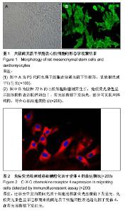
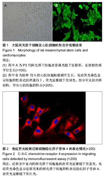
2.1 实验动物数量分析 12只SD大鼠用于分离单个核细胞,其中行心肌梗死手术6只,行假手术6只,全部进入结果分析,无脱失。 2.2 细胞形态学观察 原代间充质干细胞接种后24-72 h后完全贴壁,呈现集落状生长,传代后增殖迅速,呈放射状、螺旋状或平行生长,类似成纤维细胞形态(图1A)。原代心肌细胞接种24-48 h后基本贴壁生长,呈梭形、杆状或不规则形态,部分可见节律性收缩,频率为30-100次/min,免疫细胞化学染色示95%以上贴壁细胞肌钙蛋白Ⅰ阳性表达(图1B)。 2.3 迁移细胞计数及趋化因子受体4免疫荧光染色 DAPI标记的间充质干细胞胞核在荧光镜下发蓝光。心肌梗死组、AMD3100组及假手术组中室内均可见胞核发蓝光的迁移细胞,单位视野细胞迁移数分别为13.5±1.0,3.9±0.6及3.7±0.7,而空白对照组中室内未见迁移细胞。其中,心肌梗死组单位视野细胞迁移数显著高于假手术组(P < 0.05),加入趋化因子受体4抑制剂AMD3100后单位视野细胞迁移数明显减少,差异有显著性意义(P < 0.05)。迁移至中室的间充质干细胞阳性表达趋化因子受体4,在荧光显微镜下发红光,胞核发蓝光,细胞形态似成纤维细胞,呈梭形、短三角形等形态(图2)。"
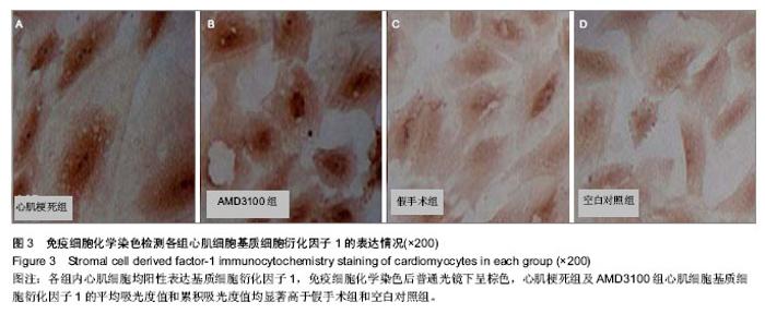
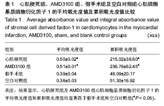
2.4 心肌细胞基质细胞衍化因子1表达 各组心肌细胞均阳性表达基质细胞衍化因子1,免疫细胞化学染色呈棕色(图3),颜色越深吸光度值越高,基质细胞衍化因子1表达量越高。其中,心肌梗死组及AMD3100组心肌细胞基质细胞衍化因子1平均吸光度值和累积吸光度值均显著高于假手术组及空白对照组(P < 0.05)(表1),而心肌梗死组与AMD3100组间,假手术组与空白对照组间比较差异均无显著性意义。"

"
| [1] Li Z, Guo J, Chang Q,et al. Paracrine role for mesenchymal stem cells in acute myocardial infarction. Biol Pharm Bull. 2009;32(8):1343-1346.[2] Buccini S, Haider KH, Ahmed RP, et al. Cardiac progenitors derived from reprogrammed mesenchymal stem cells contribute to angiomyogenic repair of the infarcted heart. Basic Res Cardiol. 2012;107(6):301.[3] Mathieu E, Lamirault G, Toquet C,et al. Intramyocardial delivery of mesenchymal stem cell-seeded hydrogel preserves cardiac function and attenuates ventricular remodeling after myocardial infarction. PLoS One. 2012;7(12):e51991.[4] Houtgraaf JH, de Jong R, Kazemi K,et al. Intracoronary infusion of allogeneic mesenchymal precursor cells directly after experimental acute myocardial infarction reduces infarct size, abrogates adverse remodeling, and improves cardiac function. Circ Res. 2013;113(2):153-166. [5] Williams AR, Hatzistergos KE, Addicott B,et al. Enhanced effect of combining human cardiac stem cells and bone marrow mesenchymal stem cells to reduce infarct size and to restore cardiac function after myocardial infarction. Circulation. 2013;127(2):213-223.[6] Barbash IM, Chouraqui P, Baron J,et al. Systemic delivery of bone marrow-derived mesenchymal stem cells to the infarcted myocardium: feasibility, cell migration, and body distribution. Circulation. 2003;108(7):863-868.[7] Wang T, Sun S, Wan Z,et al.Effects of bone marrow mesenchymal stem cells in a rat model of myocardial infarction. Resuscitation. 2012;83(11):1391-1396.[8] Mao Y, Li SN, Mao XB, et al. Therapeutic effect of transplantation of bone marrow mesenchymal stem cells over-expressing Cx43 on heart failure in post-infarction rats. Zhonghua Yi Xue Za Zhi. 2011;91(28):1982-1986.[9] Fishbein MC, Maclean D, Maroko PR. Experimental myocardial infarction in the rat: qualitative and quantitative changes during pathologic evolution. Am J Pathol. 1978;90(1):57-70.[10] 廖礼强,张晓刚,史若飞,等.心肌梗死大鼠单个核细胞分泌TNF-α对间充质干细胞迁移的影响[J].解放军医学杂志,2010,35(7): 807-810.[11] 廖礼强,张晓刚,史若飞,等.微血管内皮细胞诱导骨髓间充质干细胞心肌样分化的作用及移植可行性[J].中国组织工程研究与临床康复, 2009,13(40): 7811-7816.[12] 王佳南,张晓刚,汤为学,等.新生大鼠心肌细胞原代培养方法的改良[J].重庆医科大学学报,2009, 34(5):600-603.[13] Wollert KC, Drexler H. Mesenchymal stem cells for myocardial infarction: promises and pitfalls. Circulation. 2005; 112(2):151-153. [14] 于玲范,金丹玲,周晶.细胞间直接接触对骨髓间充质干细胞分化为心肌细胞作用的研究[J].哈尔滨医科大学学报,2006,40(2): 124-127.[15] 袁岩,陈连凤,张抒扬,等.心肌细胞裂解液对骨髓间充质干细胞向心肌细胞分化诱导作用的研究[J].中华心血管病杂志,2005; 33(2):170-173. [16] Wang TZ, Ma AQ, Xu ZY, et al. Differentiation of mesenchymal stem cells into cardiomyocytes induced by cardiomyocytes. Zhong Nan Da Xue Xue Bao Yi Xue Ban. 2005;30(3):270-275. [17] Li X, Yu X, Lin Q, et al. Bone marrow mesenchymal stem cells differentiate into functional cardiac phenotypes by cardiac microenvironment.J Mol Cell Cardiol. 2007;42(2):283-284.[18] Plotnikov EY, Khryapenkova TG, Vasileva AK, et al. Cell-to-cell cross-talk between mesenchymal stem cells and cardiomyocytes in coculture. J Cell Mol Med. 2008;50(1): 31-39.[19] 付小兵,程飚. 成体干细胞研究及其可能的临床应用[J].中华医学杂志,2005,85(27):1878-1880 [20] Roche R, Hoareau L, Mounet F, et al. Adult stem cells for cardiovascular diseases: the adipose tissue potential. Expert Opin Biol Ther. 2007;7(6):791-798. [21] Shim WS, Jiang S, Wong P, et al. Ex vivo differentiation of human adult bone marrow stem cells into cardiomyocyte-like cells. Biochem Biophys Tes Commun. 2004;324(2):481-488. [22] Tamaki T, Akatsuka A, Okada Y, et al.Cardiomyocyte formation by skeletal muscle-derived multi-myogenic stem cells after transplantation into infarcted myocardium.PLoS ONE. 2008;3(3):e1789.[23] Hahn JY, Cho HJ, Kang HJ, et al. Pre-Treatment of Mesenchymal Stem Cells With a Combination of Growth Factors Enhances Gap Junction Formation, Cytoprotective Effect on Cardiomyocytes, and Therapeutic Efficacy for Myocardial Infarction. J Am Coll Cardiol.2008;51(9):933-943. [24] Zhang Y, Liang JL, Wang S, et al. Autologous bone marrow stromal cell trans-plantation with application of granulocyte colony-stimulating factor improves rabbit cardiac performance after acute myocardial infarction. Nan Fang Yi Ke Da Xue Xue Bao. 2007;27(1):43-45, 48.[25] Wollert KC, Drexler H. Mesenchymal stem cells for myocardial infarction: promises and pitfalls. Circulation. 2005; 112(2):151-153.[26] Nakamura Y, Wang X, Xu C, et al. Xenotransplantation of Long-Term-Cultured Swine Bone Marrow-Derived Mesenchymal Stem Cells. Stem Cells. 2007;25(3): 612 - 620. [27] Forte G, Minieri M, Cossa P, et al. Hepatocyte growth factor effects on mesenchymal stem cells: proliferation, migration, and differentiation. Am J Physiol Heart Circ Physiol. 2006; 290(4):H1370-1377. [28] Wang CC, Chen CH, Lin WW, et al. Direct intramyocardial injection of mesenchymal stem cell sheet fragments improves cardiac functions after infarction. Cardiovasc Res. 2008;77: 515-524.[29] Houtgraaf JH, de Jong R, Kazemi K, et al. Intracoronary infusion of allogeneic mesenchymal precursor cells directly after experimental acute myocardial infarction reduces infarct size, abrogates adverse remodeling, and improves cardiac function. Circ Res. 2013;113(2):153-166. [30] Rodrigo SF, van Ramshorst J, Hoogslag GE,et al. Intramyocardial injection of autologous bone marrow-derived ex vivo expanded mesenchymal stem cells in acute myocardial infarction patients is feasible and safe up to 5 years of follow-up. J Cardiovasc Transl Res. 2013;6(5): 816-825. [31] Ishikane S, Hosoda H, Yamahara K,et al. Allogeneic transplantation of fetal membrane-derived mesenchymal stem cell sheets increases neovascularization and improves cardiac function after myocardial infarction in rats. Transplantation. 2013;96(8):697-706.[32] Vela DC, Silva GV, Assad JAR,et al. Histopathological Study of Healing After Allogenic Mesenchymal Stem Cell Delivery in Myocardial Infarction in Dogs. J Histochem Cytochem. 2009; 57: 167-176.[33] Ji JF, He BP, Dheen ST,et al. Interactions of chemokines and chemokine receptors mediate the migration of mesenchymal stem cells to the impaired site in the brain after hypoglossal nerve injury. Stem Cells. 2004;22(3):415-427.[34] Chen Y, Ke Q, Yang Y,et al. Cardiomyocytes overexpressing TNF-alpha attract migration of embryonic stem cells via activation of p38 and c-Jun amino-terminal kinase. FASEB J. 2003;17(15):2231-2239. [35] Kim YS, Park HJ, Hong MH, et al. TNF-alpha enhances engraftment of mesenchymal stem cells into infarcted myocardium. Front Biosci. 2009;14:2845-2856.[36] Honczarenko M, Le Y, Swierkowski M, et al. Human bone marrow stromal cells express a distinct set of biologically functional chemokine receptors. Stem Cells. 2006;24(4): 1030-1041.[37] Bobis S, Jarocha D, Majka M.Mesenchymal stem cells: characteristics and clinical applications. Folia Histochem Cytobiol. 2006;44(4):215-230.[38] Zhang M, Mal N, Kiedrowski M, et al. SDF-1 expression by mesenchymal stem cells results in trophic support of cardiac myocytes after myocardial infarction. FASEB J. 2007;21(12): 3197-3207.[39] Liu X, Duan B, Cheng Z,et al. SDF-1/CXCR4 axis modulates bone marrow mesenchymal stem cell apoptosis, migration and cytokine secretion. Protein Cell. 2011;2(10):845-854.[40] Xu X, Zhu F, Zhang M,et al. Stromal cell-derived factor-1 enhances wound healing through recruiting bone marrow-derived mesenchymal stem cells to the wound area and promoting neovascularization. Cells Tissues Organs. 2013;197(2):103-113.[41] Pasha Z, Wang Y, Sheikh R,et al. Preconditioning enhances cell survival and differentiation of stem cells during transplantation in infarcted myocardium. Cardiovasc Res. 2008;77(1):134-142.[42] Abbott JD, Huang Y, Liu D, et al.Stromal cell-derived factor-1 alpha plays a critical role in stem cell recruitment to the heart after myocardial infarction but is not sufficient to induce homing in the absence of injury. Circulation. 2004;110(21): 3300-3305.[43] Zhang D,Huang W,Dai B,et al.Genetically manipulated progenitor cell sheet with diprotin A improves myocardial function and repair of infarcted hearts. Am J Physiol Heart Circ Physiol. 2010;299(5):H1339-1347.[44] Dong F, Harvey J, Finan A,et al.Myocardial CXCR4 expression is required for mesenchymal stem cell mediated repair following acute myocardial infarction.Circulation. 2012; 126(3):314-324. |
| [1] | Pu Rui, Chen Ziyang, Yuan Lingyan. Characteristics and effects of exosomes from different cell sources in cardioprotection [J]. Chinese Journal of Tissue Engineering Research, 2021, 25(在线): 1-. |
| [2] | Lin Qingfan, Xie Yixin, Chen Wanqing, Ye Zhenzhong, Chen Youfang. Human placenta-derived mesenchymal stem cell conditioned medium can upregulate BeWo cell viability and zonula occludens expression under hypoxia [J]. Chinese Journal of Tissue Engineering Research, 2021, 25(在线): 4970-4975. |
| [3] | Hou Jingying, Yu Menglei, Guo Tianzhu, Long Huibao, Wu Hao. Hypoxia preconditioning promotes bone marrow mesenchymal stem cells survival and vascularization through the activation of HIF-1α/MALAT1/VEGFA pathway [J]. Chinese Journal of Tissue Engineering Research, 2021, 25(7): 985-990. |
| [4] | Shi Yangyang, Qin Yingfei, Wu Fuling, He Xiao, Zhang Xuejing. Pretreatment of placental mesenchymal stem cells to prevent bronchiolitis in mice [J]. Chinese Journal of Tissue Engineering Research, 2021, 25(7): 991-995. |
| [5] | Liang Xueqi, Guo Lijiao, Chen Hejie, Wu Jie, Sun Yaqi, Xing Zhikun, Zou Hailiang, Chen Xueling, Wu Xiangwei. Alveolar echinococcosis protoscolices inhibits the differentiation of bone marrow mesenchymal stem cells into fibroblasts [J]. Chinese Journal of Tissue Engineering Research, 2021, 25(7): 996-1001. |
| [6] | Fan Quanbao, Luo Huina, Wang Bingyun, Chen Shengfeng, Cui Lianxu, Jiang Wenkang, Zhao Mingming, Wang Jingjing, Luo Dongzhang, Chen Zhisheng, Bai Yinshan, Liu Canying, Zhang Hui. Biological characteristics of canine adipose-derived mesenchymal stem cells cultured in hypoxia [J]. Chinese Journal of Tissue Engineering Research, 2021, 25(7): 1002-1007. |
| [7] | Geng Yao, Yin Zhiliang, Li Xingping, Xiao Dongqin, Hou Weiguang. Role of hsa-miRNA-223-3p in regulating osteogenic differentiation of human bone marrow mesenchymal stem cells [J]. Chinese Journal of Tissue Engineering Research, 2021, 25(7): 1008-1013. |
| [8] | Lun Zhigang, Jin Jing, Wang Tianyan, Li Aimin. Effect of peroxiredoxin 6 on proliferation and differentiation of bone marrow mesenchymal stem cells into neural lineage in vitro [J]. Chinese Journal of Tissue Engineering Research, 2021, 25(7): 1014-1018. |
| [9] | Zhu Xuefen, Huang Cheng, Ding Jian, Dai Yongping, Liu Yuanbing, Le Lixiang, Wang Liangliang, Yang Jiandong. Mechanism of bone marrow mesenchymal stem cells differentiation into functional neurons induced by glial cell line derived neurotrophic factor [J]. Chinese Journal of Tissue Engineering Research, 2021, 25(7): 1019-1025. |
| [10] | Duan Liyun, Cao Xiaocang. Human placenta mesenchymal stem cells-derived extracellular vesicles regulate collagen deposition in intestinal mucosa of mice with colitis [J]. Chinese Journal of Tissue Engineering Research, 2021, 25(7): 1026-1031. |
| [11] | Pei Lili, Sun Guicai, Wang Di. Salvianolic acid B inhibits oxidative damage of bone marrow mesenchymal stem cells and promotes differentiation into cardiomyocytes [J]. Chinese Journal of Tissue Engineering Research, 2021, 25(7): 1032-1036. |
| [12] | Wang Xianyao, Guan Yalin, Liu Zhongshan. Strategies for improving the therapeutic efficacy of mesenchymal stem cells in the treatment of nonhealing wounds [J]. Chinese Journal of Tissue Engineering Research, 2021, 25(7): 1081-1087. |
| [13] | Wang Shiqi, Zhang Jinsheng. Effects of Chinese medicine on proliferation, differentiation and aging of bone marrow mesenchymal stem cells regulating ischemia-hypoxia microenvironment [J]. Chinese Journal of Tissue Engineering Research, 2021, 25(7): 1129-1134. |
| [14] | Kong Desheng, He Jingjing, Feng Baofeng, Guo Ruiyun, Asiamah Ernest Amponsah, Lü Fei, Zhang Shuhan, Zhang Xiaolin, Ma Jun, Cui Huixian. Efficacy of mesenchymal stem cells in the spinal cord injury of large animal models: a meta-analysis [J]. Chinese Journal of Tissue Engineering Research, 2021, 25(7): 1142-1148. |
| [15] | Chen Junyi, Wang Ning, Peng Chengfei, Zhu Lunjing, Duan Jiangtao, Wang Ye, Bei Chaoyong. Decalcified bone matrix and lentivirus-mediated silencing of P75 neurotrophin receptor transfected bone marrow mesenchymal stem cells to construct tissue-engineered bone [J]. Chinese Journal of Tissue Engineering Research, 2021, 25(4): 510-515. |
| Viewed | ||||||
|
Full text |
|
|||||
|
Abstract |
|
|||||