[1] 吕东来.体外三维微环境增强胶质瘤细胞“干性”和诱导化疗耐药作用及其机制[D].重庆:第三军医大学,2016.
[2] ANTUNES J, GASPAR VM, FERREIRA L, et al. In-air production of 3D co-culture tumor spheroid hydrogels for expedited drug screening. Acta Biomater. 2019;94:392-409.
[3] FRAHS SM, OXFORD JT, NEUMANN EE, et al. Extracellular Matrix Expression and Production in Fibroblast-Collagen Gels: Towards an In Vitro Model for Ligament Wound Healing. Ann Biomed Eng. 2018; 46(11):1882-1895.
[4] GIOBBE GG, CROWLEY C, LUNI C. Extracellular matrix hydrogel derived from decellularized tissues enables endodermal organoid culture. Nat Commun. 2019;10(1):5658.
[5] GJOREVSKI N, SACHS N, MANFRIN A, et al. Designer matrices for intestinal stem cell and organoid culture. Nat Commun. 2016;539 (7630):560-564.
[6] LAZZARI G, NICOLAS V, MATSUSAKI M, et al. Multicellular spheroid based on a triple co-culture: A novel 3D model to mimic pancreatic tumor complexity. Acta Biomater. 2018;78:296-307.
[7] STEVANATO L, SINDEN JD. The effects of microRNAs on human neural stem cell differentiation in two- and three-dimensional cultures. Stem Cell Res Ther. 2014;5(2):49.
[8] 吉中蛟, 郭益冰, 陆玉华. 脱细胞支架在肿瘤组织工程中的应用[J].中国组织工程研究, 2018,22(34):5571-5576.
[9] LU P, WEAVER VM, WERB Z. The extracellular matrix: a dynamic niche in cancer progression. J Cell Biol. 2012;196(4):395-406.
[10] PENCE KA, MISHRA DK, THRALL M, et al. Breast cancer cells form primary tumors on ex vivo four-dimensional lung model. J Surg Res. 2017;210:181-187.
[11] NIETZER S, BAUR F, SIEBER S, et al. Mimicking Metastases Including Tumor Stroma: A New Technique to Generate a Three-Dimensional Colorectal Cancer Model Based on a Biological Decellularized Intestinal Scaffold. Tissue Eng Part C Methods. 2016;22(7):621-35.
[12] GUHA D, SAHA T, BOSE S, et al. Integrin-EGFR interaction regulates anoikis resistance in colon cancer cells. Apoptosis. 2019;24(11-12): 958-971.
[13] STEENACKERS A, OLIVIER-VAN STICHELEN S, BALDINI SF, et al. Silencing the Nucleocytoplasmic O-GlcNAc Transferase Reduces Proliferation, Adhesion, and Migration of Cancer and Fetal Human Colon Cell Lines. Front Endocrinol (Lausanne). 2016;7:46.
[14] ZHOU M, LIU X, LI Z, et al. Caspase-3 regulates the migration, invasion and metastasis of colon cancer cells.Int J Cancer. 2018; 143(4):921-930.
[15] FERNANDEZ-MOURE JS, VAN EPS JL, RHUDY JR, et al. Porcine acellular lung matrix for wound healing and abdominal wall reconstruction: A pilot study. J Tissue Eng. 2016;7:2041731415626018.
[16] SUN X, WANG Y, GUO Z, et al. Acellular Cauda Equina Allograft as Main Material Combined with Biodegradable Chitin Conduit for Regeneration of Long-Distance Sciatic Nerve Defect in Rats. Adv Healthc Mater. 2018; 7(17):e1800276.
[17] TANG K, WU J, XIONG Z, et al. Human acellular amniotic membrane: A potential osteoinductive biomaterial for bone regeneration. J Biomater Appl. 2018;32(6):754-764.
[18] YESIL P, MICHEL M, CHWALEK K, et al. A new collagenase blend increases the number of islets isolated from mouse pancreas. Islets. 2009;1(3):185-190.
[19] MAZZA G, AL-AKKAD W, TELESE A, et al. Rapid production of human liver scaffolds for functional tissue engineering by high shear stress oscillation-decellularization.Sci Rep. 2017;7(1):5534.
[20] ALBRITTON JL, MILLER JS. 3D bioprinting: improving in vitro models of metastasis with heterogeneous tumor microenvironments.Dis Model Mech. 2017;10(1):3-14.
[21] BRODACZEWSKA KK, BIELECKA ZF, MALISZEWSKA-OLEJNICZAK K, et al. Metastatic renal cell carcinoma cells growing in 3D on poly‑D‑lysine or laminin present a stem‑like phenotype and drug resistance. Oncol Rep. 2019;42(5):1878-1892.
[22] CATTIN S, RAMONT L, RüEGG C. Characterization and In Vivo Validation of a Three-Dimensional Multi-Cellular Culture Model to Study Heterotypic Interactions in Colorectal Cancer Cell Growth, Invasion and Metastasis. Front Bioeng Biotechnol. 2018;6:97.
[23] LEE JH, KIM SK, KHAWAR IA, et al. Microfluidic co-culture of pancreatic tumor spheroids with stellate cells as a novel 3D model for investigation of stroma-mediated cell motility and drug resistance.J Exp Clin Cancer Res. 2018;37(1):4.
[24] KAJBAFZADEH AM, MASOUMI A, HOSSEINI M, et al. Sheep colon acellular matrix: Immunohistologic, biomechanical, scanning electron microscopic evaluation and collagen quantification. J Biosci Bioeng. 2014;117(2):236-241.
[25] CHEN HJ, SHULER ML. Engineering a Bioartificial Human Colon Model Through Decellularization and Recellularization. Methods Mol Biol. 2019;1907:91-102.
[26] MAYORCA-GUILIANI AE, MADSEN CD, COX TR, et al. ISDoT: in situ decellularization of tissues for high-resolution imaging and proteomic analysis of native extracellular matrix. Nat Med. 2017;23(7):890-898.
[27] SPILL F, REYNOLDS DS, KAMM RD, et al. Impact of the physical microenvironment on tumor progression and metastasis. Curr Opin Biotechnol. 2016;40:41-48.
[28] GIANSANTI L, CONDELLO M, ALTIERI B, et al. Influence of lipid composition on the ability of liposome loaded voacamine to improve the reversion of doxorubicin resistant osteosarcoma cells. Chem Phys Lipids. 2019;223:104781.
[29] YU Y, BLOKHUIS B, DERKS Y. Human mast cells promote colon cancer growth via bidirectional crosstalk: studies in 2D and 3D coculture models. Oncoimmunology. 2018;7(11):e1504729.
[30] ANGELONI V, CONTESSI N, DE MARCO C, et al. Polyurethane foam scaffold as in vitro model for breast cancer bone metastasis. Acta Biomater. 2017;63:306-316.
[31] GILL BJ, WEST JL. Modeling the tumor extracellular matrix: Tissue engineering tools repurposed towards new frontiers in cancer biology. J Biomech. 2014;47(9):1969-1978.
[32] GIRARD YK, WANG C, RAVI S, et al. A 3D fibrous scaffold inducing tumoroids: a platform for anticancer drug development. PLoS One. 2013;8(10):e75345.
[33] LU WD, SUN RF, HU YR, et al. Photooxidatively crosslinked acellular tumor extracellular matrices as potential tumor engineering scaffolds. Acta Biomater. 2018;71:460-473.
[34] LIU H, WANG N, ZHANG Z, et al. Effects of Tumor Necrosis Factor-α on Morphology and Mechanical Properties of HCT116 Human Colon Cancer Cells Investigated by Atomic Force Microscopy. 2017; 2017:2027079.
[35] WAN G, PEHLKE C, PEPERMANS R, et al. The H1047R point mutation in p110 alpha changes the morphology of human colon HCT116 cancer cells. Scanning. 2015;1:15044.
[36] FU RH, WANG YC, LIU SP, et al. Decellularization and recellularization technologies in tissue engineering. Cell Transplant. 2014;23(4-5): 621-630.
[37] JENSEN T, ROSZELL B, ZANG F, et al. A rapid lung de-cellularization protocol supports embryonic stem cell differentiation in vitro and following implantation. Tissue Eng Part C Methods. 2012;18(8): 632-646.
[38] WU J, DING Q, DUTTA A, et al. An injectable extracellular matrix derived hydrogel for meniscus repair and regeneration. Acta Biomater. 2015;16:49-59.
[39] JUNTTILA MR, DE SAUVAGE FJ. Influence of tumour micro-environment heterogeneity on therapeutic response. Nature. 2013;501(7467): 346-354.
[40] SEO JS, KIM A, SHIN JY, et al. Comprehensive analysis of the tumor immune micro-environment in non-small cell lung cancer for efficacy of checkpoint inhibitor. Sci Rep. 2018;8(1):14576.
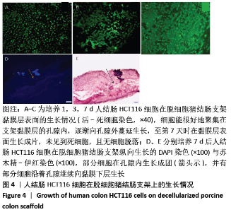
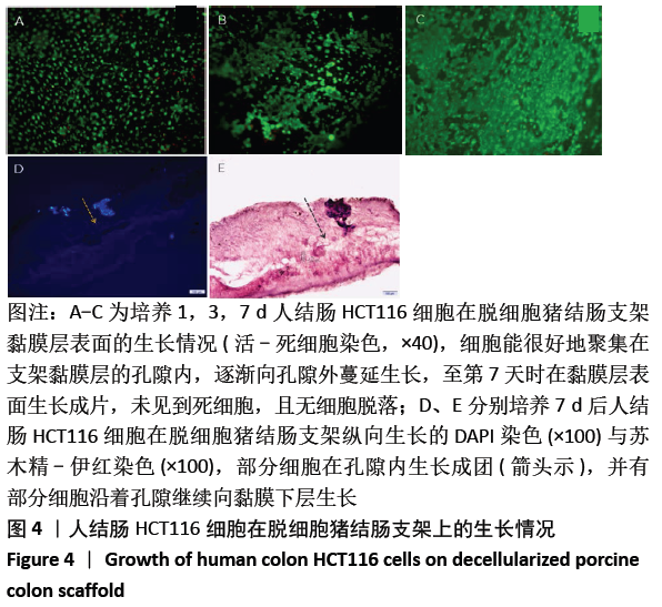
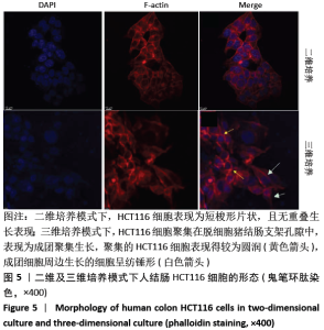
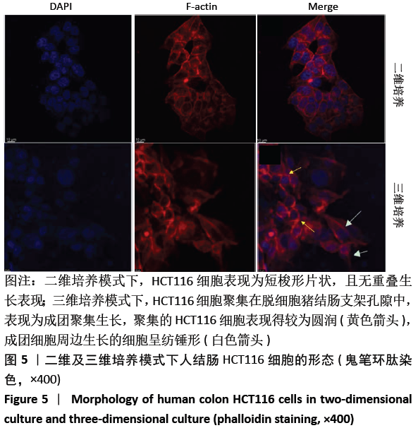
|