Chinese Journal of Tissue Engineering Research ›› 2021, Vol. 25 ›› Issue (10): 1539-1544.doi: 10.3969/j.issn.2095-4344.3061
Previous Articles Next Articles
Comparison of the impacts of two kinds of brackets on external apical root resorption in orthodontic treatment of bimaxillary protrusion patients
Wang Guangping1, Li Mingxia2, Han Yu1, Xu Xiaomei1, Xu Jie1, Huang Suhua1
- 1Department of Orthodontics, 2Department of Oral Radiology, Oral & Maxillofacial Reconstruction and Regeneration Laboratory, Hospital of Stomatology of Southwest Medical University, Luzhou 646000, Sichuan Provence, China
-
Received:2020-04-24Revised:2020-04-28Accepted:2020-07-10Online:2021-04-08Published:2020-12-18 -
Contact:Li Mingxia, Attending physician, Department of Oral Radiology, Oral & Maxillofacial Reconstruction and Regeneration Laboratory, Hospital of Stomatology of Southwest Medical University, Luzhou 646000, Sichuan Provence, China -
About author:Wang Guangping, Master, Attending physician, Department of Orthodontics, Oral & Maxillofacial Reconstruction and Regeneration Laboratory, Hospital of Stomatology of Southwest Medical University, Luzhou 646000, Sichuan Provence, China -
Supported by:the Innovation Training Program of Southwest Medical University, No. 2019338; the Youth Program in Stomatological Hospital Affiliated to Southwest Medical University, No. 201907
CLC Number:
Cite this article
Wang Guangping, Li Mingxia, Han Yu, Xu Xiaomei, Xu Jie, Huang Suhua. Comparison of the impacts of two kinds of brackets on external apical root resorption in orthodontic treatment of bimaxillary protrusion patients[J]. Chinese Journal of Tissue Engineering Research, 2021, 25(10): 1539-1544.
share this article
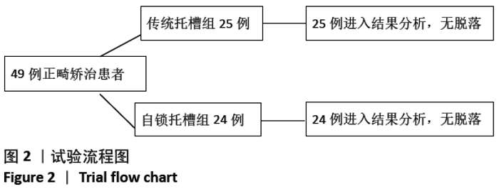
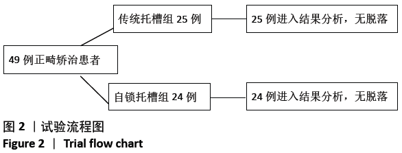
2.1 参与者数量分析 49例患者均进入结果分析。 2.2 两组基线资料比较 自锁托槽组矫治前平均年龄(14.3±0.6)岁,传统托槽组矫治前平均年龄(14.6±0.5)岁,组间比较差异无显著性意义(P > 0.05),具有可比性。 2.3 试验流程 见图2。"
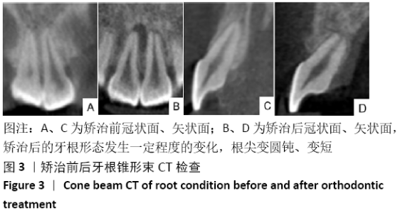
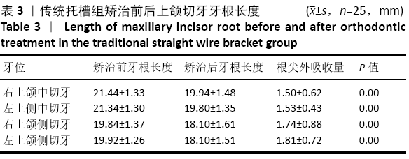
2.4 两组矫治疗程及效果 自锁托槽组、传统托槽组矫治疗程分别为(21.37±1.70),(25.4±2.48)个月,组间比较差异有非常显著性意义(P < 0.01)。两组患者矫治后牙齿排列整齐,磨牙达到中性咬牙合关系,前牙浅覆牙合浅覆盖,前牙实现整体内收,有效改善双颌前突面型及开唇露齿症状,上、下牙列中线对齐,前牙唇倾度正常、未丢失转矩,均取得了良好的临床效果。 2.5 两组矫治后的牙根形态及根尖外吸收 锥形束CT提示,经过矫治后49例患者的牙根形态发生一定程度的改变,大部分仅表现为轮廓的改变如根尖变圆钝等轻微改变,个别牙齿根尖变短,见图3。测量显示所有切牙均发生了根尖外吸收,未发现牙根吸收量超过4 mm以上的病例。自锁托槽组、传统托槽组上颌切牙矫治前后牙根长度统计情况分别见表2,3。矫治后,自锁托槽组4颗上颌切牙的平均根尖外吸收量为(1.36±0.72)mm,传统托槽组吸收量为(1.65±0.69)mm,组间比较差异有显著性意义(P < 0.05),提示高转矩自锁托槽组上切牙发生根尖外吸收少。 "


"


"
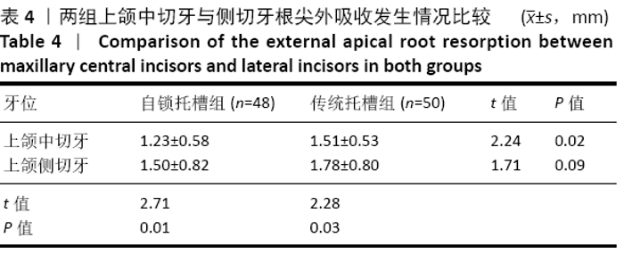
矫治后,自锁托槽与传统托槽组间上颌切牙、同组内中切牙与侧切牙根尖外吸收发生情况见表4。自锁托槽组上颌中切牙根尖外吸收量少于传统托槽组(P < 0.05);自锁托槽组上颌侧切牙根尖外吸收量稍少于传统托槽组,但差异无显著性意义(P > 0.05);两组组内,上颌中切牙根尖外吸收量少于侧切牙(P < 0.05)。 2.6 矫治材料生物相容性 矫治过程中未发生与矫治材料相关的不良反应。 "

| [1] WELTMAN B, VIG KW, FIELDS HW, et al. Root resorption associated with orthodontic tooth movement: a systematic review. Am J Orthod Dentofacial Orthop. 2010;137(4):462-476. [2] DE FREITAS JC, LYRA OCP, DE ALENCAR AHG, et al. Long-term evaluation of apical root resorption after orthodontic treatment using, periapical radiography and cone beam computed tomography. Dental Press J Orthod. 2013;18(4):104-112. [3] EISSA O, CARLYLE T, EL-BIALY T. Evaluation of root length following treatment with clear aligners and two different fixed orthodontic appliances. A pilot study. J Orthod Sci. 2018;7:11. [4] KILLIANY DM. Root resorption caused by orthodontic treatment: an evidence-based review of literature. Semin Orthod. 1999;5(2):128-133. [5] RAKHSHAN V, NATEGHIAN N, ORDOUBAZARI M. Risk factors associated with external apical root resorption of the maxillary incisors: a 15-year retrospective study. Aust Orthod J. 2012;28(1):51-56. [6] CUTRERA A, ALLAREDDY V, AZAMI N, et al. Is Short Root Anomaly (SRA) a risk factor for increased external apical root resorption in orthodontic patients? A retrospective case control study using cone beam computerized tomography. Orthod Craniofac Res. 2019;22(1):32-37. [7] DE ALMEIDA MR, MARÇAL ASB, FERNANDES TMF, et al. A comparative study of the effect of the intrusion arch and straight wire mechanics on incisor root resorption: A randomized, controlled trial. Angle Orthod. 2018;88(1):20-26. [8] MOTOKAWA M, SASAMOTO T, KAKU M, et al. Association between root resorption incident to orthodontic treatment and treatment factors. Eur J Orthod. 2012;34(3):350-356. [9] JUNG YH, CHO BH. External root resorption after orthodontic treatment: a study of contributing factors. Imaging Sci Dent. 2011;41(1):17-21. [10] MARTINS DR, TIBOLA D, JANSON G, et al. Effects of intrusion combined with anterior retraction on apical root resorption. Eur J Orthod. 2012;34(2):170-175. [11] LEVANDER E, BAJKA R, MALMGREN O. Early radiographic diagnosis of apical root resorption during orthodontic treatment: a study of maxillary incisors. Eur J Orthod. 1998;20(1):57-63. [12] PICANÇO GV, DE FREITAS KM, CANÇADO RH, et al. Predisposing factors to severe external root resorption associated to orthodontic treatment. Dental Press J Orthod. 2013;18(1):110-120. [13] SINGLA R, KELUSKAR K, SINGLA N, et al. Assessment of external root resorption-A radiographic study. J Dent. 2014;4(1):545-556. [14] NANEKRUNGSAN K, PATANAPORN V, JANHOM A, et al. External apical root resorption in maxillary incisors in orthodontic patients: associated factors and radiographic evaluation. Imaging Sci Dent. 2012;42(3):147-154. [15] MAUÉS CP, DO NASCIMENTO RR, VILELLA ODE V. Severe root resorption resulting from orthodontic treatment: prevalence and risk factors. Dental Press J Orthod. 2015;20(1):52-58. [16] ACAR A, CANYÜREK U, KOCAAGA M, et al. Continuous vs. discontinuous force application and root resorption. Angle Orthod. 1999;69(2):159-164. [17] ROSCOE MG, MEIRA JB, CATTANEO PM. Association of orthodontic force system and root resorption: A systematic review. Am J Orthod Dentofacial Orthop. 2015;147(5):610-626. [18] PARKER RJ, HARRIS EF. Directions of orthodontic tooth movements associated with external apical root resorption of the maxillary central incisor. Am J Orthod Dentofacial Orthop. 1998;114(6):677-683. [19] PATTERSON BM, DALCI O, PAPADOPOULOU AK, et al. Effect of piezocision on root resorption associated with orthodontic force: A microcomputed tomography study. Am J Orthod Dentofacial Orthop. 2017;151(1):53-62. [20] AMAN C, AZEVEDO B, BEDNAR E, et al. Apical root resorption during orthodontic treatment with clear aligners: A retrospective study using cone-beam computed tomography. Am J Orthod Dentofacial Orthop. 2018; 153(6):842-851. [21] FANG X, QI R, LIU C. Root resorption in orthodontic treatment with clear aligners: A systematic review and meta-analysis. Orthod Craniofac Res. 2019;22(4):259-269. [22] ROSCOE MG, MEIRA JB, CATTANEO PM. Association of orthodontic force system and root resorption: A systematic review. Am J Orthod Dentofacial Orthop. 2015;147(5):610-626. [23] FELLER L, KHAMMISSA RA, THOMADAKIS G, et al. Apical External Root Resorption and Repair in Orthodontic Tooth Movement: Biological Events. Biomed Res Int. 2016;2016:4864195. [24] TECCO S, DI IORIO D, NUCERA R, et al. Evaluation of the friction of self-ligating and conventional bracket systems. Eur J Dent. 2011;5(3):310-317. [25] SZCZUPAKOWSKI A, REIMANN S, DIRK C, et al. Friction behavior of self-ligating and conventional brackets with different ligature systems. Reibungsverhalten von selbstligierenden und konventionellen Brackets mit verschiedenen Ligatursystemen. J Orofac Orthop. 2016;77(4):287-295. [26] JIANG J, GUO X, QIAO Y, et al. Recent Patents on Active Self-Ligating Brackets for Orthodontic Treatment. Recent Patents on Mechan Engineering. 2017; 10(1):3-11. [27] JACOBS C, GEBHARDT PF, JACOBS V, et al. Root resorption, treatment time and extraction rate during orthodontic treatment with self-ligating and conventional brackets. Head Face Med. 2014;10(1):2. [28] ARAS I, UNAL I, HUNILER G, et al. Root resorption due to orthodontic treatment using self-ligating and conventional brackets: A cone-beam computed tomography study.J Orofac Orthop. 2018;79(3):181-190. [29] CHEN W, HAQ AA, ZHOU Y. Root resorption of self-ligating and conventional preadjusted brackets in severe anterior crowding Class I patients: a longitudinal retrospective study. BMC Oral Health. 2015;15:115. [30] 刘筠,郭宏铭.自锁托槽与传统托槽导致正畸牙根吸收的锥形束CT比较[J].上海口腔医学,2016, 25(2):238-241. [31] YI J, LI M, LI Y, et al. Root resorption during orthodontic treatment with self-ligating or conventional brackets: a systematic review and meta-analysis. BMC Oral Health. 2016;16(1):125. [32] 陆一平.自锁托槽和传统托槽矫治后切牙牙根吸收差异的影像学研究[J].口腔医学, 2017,37(7).635-637. [33] BILLS DA, HANDELMAN CS, BEGOLE EA. Bimaxillary dentoalveolar protrusion: Traits and orthodontic correction. Angle Orthod.2005;75(3):333-339. [34] SOLEM RC, MARASCO R, GUITERREZ-PULIDO L, et al. Three-dimensional soft-tissue and hard-tissue changes in the treatment of bimaxillary protrusion. Am J Orthod Dentofacial Orthop. 2013;144(2):218-228. [35] 陈双喜,唐国华,蒋雨楠,等.高转矩自锁托槽拔牙矫治上颌前突的效果评价[J].上海口腔医学, 2015,24(6):109-112. [36] 宋卫军,郑琳琳,李寒,等.安氏Ⅰ类和安氏Ⅱ类错畸形正畸后上切牙牙根吸收的临床研究[J].临床口腔医学杂志,2019,35(3):40-43. [37] MAVRAGANI M, APISARIYAKUL J, BRUDVIK P, et al. Is mild dental invagination a risk factor for apical root resorption in orthodontic patients?. Eur J Orthod. 2006;28(4):307-312. [38] MAH R, HOLLAND GR, PEHOWICH E. Periapical changes after orthodontic movement of root-filed ferret canines. J Endod.1996;22(6):298-303. [39] ESTEVES T, RAMOS AL, PEREIRA CM, et al. Orthodontic Root Resorption of Endodontically Treated Teeth. J Endod. 2007;33(2):119-122. [40] COSTOPOULOS G, NANDA R. An evaluation of root resorption incident to orthodontic intrusion. Am J Orthod Dentofacial Orthop. 1996;109(5):543-548. [41] IOANNIDOUMARATHIOTOU I, KOKKAS A, BARKA G. Orthodontic movement of endodontically treated teeth: Contemporary views. Hellenic Orthodontic Review. 2010;13(1-2):451-724. [42] OUSEHAL L, LAZRAK L, ESSMAALI FE, et al. Apical root resorption in patients wearing orthodontic appliances. Odontostomatol Trop. 2012;35(139):12-18. [43] DENG Y, SUN Y, XU T. Evaluation of root resorption after comprehensive orthodontic treatment using cone beam computed tomography (CBCT): a meta-analysis. BMC Oral Health. 2018;18(1):116. [44] 肖遥,黄素华.关闭曲法和滑动法对牙根吸收影响的比较研究[J].实用口腔医学杂志,2015, 31(1):92-95. [45] BALLARD DJ, JONES AS, PETOCZ P, et al. Physical properties of root cementum: part 11. Continuous vs intermittent controlled orthodontic forces on root resorption. A microcomputed-tomography study. Am J Orthod Dentofacial Orthop. 2009;136(1):8.e1-9. [46] SAMESHIMA GT, ASGARIFAR KO. Assessment of root resnrptinn and root shape:periapical VS panoramic films.Angle Orthod. 2001;71(3):185-189. [47] ERICSON S, KUROL PJ. Resorption of incisors after ectopic eruption of maxillary canines:a CT study. Angle Orthod. 2000;70 (6):415-423. [48] SAMANDARA A, PAPAGEORGIOU SN, IOANNIDOU-MARATHIOTOU I, et al. Evaluation of orthodontically induced external root resorption following orthodontic treatment using cone beam computed tomography (CBCT): a systematic review and meta-analysis. Eur J Orthod. 2019;41(1):67-79. |
| [1] | Wang Zhengdong, Huang Na, Chen Jingxian, Zheng Zuobing, Hu Xinyu, Li Mei, Su Xiao, Su Xuesen, Yan Nan. Inhibitory effects of sodium butyrate on microglial activation and expression of inflammatory factors induced by fluorosis [J]. Chinese Journal of Tissue Engineering Research, 2021, 25(7): 1075-1080. |
| [2] | Jiao Hui, Zhang Yining, Song Yuqing, Lin Yu, Wang Xiuli. Advances in research and application of breast cancer organoids [J]. Chinese Journal of Tissue Engineering Research, 2021, 25(7): 1122-1128. |
| [3] | Zhu Xuefen, Huang Cheng, Ding Jian, Dai Yongping, Liu Yuanbing, Le Lixiang, Wang Liangliang, Yang Jiandong. Mechanism of bone marrow mesenchymal stem cells differentiation into functional neurons induced by glial cell line derived neurotrophic factor [J]. Chinese Journal of Tissue Engineering Research, 2021, 25(7): 1019-1025. |
| [4] | Huang Dengcheng, Wang Zhike, Cao Xuewei. Intravenous, topical tranexamic acid alone or their combination in total knee arthroplasty: a meta-analysis of randomized controlled trials [J]. Chinese Journal of Tissue Engineering Research, 2021, 25(6): 948-956. |
| [5] | Zhan Fangbiao, Cheng Jun, Zou Xinsen, Long Jie, Xie Lizhong, Deng Qianrong. Intraoperative intravenous application of tranexamic acid reduces perioperative bleeding in multilevel posterior spinal surgery: a meta-analysis [J]. Chinese Journal of Tissue Engineering Research, 2021, 25(6): 977-984. |
| [6] | Yang Xin, Jin Zhe, Feng Xu, Lu Bing. The current situation of knowledge and attitudes towards organ, eye tissue, body donation of residents in Shenyang [J]. Chinese Journal of Tissue Engineering Research, 2021, 25(5): 779-784. |
| [7] | Zhang Guomei, Zhu Jun, Hu Yang, Jiao Hongwei. Stress of three-dimensional finite element models of E-MAX porcelain inlay [J]. Chinese Journal of Tissue Engineering Research, 2021, 25(4): 537-541. |
| [8] | Liu Fei, Cui Yutao, Liu He. Advantages and problems of local antibiotic delivery system in the treatment of osteomyelitis [J]. Chinese Journal of Tissue Engineering Research, 2021, 25(4): 614-620. |
| [9] | Ye Haimin, Ding Linghua, Kong Weihao, Huang Zutai, Xiong Long. Role and mechanism of hierarchical microchanneled bone scaffolds in promoting osteogenesis and angiogenesis [J]. Chinese Journal of Tissue Engineering Research, 2021, 25(4): 621-625. |
| [10] | Yu Langbo, Qing Mingsong, Zhao Chuntao, Peng Jiachen. Hot issues in clinical application of dynamic contrast-enhanced magnetic resonance imaging in orthopedics [J]. Chinese Journal of Tissue Engineering Research, 2021, 25(3): 449-455. |
| [11] | Huang Maomao, Hu Yue, Wang Binchuan, Zhang Chi, Xie Yujie, Wang Jianxiong, Wang Li, Xu Fangyuan. Bibliometric and visual analysis of international literature addressing ischemic stroke rehabilitation in recent 10 years [J]. Chinese Journal of Tissue Engineering Research, 2021, 25(23): 3725-3733. |
| [12] | Zhu Rui, Zeng Qing, Huang Guozhi. Ferroptosis and stroke [J]. Chinese Journal of Tissue Engineering Research, 2021, 25(23): 3734-3739. |
| [13] | Li Shanshan, You Ran, Guo Xiaoxiao, Zhao Lu, Wang Yanling, Chen Xi. Advances in the mechanisms of optic nerve regeneration [J]. Chinese Journal of Tissue Engineering Research, 2021, 25(23): 3740-3745. |
| [14] | Wang Zhen, Lin Haiqi, He Fei, Lin Wentao. Exercise activates skeletal muscle satellite cells: exercise prevention and treatment for age-related sarcopenia and muscle injury [J]. Chinese Journal of Tissue Engineering Research, 2021, 25(23): 3752-3759. |
| [15] | Song Shilei, Chen Yueping, Zhang Xiaoyun, Li Shibin, Lai Yu, Zhou Yi. Potential molecular mechanism of Wuling powder in treating osteoarthritis based on network pharmacology and molecular docking [J]. Chinese Journal of Tissue Engineering Research, 2021, 25(20): 3185-3193. |
| Viewed | ||||||
|
Full text |
|
|||||
|
Abstract |
|
|||||