Chinese Journal of Tissue Engineering Research ›› 2020, Vol. 24 ›› Issue (24): 3821-3827.doi: 10.3969/j.issn.2095-4344.2748
Previous Articles Next Articles
Finite element analysis of different fixation methods for mandibular defects reconstructing with fibula flaps
Ji Haihong1, 2, 3, Dong Qiang1, 2
- 1Guizhou Medical University, Guiyang 550004, Guizhou Province, China; 2School of Stomatology/ Stomatological Hospital of Guizhou Medical University, Guiyang 550004, Guizhou Province, China; 3The Second People’s Hospital of Guiyang (Jinyang Hospital), Guiyang 5500023, Guizhou Province, China
-
Received:2019-09-28Revised:2019-10-08Accepted:2019-12-05Online:2020-08-28Published:2020-08-13 -
Contact:Dong Qiang, MD, Chief physician, Professor, Master’s supervisor, Guizhou Medical University, Guiyang 550004, Guizhou Province, China; School of Stomatology/Stomatological Hospital of Guizhou Medical University, Guiyang 550004, Guizhou Province, China -
About author:Ji Haihong, Associate chief physician, Guizhou Medical University, Guiyang 550004, Guizhou Province, China; School of Stomatology/ Stomatological Hospital of Guizhou Medical University, Guiyang 550004, Guizhou Province, China; The Second People’s Hospital of Guiyang (Jinyang Hospital), Guiyang 5500023, Guizhou Province, China
CLC Number:
Cite this article
Ji Haihong, Dong Qiang. Finite element analysis of different fixation methods for mandibular defects reconstructing with fibula flaps[J]. Chinese Journal of Tissue Engineering Research, 2020, 24(24): 3821-3827.
share this article
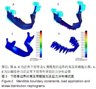
2.1 Case 1正常完整下颌骨受力有限元仿真分析 经过有限元计算得出,正常完整下颌骨及牙齿在正常咀嚼下其结构Von Mises应力分布情况,见图2a-d。 "

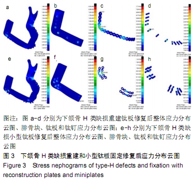
2.2 Case 2下颌骨H类缺损重建钛板修复受力有限元仿真分析 经过有限元计算得出,下颌骨H类缺损重建钛板修复下其结构Von Mises应力分布、结构位移变化等情况,见图3a-d。 2.3 Case 3下颌骨H类缺损小型钛板修复受力有限元仿真分析 经过有限元计算得出,下颌骨H类缺损小型钛板修复下其结构Von Mises应力分布变化情况,见图3e-h。 "
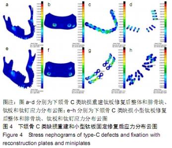
2.4 Case 4下颌骨C类缺损重建钛板修复受力有限元仿真分析 经过有限元计算得出,下颌骨C类缺损重建钛板修复下其结构Von Mises应力分布情况,见图4a-d。 2.5 Case 5下颌骨C类缺损重小型板修复受力有限元仿真分析 经过有限元计算得出,下颌骨C类缺损小型钛板修复下其结构Von Mises应力分布情况,见图4e-h。 "


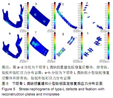
2.6 Case 6下颌骨L类缺损重建钛板修复受力有限元仿真分析 经过有限元计算得出,下颌骨L类缺损重建钛板修复下其结构Von Mises应力分布情况,见图5a-d。 2.7 Case 7下颌骨L类缺损小型钛板修复受力有限元仿真分析 经过有限元计算得出,下颌骨L类缺损小型钛板修复下其结构Von Mises应力分布情况,见图5e-h。 "
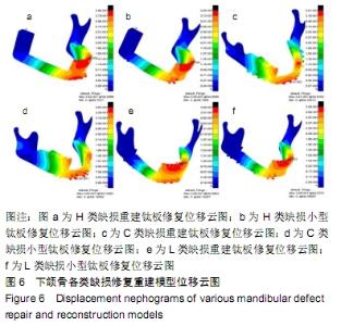
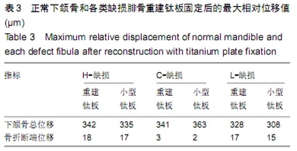
2.8 下颌骨各类缺损重建钛板和小型钛板修复后位移情况有限元仿真分析 经过有限元模拟各种缺损类型并用重建钛板和小型钛板固定腓骨瓣后再施加咬合力后的整体位移和腓骨块边缘骨断端之间的相对位移情况,见图6。 "

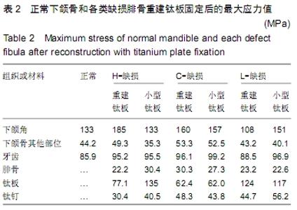
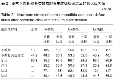
下颌骨各类缺损修复计算数据对比分析:将正常、H、C、L类缺损2种固定修复的各计算数据,汇总于表2,3中。为了更加清晰、直观对比H,C,L缺损修复前后应力及位移的变化情况,特将计算数据以柱状图形式来表示,见图7。 "
"

"
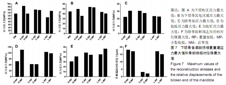

由上述图表可知,通过有限元方法对正常下颌骨、H、C、L类缺损小型钛板与重建钛板修复仿真模拟,从应力云图可以大致判断,应力较大值主要发生在髁突、髁颈、下颌角、磨牙、钛板钛钉等周围区域处,尤其以下颌角附近处Von Mises应力最大,可能跟咬肌等软组织约束有关。通过对比几组修复情况的应力最值可以发现,H类缺损重建钛板修复下颌骨时将对下颌角附近产生较大应力,局部应力集中达185 MPa,相比正常下颌骨,其应力增幅约39.10%。下颌骨非应力集中区域的应力最值为35.3-53.3 MPa,H类缺损小型钛板修复下的下颌骨非应力集中区应力比正常下颌骨受力要低,降幅约在20.14%,L类缺损重建钛板修复后非应力集中区的应力值降幅为2.26%-18.8%。H,C,L类6组下颌骨钛板修复下腓骨块、钛钉、钛板的受力也不尽相同,其中腓骨块的Von Mises应力最值在30.4 MPa以下。文章结果显示不管是重建钛板还是小型钛板固定,靠近断端的第一枚螺钉的应力值均较其他螺钉的大,应力最大值为56.2 MPa,发生在L类下颌骨缺损小型钛板修复下的远中上端第一枚螺钉。在H类小型钛板、L类小型钛板及重建钛板修复下钛板的Von Mises应力均大于100 MPa,为 117 -135 MPa。对于下颌骨最大位移来说,几组缺损修复下颌骨整体变形最值相差不大,为308-363 μm;H及L类缺损小型钛板、重建钛板修复后骨断端相对位移变化很小,为15-18 μm;相比H类和L类修复,C类缺损下颌骨钛板修复后骨断端相对位移最小,基本不产生相对位移。 "

| [1] ZHANG Q, WU W, QIAN C, et al. Advanced biomaterials for repairing and reconstruction of mandibular defects. Mater Sci Eng C Mater Biol Appl. 2019. doi: 10.1016/j.msec.2019.109858. [2] HIDALGO DA. Fibula free flap: a new method of mandible reconstruction. Plastic Reconstruct Surg. 1989;84(1):71-79. [3] OKAY D, SHETAWI AHA, MOUBAYED SP, et al. Worldwide 10-year systematic review of treatment trends in fibula free flap for mandibular reconstruction. J Oral Maxillofac Surg. 2016;74(12):2526-2531. [4] 任文豪,高岭,李少明,等.虚拟手术计划及3D打印导板辅助在游离腓骨瓣精准重建下颌骨中的价值分析[J].中华医学杂志,2018, 98(33):2666-2670. [5] WITJES MJH, SCHEPERS RH, KRAEIMA J. Impact of 3D virtual planning on reconstruction of mandibular and maxillary surgical defects in head and neck oncology. Curr Opin Otolaryngol Head Neck Surg. 2018;26(2):108-114. [6] AGGARWAL S, SINGH M, MODI P, et al. Comparison of 3D plate and locking plate in treatment of mandibular fracture-a clinical study. Oral Maxillofac Surg. 2017;21(4): 383-390. [7] KOPP RW, CROZIER DL, GOYAL P, et al. Decade review of mandible fractures and arch bar impact on outcomes of nonsubcondylar fractures. Laryngoscope.2016;126(3): 596-601. [8] PEDRAZZOLI M, AUTELITANO L, BIGLIOLI F. Prevention of bisphosphonate-related mandibular fractures. Acta Otorhinolaryngol Ital. 2016;36(4):317-320. [9] JEWER DD, BOYD JB, MANKTELOW RT, et al. Orofacial and mandibular reconstruction with the iliac crest free flap: a review of 60 cases and a new method of classification. Plast Reconstr Surg. 1989;84(3):391-405. [10] STYRANIVSKA O, KLIUCHKOVSKA N, MYKYYEVYCH N. Comparison of using different bridge prosthetic designs for partial defect restoration through mathematical modeling. Eur J Dent. 2017;11(3):345-351. [11] OKUDA S, EIRAKU M. Role of molecular turnover in dynamic deformation of a three-dimensional cellular membrane. Biomechan Model Mechanobiol. 2017;16(9):1805-1818. [12] 杨宇,汤文成.纳米压痕法测量牙周膜不同层面弹性模量(英文)[J].Journal of Southeast University (English Edition), 2017,33(1):33-38. [13] RUDDY BH, KURUPPUMULLAGE DN, CARNABY G, et al. Computational modelling of cough function and airway penetrant behavior in patients with disorders of laryngeal function. Laryngoscope Invest Otolaryngol. 2017;2(1):23-29. [14] KUMAR D, SIVARAM G, SHIVAKUMAR B, et al. Comparative evaluation of soft and hard tissue changes following endosseous implant placement using flap and flapless techniques in the posterior edentulous areas of the mandible-a randomized controlled trial. Oral Maxillofac Surg. 2018;22(2):215-223. [15] MURAKAMI K, YAMAMOTO K, SUGIURA T, et al. Computed tomography-based 3-dimensional finite element analyses of various types of plates placed for a virtually reduced unilateral condylar fracture of the mandible of a patient. J Oral Maxillofac Surg. 2017;75(6):1239.e1-e11. [16] KANAZAWA M, TANOUE M, MIYAYASU A, et al. The patient general satisfaction of mandibular single-implant overdentures and conventional complete dentures: study protocol for a randomized crossover trial. Medicine. 2018;97(20):e10721. [17] MAURER P, ECKERT AW, KRIWALSKY MS, et al. Scope and limitations of methods of mandibular reconstruction: a long-term follow-up. Br J Oral Maxillofac Surg. 2010;48(2): 100-104. [18] DISA JJ, CORDEIRO PG. Mandible reconstruction with microvascular surgery. Semin Surg Oncol. 2000;19(3): 226-234. [19] SHAYESTEH MOGHADDAM N, JAHADAKBAR A, AMERINATANZI A, et al. Metallic fixation of mandibular segmental defects: graft immobilization and orofacial functional maintenance. Plastic Reconstr Surg. 2016;4(9): e858. [20] 毛广文,辛俊彤,刘延山,等.双层腓骨移植联合牙种植重建下颌咬合功能[J].现代口腔医学杂志,2016,32(4):221-224. [21] ZOUMALAN RA, HIRSCH DL, LEVINE JP, et al. Plating in microvascular reconstruction of the mandible: can fixation be too rigid. J Craniofac Surg. 2009;20(5):1451-1454. [22] STRACKEE SD, KROON FH, BOS KE. Fixation methods in mandibular reconstruction using fibula grafts: acomparative study into the relative strength of three different types of osteosynthesis. Head Neck. 2001;23(1):1-7. [23] LIU SP, CAI ZG, ZHANG J, et al. Stability and complications of miniplates for mandibular reconstruction with a fibular graft: Outcomes for 544 patients. Br J Oral Maxillofac Surg. 2015; 54(5):496-500. [24] LI P, SHEN L, LI J, et al. Optimal design of an individual endoprosthesis for the reconstruction of extensive mandibular defects with finite element analysis. J Cranio-Maxillofac Surg. 2014;42(1):73-78. [25] ALBOGHA MH, MORI Y, TAKAHASHI I. Three-dimensional titanium miniplates for fixation of subcondylar mandibular fractures: comparison of five designs using patient-specific finite element analysis. J Craniomaxillofac Surg. 2018;46(3): 391-397. [26] STRACKEE SD, KROON FH, BOS KE. Fixation methods in mandibular reconstruction using fibula grafts: a comparative study into the relative strength of three different types of osteosynthesis. Head Neck. 2001;23(1):1-7. [27] ZHANG ZL, WANG S, SUN CF, et al. Miniplates versus reconstruction plates in vascularized osteocutaneous flap reconstruction of the mandible. J Craniofac Surg. 2019;30(2): e119-e125. [28] 陈旭兵,柳兆刚,袁建兵,等.三维模拟技术在游离腓骨瓣移植重建下颌骨缺损中的应用[J].上海口腔医学,2015,24(4):460-464. [29] KOKOSIS G, SCHMITZ R, POWERS DB, et al. Mandibular reconstruction using the free vascularized fibula graft: an overview of different modifications. Arch Plastic Surg. 2016; 43(1):3-9. [30] 孙伟桐,陈辰,蒋协远,等.合成骨在骨科生物力学研究中的应用[J].国际骨科学杂志,2018,39(2):72-75. |
| [1] | Xu Feng, Kang Hui, Wei Tanjun, Xi Jintao. Biomechanical analysis of different fixation methods of pedicle screws for thoracolumbar fracture [J]. Chinese Journal of Tissue Engineering Research, 2021, 25(9): 1313-1317. |
| [2] | Jiang Yong, Luo Yi, Ding Yongli, Zhou Yong, Min Li, Tang Fan, Zhang Wenli, Duan Hong, Tu Chongqi. Von Mises stress on the influence of pelvic stability by precise sacral resection and clinical validation [J]. Chinese Journal of Tissue Engineering Research, 2021, 25(9): 1318-1323. |
| [3] | Chen Zehua, Ye Xiangling, Chen Weijian, Du Jianping, Liu Wengang, Xu Xuemeng. Effect of pronated foot posture on proprioception and postural stability based on foot posture index [J]. Chinese Journal of Tissue Engineering Research, 2021, 25(9): 1324-1328. |
| [4] | Chen Xinmin, Li Wenbiao, Xiong Kaikai, Xiong Xiaoyan, Zheng Liqin, Li Musheng, Zheng Yongze, Lin Ziling. Type A3.3 femoral intertrochanteric fracture with augmented proximal femoral nail anti-rotation in the elderly: finite element analysis of the optimal amount of bone cement [J]. Chinese Journal of Tissue Engineering Research, 2021, 25(9): 1404-1409. |
| [5] | Zhou Jihui, Li Xinzhi, Zhou You, Huang Wei, Chen Wenyao. Multiple problems in the selection of implants for patellar fracture [J]. Chinese Journal of Tissue Engineering Research, 2021, 25(9): 1440-1445. |
| [6] | Pei Lili, Sun Guicai, Wang Di. Salvianolic acid B inhibits oxidative damage of bone marrow mesenchymal stem cells and promotes differentiation into cardiomyocytes [J]. Chinese Journal of Tissue Engineering Research, 2021, 25(7): 1032-1036. |
| [7] | Yang Weiqiang, Ding Tong, Yang Weike, Jiang Zhengang. Combined variable stress plate internal fixation affects changes of bone histiocyte function and bone mineral density at the fractured end of goat femur [J]. Chinese Journal of Tissue Engineering Research, 2021, 25(6): 890-894. |
| [8] | Song Chengjie, Chang Hengrui, Shi Mingxin, Meng Xianzhong. Research progress in biomechanical stability of lateral lumbar interbody fusion [J]. Chinese Journal of Tissue Engineering Research, 2021, 25(6): 923-928. |
| [9] | Liu Zhao, Xu Xilin, Shen Yiwei, Zhang Xiaofeng, Lü Hang, Zhao Jun, Wang Zhengchun, Liu Xuzhuo, Wang Haitao. Guiding role and prospect of staging and classification combined collapse prediction method for osteonecrosis of femoral head [J]. Chinese Journal of Tissue Engineering Research, 2021, 25(6): 929-934. |
| [10] | Xu Yulin, Shen Shi, Zhuo Naiqiang, Yang Huilin, Yang Chao, Li Yang, Zhao Heng, Zhao Lu. Biomechanical comparison of three different plate fixation methods for acetabular posterior column fractures in standing and sitting positions [J]. Chinese Journal of Tissue Engineering Research, 2021, 25(6): 826-830. |
| [11] | Cai Qunbin, Zou Xia, Hu Jiantao, Chen Xinmin, Zheng Liqin, Huang Peizhen, Lin Ziling, Jiang Ziwei. Relationship between tip-apex distance and stability of intertrochanteric femoral fractures with proximal femoral anti-rotation nail: a finite element analysis [J]. Chinese Journal of Tissue Engineering Research, 2021, 25(6): 831-836. |
| [12] | Wang Jiangna, Zheng Huifen, Sun Wei. Changes in dynamic stability, motor coordination and joint mechanics of the lower extremity during stair descent and performing phone task [J]. Chinese Journal of Tissue Engineering Research, 2021, 25(6): 837-843. |
| [13] | Xie Chongxin, Zhang Lei. Comparison of knee degeneration after anterior cruciate ligament reconstruction with or without remnant preservation [J]. Chinese Journal of Tissue Engineering Research, 2021, 25(5): 735-740. |
| [14] | Liu Bo, Chen Xianghe, Yang Kang, Yu Huilin, Lu Pengcheng. Mechanism of DNA methylation in exercise intervention for osteoporosis [J]. Chinese Journal of Tissue Engineering Research, 2021, 25(5): 791-797. |
| [15] | Zhang Guomei, Zhu Jun, Hu Yang, Jiao Hongwei. Stress of three-dimensional finite element models of E-MAX porcelain inlay [J]. Chinese Journal of Tissue Engineering Research, 2021, 25(4): 537-541. |
| Viewed | ||||||
|
Full text |
|
|||||
|
Abstract |
|
|||||