[1] LU T, LU Y. Comparison of biomechanical performance among posterolateral fusion and transforaminal, extreme, and oblique lumbar interbody fusion: a finite element analysis. World Neurosurg. 2019; 129:e890-e899.
[2] LUCA A, OTTARDI C, LOVI A, et al. Anterior support reduces the stresses on the posterior instrumentation after pedicle subtraction osteotomy: a finite-element study. Eur Spine J. 2017;26:450-456.
[3] 林伟文,赖茂松,熊浩,等.单侧与双侧椎弓根螺钉联合微创经椎间孔椎体间融合术治疗腰椎退行性疾病的临床疗效和影像学比较[J]. 中国实用医药, 2019,14(20):62-65.
[4] WU JC, HUANG WC, TSAI HW, et al. Pedicle screw loosening in dynamic stabilization: incidence, risk, and outcome in 126 patients. Neurosurg Focus. 2011;31:E9.
[5] GALBUSERA F, VOLKHEIMER D, REITMAIER S, et al. Pedicle screw loosening: a clinically relevant complication?. Eur Spine J. 2015;24: 1005-1016.
[6] HOPPE S, KEEL MJ. Pedicle screw augmentation in osteoporotic spine: indications, limitations and technical aspects. Eur J Trauma Emerg Surg. 2017;43:3-8.
[7] 姚珍松,唐永超,陈康,等.骨水泥螺钉强化固定伴骨质疏松腰椎滑脱症的稳定性及椎间融合[J].中国组织工程研究, 2016,20(4):517-521.
[8] 张大鹏,强晓军,王振江,等.骨水泥强化椎弓根螺钉在治疗强直性脊柱炎胸腰椎应力骨折的临床应用[J].中国矫形外科杂志,2016, 24(10):946-949.
[9] QIAN L, JIANG C, SUN P, et al. A comparison of the biomechanical stability of pedicle-lengthening screws and traditional pedicle screws: an in vitro instant and fatigue-resistant pull-out test. Bone Joint J. 2018;100-B:516-521.
[10] MARTÍN-FERNÁNDEZ M, LÓPEZ-HERRADÓN A, PIÑERA AR, et al. Potential risks of using cement-augmented screws for spinal fusion in patients with low bone quality. Spine J. 2017;17:1192-1199.
[11] MUELLER JU, BALDAUF J, MARX S, et al. Cement leakage in pedicle screw augmentation: a prospective analysis of 98 patients and 474 augmented pedicle screws. J Neurosurg Spine. 2016;25:103-109.
[12] YUAN Q, ZHANG G, WU J, et al. Clinical evaluation of the polymethylmethacrylate-augmented thoracic and lumbar pedicle screw fixation guided by the three-dimensional navigation for the osteoporosis patients. Eur Spine J.2015;24:1043-1050.
[13] HATZANTONIS C, CZYZ M, PYZIK R, et al. Intracardiac bone cement embolism as a complication of vertebroplasty: management strategy. Eur Spine J. 2017;26:3199-3205.
[14] WEISER L, HUBER G, SELLENSCHLOH K, et al. Time to augment?! Impact of cement augmentation on pedicle screw fixation strength depending on bone mineral density. Eur Spine J.2018;27:1964-1971.
[15] SCHMID SL, BACHMANN E, FISCHER M, et al. Pedicle screw augmentation with bone cement enforced Vicryl mesh. J Orthop Res. 2018;36:212-216.
[16] GUO HZ, TANG YC, LI YX, et al. The effect and safety of polymethylmethacrylate-augmented sacral pedicle screws applied in osteoporotic spine with lumbosacral degenerative disease: a 2-year follow-up of 25 patients. World Neurosurg. 2019;121:e404-e410.
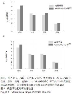
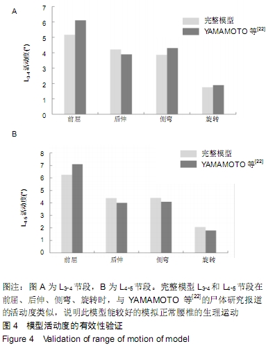
[17] 闫家智,吴志宏,汪学松,等.腰椎三维有限元模型建立和应力分析[J].中华医学杂志, 2009, 89(17):1162-1165.
[18] SHIM CS, PARK SW, LEE SH, et al. Biomechanical evaluation of an interspinous stabilizing device, Locker. Spine (Phila Pa 1976). 2008;33: E820-827.
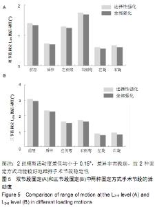
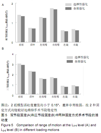
[19] 费琦,赵凡,杨雍,等.腰椎后路融合手术对失稳模型节段稳定性及相邻节段力学的影响[J].中华医学杂志, 2015,95(45):3681-3686.
[20] XU H, JU W, XU N, et al. Biomechanical comparison of transforaminal lumbar interbody fusion with 1 or 2 cages by finite-element analysis. Neurosurgery. 2013;73:ons198-205; discussion ons205.
[21] WANG T, ZHAO Y, CAI Z, et al. Effect of osteoporosis on internal fixation after spinal osteotomy: A finite element analysis. Clin Biomech (Bristol, Avon). 2019;69:178-183.
[22] YAMAMOTO I, PANJABI MM, CRISCO T, et al. Three-dimensional movements of the whole lumbar spine and lumbosacral joint. Spine (Phila Pa 1976). 1989;14:1256-1260.
[23] 《中国老年骨质疏松症诊疗指南》(2018)工作组, 中国老年学和老年医学学会骨质疏松分会,马远征,等. 中国老年骨质疏松症诊疗指南(2018) [J].中国骨质疏松杂志, 2018,24(12):1541-1567.
[24] WEISER L, DREIMANN M, HUBER G, et al. Cement augmentation versus extended dorsal instrumentation in the treatment of osteoporotic vertebral fractures: a biomechanical comparison. Bone Joint J. 2016;98-B:1099-1105.
[25] GUO HZ, TANG YC, GUO DQ, et al. The cement leakage in cement-augmented pedicle screw instrumentation in degenerative lumbosacral diseases: a retrospective analysis of 202 cases and 950 augmented pedicle screws. Eur Spine J. 2019;28:1661-1669.
[26] 唐永超,梁德,陈博来,等.骨水泥钉道强化与否治疗伴骨质疏松的单节段腰椎退行性疾病的临床对照研究[J]. 中国脊柱脊髓杂志, 2017,27(12): 1092-1098.
[27] LI T, SHI L, LUO Y, et al. One-Level or multilevel interbody fusion for multilevel lumbar degenerative diseases: a prospective randomized control study with a 4-year follow-up. World Neurosurg. 2018;110: e815-e822.
|