Chinese Journal of Tissue Engineering Research ›› 2020, Vol. 24 ›› Issue (11): 1671-1676.doi: 10.3969/j.issn.2095-4344.2535
Previous Articles Next Articles
3-Methyladenine improves the efficiency of sciatic nerve allograft in mice
Xu Zhuqiu, Lu Haibin, Feng Weifeng, Yang Xiaonan, Qi Zuoliang
- Plastic Surgery Hospital, Chinese Academy of Medical Science & Peking Union Medical College, Beijing 100041, China
-
Received:2019-05-21Revised:2019-05-28Accepted:2019-07-10Online:2020-04-18Published:2020-02-21 -
Contact:Yang Xiaonan, Associate chief physician, Associate professor, Plastic Surgery Hospital, Chinese Academy of Medical Science & Peking Union Medical College, Beijing 100041, China Qi Zuoliang, Chief physician, Professor, Plastic Surgery Hospital, Chinese Academy of Medical Science & Peking Union Medical College, Beijing 100041, China -
About author:Xu Zhuqiu, MD candidate, Plastic Surgery Hospital, Chinese Academy of Medical Science & Peking Union Medical College, Beijing 100041, China -
Supported by:the National Natural Science Foundation of China, No. 81571921 and 81671908; the Innovation Fund of Graduate Students, CAMS, PUMC, No. 2017-1002-1-10
CLC Number:
Cite this article
Xu Zhuqiu, Lu Haibin, Feng Weifeng, Yang Xiaonan, Qi Zuoliang. 3-Methyladenine improves the efficiency of sciatic nerve allograft in mice[J]. Chinese Journal of Tissue Engineering Research, 2020, 24(11): 1671-1676.
share this article
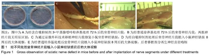
2.1 自噬抑制剂处理后的标本及手术大体观察 经过72 h 3-甲基腺嘌呤处理后的神经片段在大体观察下和普通培养基处理组没有明显区别,见图1A,B;将3 mm处理后的神经段移植入损伤动物模型(图1C)饲养8周后,大体观察对照组移植部分再生神经直径略细,其余无明显差异,见图1D,E。 "

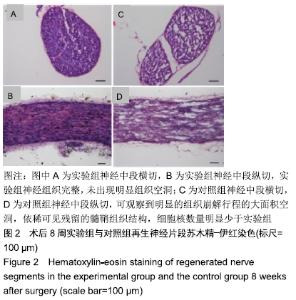
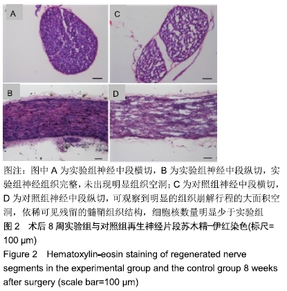
2.2 组织学观察 2.2.1 苏木精-伊红染色结果 将术后8周再生神经片段中段横切面及纵切面冰冻切片进行苏木精-伊红染色,横切面中可观察到实验组神经组织完整,未出现明显组织空洞,见图2A,纵切面中可观察到完整髓鞘结构及大量细胞核,见图2B;对照组可观察到明显的组织崩解行程的大面积空洞,依稀可见残留的髓鞘组织结构,细胞核数量明显少于实验组,见图2C,D。 "
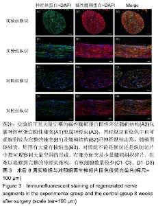
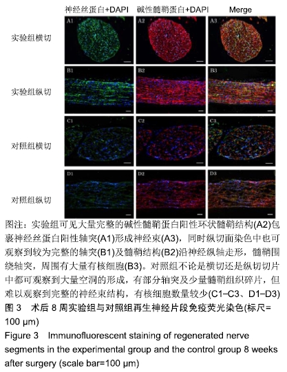
2.2.2 免疫荧光染色结果 将术后8周神经中段切面及纵切面冰冻切片进行免疫荧光染色,神经丝蛋白标记轴突,碱性髓鞘蛋白标记髓鞘。 实验组可观察到大量完整的碱性髓鞘蛋白阳性环状髓鞘结构(图3A2)包裹神经丝蛋白阳性轴突(图3A1)形成神经束(图3A3),同时在纵切面染色中也可观察到较为完整的轴突(图3B1)及髓鞘结构(图3B2)沿神经纵轴走形,髓鞘围绕轴突,周围有大量有核细胞(图3B3)。对照组不论是横切还是纵切切片中都可观察到大量空洞的形成,有部分轴突及少量髓鞘组织碎片,但难以观察到完整的神经束结构,有核细胞数量较少,见图3C1-C3、D1-D3。 "
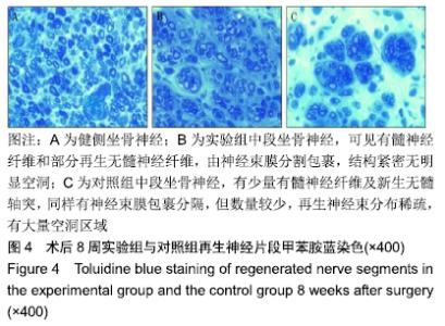
2.2.3 甲苯胺蓝染色结果 取术后8周的术侧及健侧神经组织,截取神经中段做半薄切片进行甲苯胺蓝染色。健侧结果可见均匀分布的有髓神经纤维,髓鞘厚度及轴突直径也较为均一,未见明显神经束膜,见图4A。实验组可见有髓神经纤维和部分再生无髓神经纤维,由神经束膜分割包裹,结构紧密无明显空洞,见图4B。 对照组有少量有髓神经纤维及新生无髓轴突,同样有神经束膜包裹分隔,但数量较少,再生神经束分布稀疏,有大量空洞区域,见图4C。 "
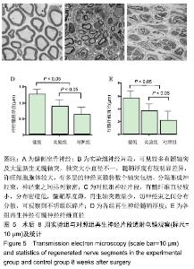
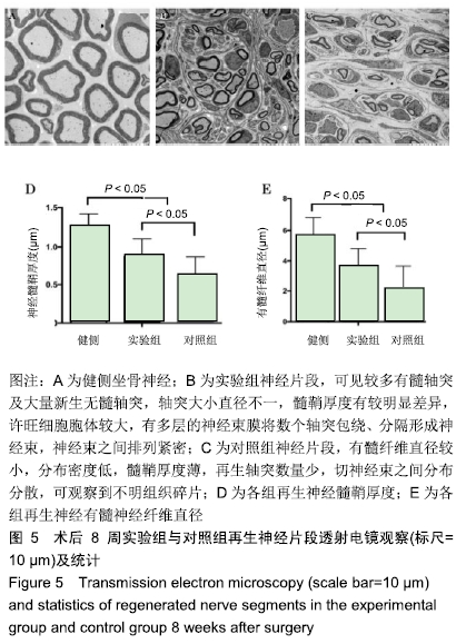
2.2.4 透射电镜观察 将术后8周双侧神经组织做超薄切片,柠檬酸铅染色后透射电镜下观察。健侧神经组织有髓神经纤维直径大,髓鞘较厚,分布均匀,许旺细胞胞体较小,未见神经束膜结构,见图5A。 实验组可见较多有髓轴突及大量新生无髓轴突,轴突大小直径不一,髓鞘厚度有较明显差异,许旺细胞胞体较大,有多层的神经束膜将数个轴突包绕、分隔形成神经束,神经束之间排列紧密,见图5B。对照组有髓纤维直径较小,分布密度低,髓鞘厚度薄,再生轴突数量少,切神经束之间分布分散,可观察到不明组织碎片,见图5C。 应用ImageJ软件对电镜下的髓鞘厚度、有髓神经纤维直径进行测量,见图5D,E,3组髓鞘厚度分别为(1.267± 0.151),(0.877±0.231),(0.628±0.242) μm,健侧髓鞘厚于实验组、对照组,差异有显著性意义(P < 0.05),实验组厚于对照组(P < 0.05)。3组有髓神经纤维直径分别为(5.590±1.170),(3.580±1.233),(2.112±1.546) μm,健侧直径大于实验组、对照组,差异有显著性意义(P < 0.05),实验组直径大于对照组(P < 0.05)。 "
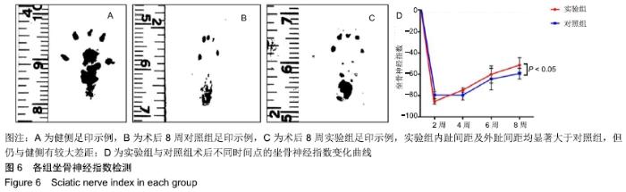
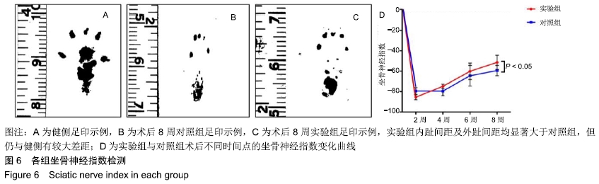
2.3 功能学检测 通过术后2,4,6,8周小鼠的足印测量计算出小鼠的坐骨神经指数变化,评估其功能恢复情况,见图6。 图6A-C分别为术后8周时健侧、对照组及实验组的足印示例,可见实验组内趾间距及外趾间距均显著大于对照组,但仍与健侧有较大差距。图6D为两组小鼠坐骨神经指数变化曲线,均随时间延长逐渐增高,前6周两组之间并无差异,第8周时实验组坐骨神经指数高于对照组(-48.51± 4.36,-60.84±4.40,P < 0.05)。 "
| [1] RIVLIN M, SHEIKH E, ISAAC R, et al.The role of nerve allografts and conduits for nerve injuries. Hand Clin.2010; 26(3):435-446. [2] RAY WZ, MACKINNON SE.Management of nerve gaps: autografts, allografts, nerve transfers, and end-to-side neurorrhaphy.Exp Neurol.2010;223(1):77-85. [3] GRINSELL D, KEATING CP. Peripheral nerve reconstruction after injury: a review of clinical and experimental therapies. Biomed Res Int.2014;2014:698256. [4] CASTILLO-GALVAN ML, MARTINEZ-RUIZ FM, DE LA GARZA-CASTRO O, et al.[Study of peripheral nerve injury in trauma patients].Gac Med Mex.2014;150(6):527-532. [5] SCHMID AB, COPPIETERS MW, RUITENBERG MJ, et al. Local and remote immune-mediated inflammation after mild peripheral nerve compression in rats.J Neuropathol Exp Neurol.2013;72(7):662-680. [6] CONFORTI L, GILLEY J, COLEMAN MP. Wallerian degeneration: an emerging axon death pathway linking injury and disease.Nat Rev Neurosci.2014;15(6):394-409. [7] GOMEZ-SANCHEZ JA, CARTY L, IRUARRIZAGA-LEJARRETA M, et al.Schwann cell autophagy, myelinophagy, initiates myelin clearance from injured nerves.J Cell Biol.2015;210(1):153-168. [8] HUANG HC, CHEN L, ZHANG HX, et al.Autophagy Promotes Peripheral Nerve Regeneration and Motor Recovery Following Sciatic Nerve Crush Injury in Rats.J Mol Neurosci. 2016;58(4):416-423. [9] CAI Y, ARIKKATH J, YANG L, et al.Interplay of endoplasmic reticulum stress and autophagy in neurodegenerative disorders.Autophagy.2016;12(2):225-244. [10] 徐筑秋,杨晓楠,祁佐良.细胞自噬在周围神经损伤及再生中研究进展[J].中国修复重建外科杂志, 2017,31(1):122-125. [11] INSERRA MM, BLOCH DA, TERRIS DJ.Functional indices for sciatic, peroneal, and posterior tibial nerve lesions in the mouse.Microsurgery.1998;18(2):119-124. [12] GAUDET AD, POPOVICH PG, RAMER MS.Wallerian degeneration: gaining perspective on inflammatory events after peripheral nerve injury.J Neuroinflammation.2011;8:110. [13] LIU JH, TANG Q, LIU XX, et al.Analysis of transcriptome sequencing of sciatic nerves in Sprague-Dawley rats of different ages.Neural Regen Res. 2018;13(12):2182-2190. [14] CASTANO A, BELL MD, PERRY VH.Unusual aspects of inflammation in the nervous system: Wallerian degeneration. Neurobiol Aging.1996;17(5):745-751. [15] RUSSELL RC, YUAN HX, GUAN KL.Autophagy regulation by nutrient signaling.Cell Res.2014;24(1):42-57. [16] MIZUSHIMA N, YOSHIMORI T, OHSUMI Y.The role of Atg proteins in autophagosome formation.Annu Rev Cell Dev Biol.2011;27:107-132. [17] XIAO X, ZHU Y, BU J, et al.The autophagy inhibitor 3-methyladenine restores sevoflurane anesthesiainduced cognitive dysfunction and neurons apoptosis. Pharmazie. 2017;72(4):214-218. [18] BUTTEMEYER R, RAO U, JONES NF. Peripheral nerve allograft transplantation with FK506: functional, histological, and immunological results before and after discontinuation of immunosuppression.Ann Plast Surg.1995;35(4):396-401. [19] ZALEWSKI AAGULATI AK. Evaluation of histocompatibility as a factor in the repair of nerve with a frozen nerve allograft.J Neurosurg.1982;56(4):550-554. [20] PATEL NP, LYON KA, HUANG JH.An update-tissue engineered nerve grafts for the repair of peripheral nerve injuries.Neural Regen Res. 2018;13(5):764-774. [21] KARANTH S, YANG G, YEH J, et al.Nature of signals that initiate the immune response during Wallerian degeneration of peripheral nerves.Exp Neurol.2006;202(1):161-166. [22] DEFRANCESCO-LISOWITZ A, LINDBORG JA, NIEMI JP, et al.The neuroimmunology of degeneration and regeneration in the peripheral nervous system.Neuroscience. 2015;302: 174-203. [23] RAIMONDO S, FORNARO M, TOS P, et al.Perspectives in regeneration and tissue engineering of peripheral nerves.Ann Anat.2011;193(4):334-340. [24] MASAKI T, MATSUMURA K, SAITO F, et al.Expression of dystroglycan and laminin-2 in peripheral nerve under axonal degeneration and regeneration.Acta Neuropathol. 2000;99(3): 289-295. [25] CHEN P, PIAO X, BONALDO P. Role of macrophages in Wallerian degeneration and axonal regeneration after peripheral nerve injury.Acta Neuropathol. 2015;130(5): 605-618. [26] PULESTON DJ, SIMON AK. Autophagy in the immune system. Immunology.2014;141(1): 1-8. [27] CHOI SH, GONEN A, DIEHL CJ, et al.SYK regulates macrophage MHC-II expression via activation of autophagy in response to oxidized LDL.Autophagy.2015;11(5):785-795. [28] OKANO M, SAKATA N, UEDA S, et al.Appropriate modulation of autophagy sensitizes malignant peripheral nerve sheath tumor cells to treatment with imatinib mesylate. J Pediatr Hematol Oncol. 2014;36(3):200-211. [29] 范洪伟,殷钢,马玉林,.枸杞多糖对大鼠坐骨神经离断后创伤性神经瘤形成及其疼痛影响[J].中国修复重建外科杂志, 2010,24(1): 1298-1301. [30] MARINELLI S, NAZIO F, TINARI A, et al.Schwann cell autophagy counteracts the onset and chronification of neuropathic pain.Pain.2014;155(1):93-107. |
| [1] | Zhang Tongtong, Wang Zhonghua, Wen Jie, Song Yuxin, Liu Lin. Application of three-dimensional printing model in surgical resection and reconstruction of cervical tumor [J]. Chinese Journal of Tissue Engineering Research, 2021, 25(9): 1335-1339. |
| [2] | Zeng Yanhua, Hao Yanlei. In vitro culture and purification of Schwann cells: a systematic review [J]. Chinese Journal of Tissue Engineering Research, 2021, 25(7): 1135-1141. |
| [3] | Ma Zetao, Zeng Hui, Wang Deli, Weng Jian, Feng Song. MicroRNA-138-5p regulates chondrocyte proliferation and autophagy [J]. Chinese Journal of Tissue Engineering Research, 2021, 25(5): 674-678. |
| [4] | Xie Yang, Zhang Shujiang, Liu Menglan, Luo Ying, Yang Yang, Li Zuoxiao. Mechanism by which rapamycin protects spinal cord neurons in experimental autoimmune encephalomyelitis mice [J]. Chinese Journal of Tissue Engineering Research, 2021, 25(5): 695-700. |
| [5] | Xu Dongzi, Zhang Ting, Ouyang Zhaolian. The global competitive situation of cardiac tissue engineering based on patent analysis [J]. Chinese Journal of Tissue Engineering Research, 2021, 25(5): 807-812. |
| [6] | Wu Zijian, Hu Zhaoduan, Xie Youqiong, Wang Feng, Li Jia, Li Bocun, Cai Guowei, Peng Rui. Three-dimensional printing technology and bone tissue engineering research: literature metrology and visual analysis of research hotspots [J]. Chinese Journal of Tissue Engineering Research, 2021, 25(4): 564-569. |
| [7] | Chang Wenliao, Zhao Jie, Sun Xiaoliang, Wang Kun, Wu Guofeng, Zhou Jian, Li Shuxiang, Sun Han. Material selection, theoretical design and biomimetic function of artificial periosteum [J]. Chinese Journal of Tissue Engineering Research, 2021, 25(4): 600-606. |
| [8] | Liu Fei, Cui Yutao, Liu He. Advantages and problems of local antibiotic delivery system in the treatment of osteomyelitis [J]. Chinese Journal of Tissue Engineering Research, 2021, 25(4): 614-620. |
| [9] | Li Xiaozhuang, Duan Hao, Wang Weizhou, Tang Zhihong, Wang Yanghao, He Fei. Application of bone tissue engineering materials in the treatment of bone defect diseases in vivo [J]. Chinese Journal of Tissue Engineering Research, 2021, 25(4): 626-631. |
| [10] | Zhang Zhenkun, Li Zhe, Li Ya, Wang Yingying, Wang Yaping, Zhou Xinkui, Ma Shanshan, Guan Fangxia. Application of alginate based hydrogels/dressings in wound healing: sustained, dynamic and sequential release [J]. Chinese Journal of Tissue Engineering Research, 2021, 25(4): 638-643. |
| [11] | Chen Jiana, Qiu Yanling, Nie Minhai, Liu Xuqian. Tissue engineering scaffolds in repairing oral and maxillofacial soft tissue defects [J]. Chinese Journal of Tissue Engineering Research, 2021, 25(4): 644-650. |
| [12] | Xing Hao, Zhang Yonghong, Wang Dong. Advantages and disadvantages of repairing large-segment bone defect [J]. Chinese Journal of Tissue Engineering Research, 2021, 25(3): 426-430. |
| [13] | Chen Siqi, Xian Debin, Xu Rongsheng, Qin Zhongjie, Zhang Lei, Xia Delin. Effects of bone marrow mesenchymal stem cells and human umbilical vein endothelial cells combined with hydroxyapatite-tricalcium phosphate scaffolds on early angiogenesis in skull defect repair in rats [J]. Chinese Journal of Tissue Engineering Research, 2021, 25(22): 3458-3465. |
| [14] | Wang Hao, Chen Mingxue, Li Junkang, Luo Xujiang, Peng Liqing, Li Huo, Huang Bo, Tian Guangzhao, Liu Shuyun, Sui Xiang, Huang Jingxiang, Guo Quanyi, Lu Xiaobo. Decellularized porcine skin matrix for tissue-engineered meniscus scaffold [J]. Chinese Journal of Tissue Engineering Research, 2021, 25(22): 3473-3478. |
| [15] | Mo Jianling, He Shaoru, Feng Bowen, Jian Minqiao, Zhang Xiaohui, Liu Caisheng, Liang Yijing, Liu Yumei, Chen Liang, Zhou Haiyu, Liu Yanhui. Forming prevascularized cell sheets and the expression of angiogenesis-related factors [J]. Chinese Journal of Tissue Engineering Research, 2021, 25(22): 3479-3486. |
| Viewed | ||||||
|
Full text |
|
|||||
|
Abstract |
|
|||||