Chinese Journal of Tissue Engineering Research ›› 2017, Vol. 21 ›› Issue (34): 5423-5429.doi: 10.3969/j.issn.2095-4344.2017.34.003
Previous Articles Next Articles
Feasibility of constructing a scaffold for osteochondral tissue engineering using poly(lactide-co-glycolide) alone
- 1Department of Orthopedics, First Affiliated Hospital of Nanchang University, Nanchang 330006, Jiangxi Province, China; 2Medical School of Nanchang University, Nanchang 330006, Jiangxi Province, China; 3Department of Polymer Science, Fudan University, Shanghai 200433, China
-
Received:2017-09-19Online:2017-12-08Published:2018-01-04 -
Contact:Yao Hao-qun, M.D., Associate professor, Department of Orthopedics, First Affiliated Hospital of Nanchang University, Nanchang 330006, Jiangxi Province, China -
About author:Duan Ping-guo, M.D., Attending physician, Department of Orthopedics, First Affiliated Hospital of Nanchang University, Nanchang 330006, Jiangxi Province, China -
Supported by:the National Natural Science Foundation of China, No. 81401790; the Natural Science Foundation for the Youth in Jiangxi Province, No. 20171ACB21057; the Natural Science Foundation of Jiangxi Province, No. 20151BAB205053, 20161BAB205235; the Science and Technology Project of Jiangxi Provincial Education Department, No. GJJ160028
CLC Number:
Cite this article
Duan Ping-guo, Guo Run-sheng, Yu Xing-yuan, Pan Zhen, Li Xiao-feng, Li Hu1, Liu Jun, Yao Hao-qun.
share this article
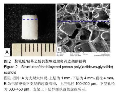
2.1 PLGA双层多孔支架的大体与微观结构 成功制备一体化PLGA双层多孔支架(图2A),扫描电镜观察其超微结构,可见支架内部孔壁相互连通,孔大小不一,形态良好,支架上下层界面相通,但整合紧密融为一体;经测算得出支架上层(软骨层)孔径100-200 μm,下层(软骨下骨层)孔径为300-450 μm,孔隙率均为85% (图2B)。 "

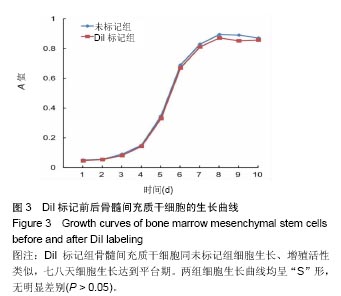
2.2 骨髓间充质干细胞的活性及DiI标识后状态 第3代骨髓间充质干细胞贴壁生长,倒置相差显微镜下观察,可见细胞呈鱼群样或漩涡状排列,具有明显方向性,形态为典型的梭形细胞;用DiI标记后细胞继续生长良好,与未经标记的细胞比较形态上无明显改变;倒置荧光显微镜下观察,可见大量细胞胞浆及细胞膜呈现红色荧光,对照组未见红色荧光。 DiI标记的髓间充质干细胞同未标记的细胞生长、增殖活性类似,接种贴壁二三天潜伏期后呈现对数生长, 七八天细胞生长达到平台期,增殖速度减慢。两组细胞生长曲线均呈“S”形,无明显差别(图3)。"
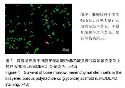
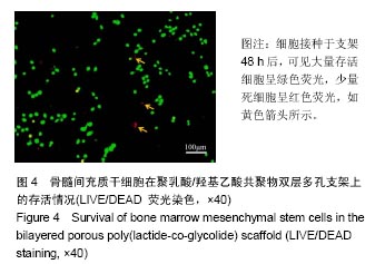
2.3 体外实验结果 2.3.1 细胞在支架内的活性 细胞接种于支架48 h后,通过LIVE/DEAD试剂盒染色后荧光显微镜下观察,在支架内部及表面发现大量发出绿色荧光的活细胞,少量发出红色荧光的死细胞(图4)。"
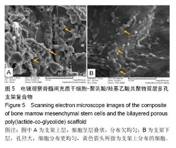
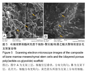
2.3.2 电镜观察细胞-支架复合物 通过扫描电镜观察细胞在支架内部的生长状态,结果发现细胞黏附在支架孔壁上,并可见大量沉积的细胞外基质;在支架上层100-200 μm小孔径的孔壁上,发现细胞迁移能力较差,细胞立体感强,伸展性差,在孔壁上分布不均匀,聚集层叠(图5A);而在下层300-450 μm大孔径的孔壁上,细胞迁移能力较强,细胞扁平,伸展性较好,细胞分布更均匀(图5B)。"
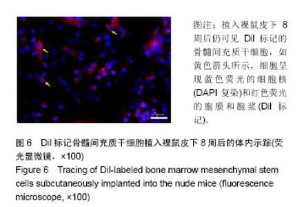
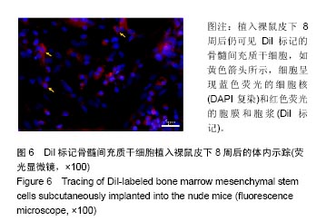
2.4 体内实验结果 2.4.1 骨髓间充质干细胞在裸鼠体内的示踪 双层支架负载细胞埋植入裸鼠皮下8周后取出,行冰冻切片并在荧光显微镜下观察,结果发现对照组可见大量呈蓝色荧光的细胞核,但未见红色荧光;而实验组也可见大量蓝色荧光的细胞核,此外还可见呈红色荧光的细胞膜和细胞浆,将同一视野下两种荧光的图片拼合,结果发现有些蓝色荧光的细胞核正好被红色荧光的细胞膜和细胞浆所包裹,提示这些细胞就是DiI标记的骨髓间充质干细胞(图6)。"
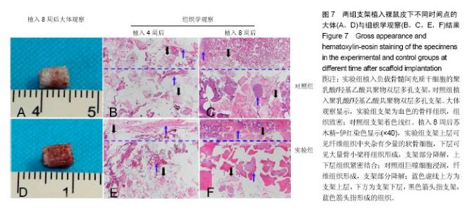
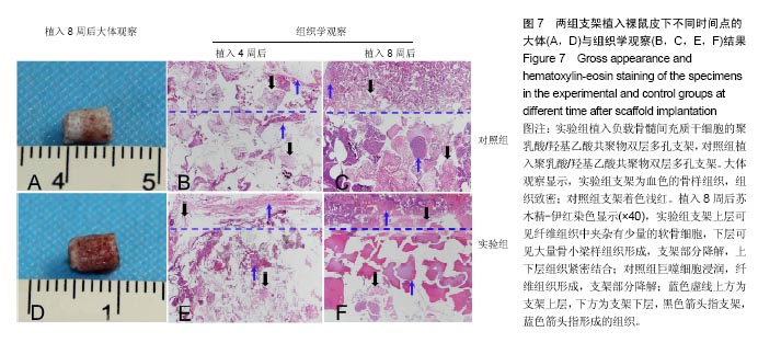
2.4.2 植入物的大体与组织学染色观察结果 裸鼠皮下埋植4,8周后,取出植入物,两组皮下未见明显炎症反应。 实验组:植入4周后,标本大体观未见明显改变,但淡红色中可见原支架白色;苏木精-伊红染色发现支架上层可见少量巨噬细胞浸润,下层可见支架中夹杂有纤维组织、少量的骨小梁样及血管样组织形成,支架上下层结构疏松。植入8周后,标本大体观呈现为血红色的骨样组织,组织致密,硬度增大;苏木精-伊红染色发现支架上层可见纤维组织中夹杂有少量的软骨细胞,下层可见大量骨小梁样组织形成,支架部分降解,上下层组织紧密结合,见图7。 对照组:植入4周后,标本大体观未见明显改变,保留着植入支架的原貌;苏木精-伊红染色显示支架周边可见少量的纤维组织,结构疏松。植入8周后,标本大体观可见支架着色浅红;苏木精-伊红染色显示巨噬细胞浸润,纤维组织形成,支架部分降解,见图7。"
| [1]Causa F,Netti PA,Ambrosio L.A multi-functional scaffold for tissue regeneration: the need to engineer a tissue analogue. Biomaterials.2007;28(34):5093-5099.[2]Frenkel SR,Di Cesare PE.Scaffolds for articular cartilage repair.Ann Biomed Eng.2004;32(1):26-34.[3]Xia W,Liu W,Cui L,et al.Tissue engineering of cartilage with the use of chitosan-gelation complex scaffolds.Biomed MaterRes B Appl Biomater.2004;71(2):373-380.[4]Wang W,Li B,Yang J,et al.The restoration of full-thickness cartilage defects with BMSCs and TGF-beta 1 loaded PLGA/fibrin gel constructs.Biomaterials.2010;31:8964-8973.[5]Fan H,Hu Y,Zhang C,et al.Cartilage regeneration using mesenchymal stem cells and a PLGA-gelatin/ chondroitin/hyaluronate hybrid scaffold. Biomaterials. 2006;27(26):4573-4580.[6]Dai W,Kawazoe N,Lin X,et al.The influence of structural design of PLGA/collagen hybrid scaffolds in cartilage tissue engineering.Biomaterials.2010;31(8):2141-2152.[7]Zhou H,Lawrence JG,Bhaduri SB.Fabrication aspects of PLA-CaP/PLGA-CaP composites for orthopedic applications: a review.Acta Biomater.2012;8(6):1999-2016.[8]Li WJ,Tuan RS. Polymeric scaffolds for cartilage tissue engineering[C].Macromolecular Symposia, 2005;227:65.[9]Tanaka Y,Yamaoka H,Nishizawa S,et al.The optimization of porous polymeric scaffolds for chondrocyte/atelocollagen based tissue-engineered cartilage.Biomaterials. 2010;31(16): 4506-4516.[10]Hollister SJ.Porous scaffold design for tissue engineering.Nat Mater.2005;4:518-524.[11]McMahon LA,O'Brien FJ,Prendergast PJ.Biomechanics and mechanobiology in osteochondral tissues. Regen Med. 2008; 3:743-759.[12]Schek RM,Taboas JM,Segvich SJ,et al.Engineered osteochondral grafts using biphasic composite solid free-form fabricated scaffolds.Tissue Eng.2004;10(9-10):1376-1385.[13]Chen GP,Sato T,Tanaka J,et al.Preparation of a biphasic scaffold for osteochondral tissue engineering.Mater Sci Eng C.2006;26(1):118-123.[14]Ho STB,Hons BE,Hutmacher DW,et al.The evaluation of a biphasic osteochondra implant coupled with an electrospun membrane in a large animal model.Tissue Eng Part A. 2010; 16(4):1123-1141.[15]Nava MM,Draghi L,Giordano C,et al.The effect of scaffold pore size in cartilage tissue engineering. J Appl Biomater Funct Mater.2016;14(3):e223-229. [16]Murphy CM,Haugh MG,O'Brien FJ.The effect of mean pore size on cell attachment, proliferation and migration in collagen-glycosaminoglycan scaffolds for bone tissue engineering.Biomaterials.2010;31(3):461-466.[17]Dai Y,Li X,Wu R,et al.Macrophages of Different Phenotypes Influence the Migration of BMSCs in PLGA Scaffolds with Different Pore Size.Biotechnol J.2017.doi: 10.1002/biot.201700297.[Epub ahead of print][18]Murphy CM,Duffy GP,Schindeler A,et al.Effect of collagen- glycosaminoglycan scaffold pore size on matrix mineralization and cellular behavior in different cell types.J Biomed Mater Res A. 2016;104(1):291-304.[19]Pan Z,Duan P,Liu X,et al.Effect of porosities of bilayered porous scaffolds on spontaneous osteochondral repair in cartilage tissue engineering.Regen Biomater. 2015;2(1): 9-19.[20]Jing D,Wu L,Ding J.Solvent-assisted room-temperature compression molding approach to fabricate porous scaffolds for tissue engineering.Macromol Biosci. 2006;6:747-757.[21]Wu LB,Jing DY,Ding JD.A “room-temperature” injection molding/ particulate leaching approach for fabrication of biodegradable three-dimensional porous scaffolds. Biomaterials.2006;27(2):185-191. [22]Pan Z,Ding J.Poly(lactide-co-glycolide) porous scaffolds for tissue engineering and regenerative medicine.Interface Focus.2012;2(3):366-377. [23]Shao XX,Goh J,Hutmacher DW,et al.Repair of large articular osteochondral defects using hybrid scaffolds and bone marrow-derived mesenchymal stem cells in a rabbit model. Tissue Eng.2006;12(6): 1539-1551.[24]Xue DT,Zheng Q,Zong C,et al.Osteochondral repair using porous poly(lactide-co-glycolide)/nano-hydroxyapatite hybrid scaffolds with undifferentiated mesen- chymal stem cells in a rat model.J Biomed Mater Res A. 2010;94(1): 259-270.[25]段平国,董健.双层支架构建组织工程骨软骨修复关节软骨缺损[J].中华创伤杂志,2012,18(2):189-191.[26]Conoscenti G,Schneider T,Stoelzel K,et al.PLLA scaffolds produced by thermally induced phase separation (TIPS) allow human chondrocyte growth and extracellular matrix formation dependent on pore size.Mater Sci Eng C Mater Biol Appl. 2017;80:449-459.[27]Nuernberger S,Cyran N,Albrecht C,et al.The influence of scaffold architecture on chondrocyte distribution and behavior in matrix-associated chondrocyte transplantation grafts. Biomaterials.2011;32(4):1032-1040.[28]Zeltinger J,Sherwood JK,Graham DA,et al.Effect of pore size and void fraction on cellular adhesion, proliferation, and matrix deposition.Tissue Eng.2001;7(5):557-572.[29]Uematsu K,Hattori K,Ishimoto Y,et al.Cartilage regeneration using mesenchymal stem cells and a three-dimensional poly-lactic- glycolic acid (PLGA) scaffold. Biomaterials. 2005;26(20):4273-4279.[30]Koga H,Muneta T,Ju YJ,et al.Synovial stem cells are regionally specified according to local microenvironments after implantation for cartilage regeneration.Stem Cells. 2007;25(3):689-696.[31]Qu D,Li J,Li Y,et al.Ectopic osteochondral formation of biomimetic porous PVA-n-HA/PA6 bilayered scaffold and BMSCs construct in rabbit.J Biomed Mater Res Part B. 2011;96(1):9-15.[32]周勇,贾兆锋,刘威,等.聚乙烯醇/壳聚糖多孔水凝胶复合骨髓间充质干细胞修复膝关节软骨缺损[J].中国组织工程研究, 2017,21(18):2881-2889. [33]Di Luca A,Szlazak K,Lorenzo-Moldero I,et al.Influencing chondrogenic differentiation of human mesenchymal stromal cells in scaffolds displaying a structural gradient in pore size. Acta Biomater. 2016;36:210-219. |
| [1] | Tan Xinfang, Guo Yanxing, Qin Xiaofei, Zhang Binqing, Zhao Dongliang, Pan Kunkun, Li Yuzhuo, Chen Haoyu. Effect of uniaxial fatigue exercise on patellofemoral cartilage injury in a rabbit [J]. Chinese Journal of Tissue Engineering Research, 2022, 26(在线): 1-6. |
| [2] | Yao Xiaoling, Peng Jiancheng, Xu Yuerong, Yang Zhidong, Zhang Shuncong. Variable-angle zero-notch anterior interbody fusion system in the treatment of cervical spondylotic myelopathy: 30-month follow-up [J]. Chinese Journal of Tissue Engineering Research, 2022, 26(9): 1377-1382. |
| [3] | Wu Cong, Jia Quanzhong, Liu Lun. Relationship between transforming growth factor beta1 expression and chondrocyte migration in adult articular cartilage after fragmentation [J]. Chinese Journal of Tissue Engineering Research, 2022, 26(8): 1167-1172. |
| [4] | Wang Jing, Xiong Shan, Cao Jin, Feng Linwei, Wang Xin. Role and mechanism of interleukin-3 in bone metabolism [J]. Chinese Journal of Tissue Engineering Research, 2022, 26(8): 1260-1265. |
| [5] | Xiao Hao, Liu Jing, Zhou Jun. Research progress of pulsed electromagnetic field in the treatment of postmenopausal osteoporosis [J]. Chinese Journal of Tissue Engineering Research, 2022, 26(8): 1266-1271. |
| [6] | An Weizheng, He Xiao, Ren Shuai, Liu Jianyu. Potential of muscle-derived stem cells in peripheral nerve regeneration [J]. Chinese Journal of Tissue Engineering Research, 2022, 26(7): 1130-1136. |
| [7] | Fan Yiming, Liu Fangyu, Zhang Hongyu, Li Shuai, Wang Yansong. Serial questions about endogenous neural stem cell response in the ependymal zone after spinal cord injury [J]. Chinese Journal of Tissue Engineering Research, 2022, 26(7): 1137-1142. |
| [8] | Wen Dandan, Li Qiang, Shen Caiqi, Ji Zhe, Jin Peisheng. Nocardia rubra cell wall skeleton for extemal use improves the viability of adipogenic mesenchymal stem cells and promotes diabetes wound repair [J]. Chinese Journal of Tissue Engineering Research, 2022, 26(7): 1038-1044. |
| [9] | Zhu Bingbing, Deng Jianghua, Chen Jingjing, Mu Xiaoling. Interleukin-8 receptor enhances the migration and adhesion of umbilical cord mesenchymal stem cells to injured endothelium [J]. Chinese Journal of Tissue Engineering Research, 2022, 26(7): 1045-1050. |
| [10] | Luo Xiaoling, Zhang Li, Yang Maohua, Xu Jie, Xu Xiaomei. Effect of naringenin on osteogenic differentiation of human periodontal ligament stem cells [J]. Chinese Journal of Tissue Engineering Research, 2022, 26(7): 1051-1056. |
| [11] | Wang Xinmin, Liu Fei, Xu Jie, Bai Yuxi, Lü Jian. Core decompression combined with dental pulp stem cells in the treatment of steroid-associated femoral head necrosis in rabbits [J]. Chinese Journal of Tissue Engineering Research, 2022, 26(7): 1074-1079. |
| [12] | Fang Xiaolei, Leng Jun, Zhang Chen, Liu Huimin, Guo Wen. Systematic evaluation of different therapeutic effects of mesenchymal stem cell transplantation in the treatment of ischemic stroke [J]. Chinese Journal of Tissue Engineering Research, 2022, 26(7): 1085-1092. |
| [13] | Guo Jia, Ding Qionghua, Liu Ze, Lü Siyi, Zhou Quancheng, Gao Yuhua, Bai Chunyu. Biological characteristics and immunoregulation of exosomes derived from mesenchymal stem cells [J]. Chinese Journal of Tissue Engineering Research, 2022, 26(7): 1093-1101. |
| [14] | Zhang Jinglin, Leng Min, Zhu Boheng, Wang Hong. Mechanism and application of stem cell-derived exosomes in promoting diabetic wound healing [J]. Chinese Journal of Tissue Engineering Research, 2022, 26(7): 1113-1118. |
| [15] | Huang Chenwei, Fei Yankang, Zhu Mengmei, Li Penghao, Yu Bing. Important role of glutathione in stemness and regulation of stem cells [J]. Chinese Journal of Tissue Engineering Research, 2022, 26(7): 1119-1124. |
| Viewed | ||||||
|
Full text |
|
|||||
|
Abstract |
|
|||||