Chinese Journal of Tissue Engineering Research ›› 2017, Vol. 21 ›› Issue (21): 3382-3387.doi: 10.3969/j.issn.2095-4344.2017.21.017
Previous Articles Next Articles
Human amniotic epithelial cells transfected by enhanced green fluorescent protein gene mediated by adenovirus vector
Jin Ling1, Liu Xiao-yong2, Xu Wei2, Hao Xiao-ning1, Niu Jing-yi1, Wang Yi-ting1, Cao Duan-rong1
- 1Department of Ophthalmology, People’s Hospital of Baoan, Shenzhen 518101, Guangdong Province, China; 2Department of Ophthalmology, Affiliated First Hospital of Jinan University, Guangzhou 510632, Guangdong Province, China
-
Revised:2017-06-09Online:2017-07-28Published:2017-08-02 -
Contact:Jin Ling, Department of Ophthalmology, People’s Hospital of Baoan, Shenzhen 518101, Guangdong Province, China -
About author:Jin Ling, M.D., Associate chief physician, Department of Ophthalmology, People’s Hospital of Baoan, Shenzhen 518101, Guangdong Province, China. Liu Xiao-yong, M.D., Associate chief physician, Department of Ophthalmology, Affiliated First Hospital of Jinan University, Guangzhou 510632, Guangdong Province, China. Jin Ling and Liu Xiao-yong contributed equally to this work. -
Supported by:the National Natural Science Foundation of China, No. 30872808, 81100637; the Medical Science and Technology Research Foundation of Guangdong Province, No. 201512902216517; the Science and Technology Plan Project of Guangdong Province, No. 2015A020212026; the Scientific Research Project of Guangdong Provincial Administration of Traditional Chinese Medicine, No. 20162044; the Science and Technology Plan Project of Shenzhen City, No. JCYJ20140414105820176, CXZZ20140418182638764
CLC Number:
Cite this article
Jin Ling, Liu Xiao-yong, Xu Wei, Hao Xiao-ning, Niu Jing-yi, Wang Yi-ting, Cao Duan-rong. Human amniotic epithelial cells transfected by enhanced green fluorescent protein gene mediated by adenovirus vector[J]. Chinese Journal of Tissue Engineering Research, 2017, 21(21): 3382-3387.
share this article
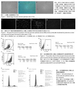
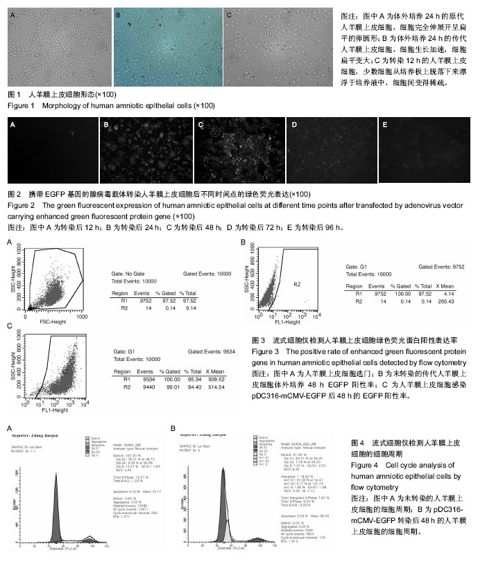
2.1 人羊膜上皮细胞形态 在接种12 h内细胞可下沉贴壁,24 h后细胞逐渐变大,折光性降低,整个细胞轮廓变暗,完全伸展的细胞呈扁平的卵圆形(图1A);2 d后,进入指数生长期,细胞数量明显增多, 呈不规则的多角形,四五天后90%细胞融合成片,形成单层。传代后人羊膜上皮细胞能较快贴壁,接种4-6 h后即可贴壁并开始生长,呈多形性;接种24 h后细胞生长加速,细胞扁平变大(图1B);三四天后细胞呈90%融合,呈典型的铺路石外观,呈多形性,与原代无明显区别。培养的人羊膜上皮细胞加入转染液12 h后,有少数细胞从培养板上脱落下来漂浮于培养液中,细胞间变得稀疏(图1C),终止转染后细胞形态和生长特性逐渐恢复正常,终止转染后48 h,细胞大多数转染细胞均与正常细胞形态上无显著差异。 2.2 荧光显微镜下观察EGFP的阳性表达 转染后12 h即可见发绿色荧光的人羊膜上皮细胞,但数量较少,强度较弱,每个低倍视野下可见0-2个细胞(图2A);转染后24 h发绿色荧光的人羊膜上皮细胞逐渐增多、荧光强度增强(图2B);转染后48 h发绿色荧光的人羊膜上皮细胞继续增多,几乎所有存活细胞均发出绿色荧光(图2C);转染后72 h发绿色荧光的人羊膜上皮细胞有所减少,荧光强度开始减弱(图2D);转染后96 h发绿色荧光的人羊膜上皮细胞继续减少,荧光强度继续减弱(图2E)。 2.3 流式细胞仪检测活细胞比例及人羊膜上皮细胞pDC316-mCMV-EGFP基因转染效率 流式细胞仪检测结果显示:未转染组人羊膜上皮细胞中,绿色荧光蛋白阳性细胞数为0.14%(图3B),瞬时转染后12,24,48,72,96 h的人羊膜上皮细胞均可见绿色荧光蛋白阳性表达,其中瞬时转染后48 h的人羊膜上皮细胞绿色荧光蛋白阳性表达率最高,达99.01%(图3C),但随着体外培养时间的延长和传代次数的增加其荧光表达率逐渐下降。 2.4 流式细胞仪检测细胞周期 转染后48 h的G0/G1期人羊膜上皮细胞占总细胞数的85.51%,G2/M期占7.48%,S期占7.02%,G2/G1为2.0%,未转染的人羊膜上皮细胞G0/G1期占总细胞数的78.21%,G2/M期占8.58%,S期占13.21%,与未转染细胞的细胞周期相比,转染后人羊膜上皮细胞G0/G1期细胞百分比大于未转染组。转染后人羊膜上皮细胞S期的细胞百分比小于未转染组,说明经腺病毒介导EGFP转染的人羊膜上皮细胞的细胞周期有所减慢(图4)。"
| [1] Amin HD, Brady MA, St-Pierre JP, et al. Stimulation of chondrogenic differentiation of adult human bone marrow-derived stromal cells by a moderate-strength static magnetic field. Tissue Eng Part A. 2014;20(11-12):1612-1620.[2] Li G, Zheng B, Meszaros LB, et al. Identification and characterization of chondrogenic progenitor cells in the fascia of postnatal skeletal muscle. J Mol Cell Biol. 2011;3(6): 369-377.[3] 刘峰舟,尹纯,孙夏承,等.AKAP1过表达腺病毒载体构建及其在心肌细胞中的功能初步研究[J].心脏杂志,2017,29(4):405-410.[4] Fernandes P, Simão D, Guerreiro MR, et al. Impact of adenovirus life cycle progression on the generation of canine helper-dependent vectors. Gene Ther. 2015;22(1):40-49.[5] Reddy VS, Nemerow GR. Structures and organization of adenovirus cement proteins provide insights into the role of capsid maturation in virus entry and infection. Proc Natl Acad Sci U S A. 2014;111(32):11715-11720.[6] Chalfie M, Tu Y, Euskirchen G, et al. Green fluorescent protein as a marker for gene expression. Science. 1994;263(5148): 802-805.[7] Listwan P, Martin JL, Kobe B,et al. Methods for high-throughput protein expression, purification and structure determination adapted for structural genomics. Australian Biochemist. 2004; 35(2):43-46.[8] Feilmeier BJ, Iseminger G, Schroeder D, et al. Green fluorescent protein functions as a reporter for protein localization in Escherichia coli. J Bacteriol. 2000;182(14): 4068-4076.[9] Shaner NC, Steinbach PA, Tsien RY. A guide to choosing fluorescent proteins. Nat Methods. 2005;2(12):905-909.[10] 金玲,陈剑,吴静,等.慢病毒介导EGFP基因修饰的人羊膜上皮细胞重建角膜表层的实验研究[J].中华实验眼科杂志,2011,29(8): 685-689.[11] 刘小勇,陈剑,吴静,等.人羊膜上皮细胞体外培养重建角膜上皮的实验研究[J].中国病理生理杂志,2012,28(4):689-693.[12] Liu XY, Chen J, Zhou Q, et al. In vitro tissue engineering of lamellar cornea using human amniotic epithelial cells and rabbit cornea stroma. Int J Ophthalmol. 2013;6(4):425-429.[13] Zhou Q, Liu XY, Ruan YX, et al. Construction of corneal epithelium with human amniotic epithelial cells and repair of limbal deficiency in rabbit models. Hum Cell. 2015;28(1): 22-36.[14] 刘小勇,陈剑,吴静,等.人羊膜上皮细胞的原代及传代培养[J].广东医学,2007,28 (2):181-183.[15] 金玲,陈剑,吴静,等.人羊膜上皮细胞的分离、纯化及其生物学特性研究[J].广东医学,2010,31(16):2055-2057.[16] 刘小勇,周清,张晓玲,等.人羊膜上皮细胞的体外培养及标记分子的检测分析[J].细胞与分子免疫学杂志, 2014, 30(12): 1318-1321.[17] Lefebvre S, Adrian F, Moreau P, et al. Modulation of HLA-G expression in human thymic and amniotic epithelial cells. Hum Immunol. 2000;61(11):1095-1101.[18] Li H, Niederkorn JY, Neelam S, et al. Immunosuppressive factors secreted by human amniotic epithelial cells. Invest Ophthalmol Vis Sci. 2005;46(3):900-907.[19] Banas RA, Trumpower C, Bentlejewski C, et al. Immunogenicity and immunomodulatory effects of amnion-derived multipotent progenitor cells. Hum Immunol. 2008;69(6):321-328.[20] Miki T, Strom SC. Amnion-derived pluripotent/multipotent stem cells. Stem Cell Rev. 2006;2(2):133-142.[21] Ilancheran S, Michalska A, Peh G, et al. Stem cells derived from human fetal membranes display multilineage differentiation potential. Biol Reprod. 2007;77(3):577-588.[22] Shinya M, Komuro H, Saihara R, et al. Neural differentiation potential of rat amniotic epithelial cells. Fetal Pediatr Pathol. 2010;29(3):133-143. [23] Lin JS, Zhou L, Sagayaraj A, et al. Hepatic differentiation of human amniotic epithelial cells and in vivo therapeutic effect on animal model of cirrhosis. J Gastroenterol Hepatol. 2015; 30(11):1673-1682.[24] Ji H,Yang Q,Ji F,et al.Human amniotic epithelial cell transplantation for the repair of injured brachial plexus nerve: evaluation of nerve viscoelastic properties. Neural Regen Res. 2015;10(2): 260-265.[25] Wang TG,Xu J,Zhu AH,et al.Human amniotic epithelial cells combined with silk fbroin sca?old in the repair of spinal cord injury.Neural Regen Res.2016;11(10): 1670-1677.[26] Mahmood R, Choudhery MS, Mehmood A, et al. In Vitro Differentiation Potential of Human Placenta Derived Cells into Skin Cells. Stem Cells Int. 2015;2015:841062.[27] Fliniaux I, Viallet JP, Dhouailly D, et al. Transformation of amnion epithelium into skin and hair follicles. Differentiation. 2004;72(9-10):558-565.[28] Okere B, Alviano F, Costa R, et al. In vitro differentiation of human amniotic epithelial cells into insulin-producing 3D spheroids. Int J Immunopathol Pharmacol. 2015; 28(3): 390-402.[29] Ginsberg M, Schachterle W, Shido K, et al. Direct conversion of human amniotic cells into endothelial cells without transitioning through a pluripotent state. Nat Protoc. 2015; 10(12):1975-1985.[30] Yang SP, Yang XZ, Cao GP. Conjunctiva reconstruction by induced differentiation of human amniotic epithelial cells. Genet Mol Res. 2015;14(4):13823-13834. [31] Tabatabaei M, Mosaffa N, Nikoo S, et al. Isolation and partial characterization of human amniotic epithelial cells: the effect of trypsin. Avicenna J Med Biotechnol. 2014;6(1):10-20.[32] Wolbank S, van Griensven M, Grillari-Voglauer R, et al. Alternative sources of adult stem cells: human amniotic membrane. Adv Biochem Eng Biotechnol. 2010;123:1-27.[33] Pappa KI, Anagnou NP. Novel sources of fetal stem cells: where do they fit on the developmental continuum. Regen Med. 2009;4(3):423-433.[34] Jiang TS, Cai L, Ji WY, et al. Reconstruction of the corneal epithelium with induced marrow mesenchymal stem cells in rats. Mol Vis. 2010;16:1304-1316.[35] Tsai CL, Chuang PC, Kuo HK, et al. Differentiation of Stem Cells From Human Exfoliated Deciduous Teeth Toward a Phenotype of Corneal Epithelium In Vitro. Cornea. 2015; 34(11): 1471-1477.[36] Ahmad S, Stewart R, Yung S, et al. Differentiation of human embryonic stem cells into corneal epithelial-like cells by in vitro replication of the corneal epithelial stem cell niche. Stem Cells. 2007;25(5):1145-1155.[37] Blazejewska EA, Schlötzer-Schrehardt U, Zenkel M, et al. Corneal limbal microenvironment can induce transdifferentiation of hair follicle stem cells into corneal epithelial-like cells. Stem Cells. 2009;27(3):642-652. [38] Nishida K, Yamato M , Hayashida Y, et al. Corneal reconstruction with tissue- engineered cell sheets composed of autologous oral mucosal epithelium. N Engl J Med. 2004; 351(12):1187-1196.[39] Inoshita S, Koizumi N, Nakamura T, et al.Transplantable cultivated mucosal epithelial sheet for ocular surface reconstruction. Exp Eye Research. 2004;78(3):483-491.[40] Noshita S, Nakamura T, et al.Development of cultivated mucosal epithelial transplantation for Ocular surface reconstruction. Artificial Organs. 2004;28(1): 22-27.[41] Inatomi T, Nakamura T, Koizumi N, et al. Midterm results on ocular surface reconstruction using cultivated autologous oral mucosal epithelial transplantation .Am J Ophthalmol. 2006; 141(2):267-275. |
| [1] | Yao Xiaoling, Peng Jiancheng, Xu Yuerong, Yang Zhidong, Zhang Shuncong. Variable-angle zero-notch anterior interbody fusion system in the treatment of cervical spondylotic myelopathy: 30-month follow-up [J]. Chinese Journal of Tissue Engineering Research, 2022, 26(9): 1377-1382. |
| [2] | Wang Jifang, Bao Zhen, Qiao Yahong. miR-206 regulates EVI1 gene expression and cell biological behavior in stem cells of small cell lung cancer [J]. Chinese Journal of Tissue Engineering Research, 2022, 26(7): 1027-1031. |
| [3] | Zhang Jinglin, Leng Min, Zhu Boheng, Wang Hong. Mechanism and application of stem cell-derived exosomes in promoting diabetic wound healing [J]. Chinese Journal of Tissue Engineering Research, 2022, 26(7): 1113-1118. |
| [4] | An Weizheng, He Xiao, Ren Shuai, Liu Jianyu. Potential of muscle-derived stem cells in peripheral nerve regeneration [J]. Chinese Journal of Tissue Engineering Research, 2022, 26(7): 1130-1136. |
| [5] | He Yunying, Li Lingjie, Zhang Shuqi, Li Yuzhou, Yang Sheng, Ji Ping. Method of constructing cell spheroids based on agarose and polyacrylic molds [J]. Chinese Journal of Tissue Engineering Research, 2022, 26(4): 553-559. |
| [6] | He Guanyu, Xu Baoshan, Du Lilong, Zhang Tongxing, Huo Zhenxin, Shen Li. Biomimetic orientated microchannel annulus fibrosus scaffold constructed by silk fibroin [J]. Chinese Journal of Tissue Engineering Research, 2022, 26(4): 560-566. |
| [7] | Chen Xiaoxu, Luo Yaxin, Bi Haoran, Yang Kun. Preparation and application of acellular scaffold in tissue engineering and regenerative medicine [J]. Chinese Journal of Tissue Engineering Research, 2022, 26(4): 591-596. |
| [8] | Kang Kunlong, Wang Xintao. Research hotspot of biological scaffold materials promoting osteogenic differentiation of bone marrow mesenchymal stem cells [J]. Chinese Journal of Tissue Engineering Research, 2022, 26(4): 597-603. |
| [9] | Shen Jiahua, Fu Yong. Application of graphene-based nanomaterials in stem cells [J]. Chinese Journal of Tissue Engineering Research, 2022, 26(4): 604-609. |
| [10] | Zhang Tong, Cai Jinchi, Yuan Zhifa, Zhao Haiyan, Han Xingwen, Wang Wenji. Hyaluronic acid-based composite hydrogel in cartilage injury caused by osteoarthritis: application and mechanism [J]. Chinese Journal of Tissue Engineering Research, 2022, 26(4): 617-625. |
| [11] | Li Hui, Chen Lianglong. Application and characteristics of bone graft materials in the treatment of spinal tuberculosis [J]. Chinese Journal of Tissue Engineering Research, 2022, 26(4): 626-630. |
| [12] | Gao Cangjian, Yang Zhen, Liu Shuyun, Li Hao, Fu Liwei, Zhao Tianyuan, Chen Wei, Liao Zhiyao, Li Pinxue, Sui Xiang, Guo Quanyi. Electrospinning for rotator cuff repair [J]. Chinese Journal of Tissue Engineering Research, 2022, 26(4): 637-642. |
| [13] | Guan Jian, Jia Yanfei, Zhang Baoxin , Zhao Guozhong. Application of 4D bioprinting in tissue engineering [J]. Chinese Journal of Tissue Engineering Research, 2022, 26(3): 446-455. |
| [14] | Liu Jiali, Suo Hairui, Yang Han, Wang Ling, Xu Mingen. Influence of lay-down angles on mechanical properties of three-dimensional printed polycaprolactone scaffolds [J]. Chinese Journal of Tissue Engineering Research, 2022, 10(16): 2612-2617. |
| [15] | Huang Bo, Chen Mingxue, Peng Liqing, Luo Xujiang, Li Huo, Wang Hao, Tian Qinyu, Lu Xiaobo, Liu Shuyun, Guo Quanyi . Fabrication and biocompatibility of injectable gelatin-methacryloyl/cartilage-derived matrix particles composite hydrogel scaffold [J]. Chinese Journal of Tissue Engineering Research, 2022, 10(16): 2600-2606. |
| Viewed | ||||||
|
Full text |
|
|||||
|
Abstract |
|
|||||