Chinese Journal of Tissue Engineering Research ›› 2016, Vol. 20 ›› Issue (30): 4469-4475.doi: 10.3969/j.issn.2095-4344.2016.30.009
Previous Articles Next Articles
Autologous oxygen-delivering biomimetic nanoscaffold composited with chondrocytes reconstructs the temporomandibular joint
Wang Hong, Liao Tian-an, Wang Tao, Fu Liang-bin, Hu Guang-wei, Deng Wei
- Department of Stomatology, Hainan Provincial People’s Hospital, Haikou 570311, Hainan Province, China
-
Online:2016-07-15Published:2016-07-15 -
Contact:Liao Tian-an, Chief physician, Professor, Master’s supervisor, Department of Stomatology, Hainan Provincial People’s Hospital, Haikou 570311, Hainan Province, China -
About author:Wang Hong, Associate chief physician, Department of Stomatology, Hainan Provincial People’s Hospital, Haikou 570311, Hainan Province, China
CLC Number:
Cite this article
Wang Hong, Liao Tian-an, Wang Tao, Fu Liang-bin, Hu Guang-wei, Deng Wei . Autologous oxygen-delivering biomimetic nanoscaffold composited with chondrocytes reconstructs the temporomandibular joint[J]. Chinese Journal of Tissue Engineering Research, 2016, 20(30): 4469-4475.
share this article
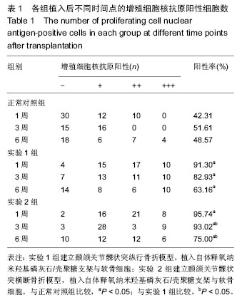
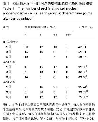
2.1 各组增殖细胞核抗原阳性细胞数表达情况 实验1组、实验2组增殖细胞核抗原阳性细胞数高于正常对照组(P < 0.05);植入1周时,实验1组与实验2组增殖细胞核抗原阳性细胞数无差异(P > 0.05);植入3,6周时,实验1组增殖细胞核抗原阳性细胞数低于实验2组 (P < 0.05),见表1。"
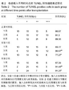
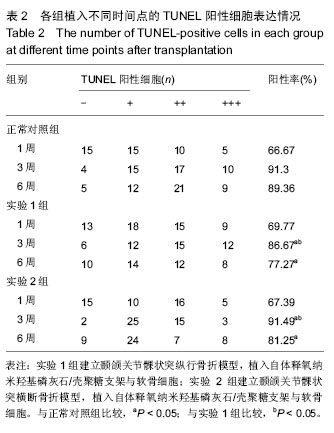
2.2 各组TUNEL检测细胞凋亡结果 实验1组、实验2组凋亡细胞数高于正常对照组(P < 0.05);植入1,6周时,实验1组与实验2组凋亡细胞数无差异(P > 0.05);植入3周时,实验1组凋亡细胞数低于实验2组(P < 0.05),见表2。"
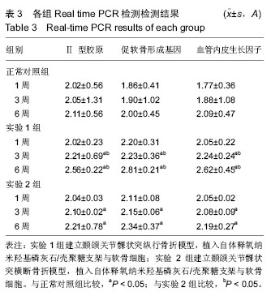
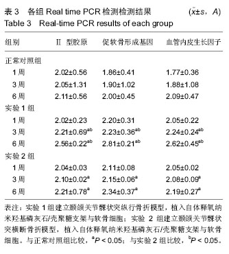
2.3 各组Real time PCR检测结果 结果显示,实验1组、实验2组髁突软骨内成骨各标志物表达量随时间的延长呈现上升的趋势,说明软骨内成骨能力升高,实验1组各标志物升高更为明显(P < 0.05),见表3。"
| [1] 杨驰,何冬梅,陈敏洁,等.課突囊内骨折的临床特点和分类研究[J].中国口腔颌面外科杂志, 2010,8(3): 208-211. [2] He D,Yang C,Chen M,et al.Intracapsular condylar fracture of the mandible:our classification and open treatment experience.J Oral Maxillofac Surg. 2009; 67(8):1672-1679. [3] 徐婷.生物机械应力对下颌髁突适应性改建的影响[J].国际口腔医学杂志,2009,36(1):52-54. [4] Uckan S,Bayram B,Kecik D,et al.Effects of titanium plate fixation on mandibular growth in a rabbit model.J Oral Maxillofac Surg.2009;67(2):318-322. [5] 杨驰,何冬梅,陈敏洁,等.下颌骨課突囊内骨折的治疗探讨[J].中国口腔颂面外科杂志,2010,8(2):112-118. [6] Kanayama H,Masuda Y,Adachi T,et al.Temporal alteration of chewing jaw movements after a reversible bite-raising in guinea pigs.Arch Oral Biol. 2010;55(1): 89-94. [7] 李毅,王东.两种手术入路治疗下颂骨髁突中位骨折效果评价[J].武警后勤学院学报(医学版), 2012,21(3):193-195. [8] Abdel-Galil K,Loukota R.Fractures of the mandibular condyle:evidence base and current concepts of management.Br J Oral Maxillofac Surg. 2010;48(7): 520-526. [9] Nishiyama KK,Graeme M.Reproducibility of bone micro-architecture measurements in rodents by in vivo micro-computed tomography is maximized with three-dimensional image registration.J Bone. 2010;46: 155-161. [10] Jiao K,Dai J,Wang MQ,et al.Age-and sex-related changes of mandibular condylar cartilage and subchondral bone:a histomorphometric and micro-CT study in rats.Arch Oral Biol.2010;55:155-163. [11] 郭家平,李志进,董青山,等.125例縣突骨折临床回顾性研究?[J].临床口腔医学杂志,2011,27(9):540-541. [12] Prasad S,Kuracina J,Edward A,et al.Altering occlusal vertical dimension provisionally with metal onlays:A clinic report.J Pros Dent. 2008;100(5): 338-342. [13] Jiao K,Dai J,Wang MQ,et al.Subchondral bone loss following orthodontically induced cartilage degradation in the mandibular condyles of rats. J.Bone. 2010;9:10-16. [14] Soares CJ,Castro CG,Neiva NA.Effect of gamma irradiation on ultimate tensile strength of enamel and dentin.J Dent Res.2010;89(2):159-164. [15] Pegado RE,Do AF,Florio FM,et al.Effect of different bonding strategies on adhesion to deep and superficial permanent dentin.Eur J Dent.2010;4(2):110-117. [16] Beloica M,Goracci C,Carvalho CA,et al.Microtensile vs microshear bond strength of all-in-one adhesives to unground enamel.J Adhes Dent. 2010;12(6): 427-433. [17] Lin J,Shinya A,Gomi H,et al.Bonding of self-adhesive resin cements to enamel using different surface treatments:bond strength and etching pattern evaluations. Dent Mater.2010;29(4):425-432. [18] Osorio R,Aguilera FS,Otero PR,et al.Primary dentin etching time,bond strength and ultra-structure characterization of dentin surfaces.J Dent. 2010;38(3): 222-231. [19] Sauro S,Di Renzo S,Castagnola R,et al.Comparison between water and ethanol wet bonding of resin composite to root canal dentin.Am J Dent. 2011;24(1): 25-30. [20] Zhao SJ,Zhang L,Tang LH,et al.Nanoleakage and microtensile bond strength at the adhesive-dentin interface after different etching times.Am J Dent. 2010;23(6):335-340. [21] Vinay S,Shivanna V.Comparative evaluation of microleakage of fifth,sixth,and seventh generation dentin bonding agents:An in vitro study.J Conserv Dent.2010;13(3):136-140. [22] Hamouda IM,Samra NR,Badawi MF.Microtensile bond strength of etch and rinse versus self-etch adhesive systems.J Mech Behav Biomed Mater. 2011;4(3): 461-466. [23] Albaladejo A,Osorio R,Toledano M,et al.Hybrid layers of etch-and-rinse versus self-etching adhesive systems.Med Oral Patol Oral Cir Bucal. 2010;15(1): 112-118. [24] Zanatta FB,Pinto TM,Kantorski KZ,et al.Plaque, gingival bleeding and calculus formation after supragingival scaling with and without polishing:a randomised clinical trial.Oral Health Prev Dent. 2011; 9(3):275-280. [25] Leung CC,Palomo L,Griffith R,et al.Accuracy and reliability ofone-beam computed tomography for measuring alveolar bone height and detecting bony dehiscences and fenestrations.Am J Orthod Dentofacial Orthop.2010;137(4):109-119. [26] Kassab MM,Badaw i H,Dentino AR.Treatment of gingival Recession.Dent Clin North Am. 2010;54(1): 129-140. [27] Lei WY,Rabie AB,Wong RW.Repair of a defect following the removal of an impacted maxillary canine by orthodontic tooth movement:a case report.Cases J.2010;3(2):62-69. [28] Liu D,Zhou Y,Li C,et al.Denaturing gradient gel electrophoresis analysis with different primers of subgingival bacterial communities under mechanical debridement.Microbiol Immunol.2010;54(11):702-706. [29] Tetradis S,Anstey P,Graff-Radford S.Cone beam computed tomography in the diagnosis of dental disease.J Calif Dent Assoc.2010;38(1):27-32. [30] Viotti RG,Kasaz A,Pena CE,et al.Microtensile bond strength of new self-adhesive luting agents and conventional multistep systems.J Prosthet Dent.2009; 102(5):306-312. [31] Levin L,Einy S,Zigdon H,et al.Guidelines for periodontal care and follow-up during orthodontic treatment in adolescents and young adults.J Appl Oral Sci.2012;20(4):399-403. [32] Boyer S,Fontanel F,Danan M,et al.Severe periodontitis and orthodontics:evaluation of long-term results.Int Orthod.2011;9(3):259-273. [33] Shrout PE,Fleiss JL.Intraclass correlations:uses in assessing rater reliability.Psychol Bull. 1979;86(2): 420-428. [34] Shoreibah EA,Ibrahim SA,Attia MS,et al.Clinical and radiographic evaluation of bone grafting in corticotomy-facilitated orthodontics in adults.J Int Acad Periodontol.2012;14(4):105-113. [35] Corbacho de Melo MM,Cardoso MG,Faber J,et al.Risk factors for periodontal changes in adult patients with banded second molars during orthodontic treatment. Angle Orthod.2012;82(2):224-228. [36] Fung K,Chandhoke TK,Uribe F,et al.Periodontal regeneration and orthodontic intrusion of a pathologically migrated central incisor adjacent to an infrabony defect.J Clin Orthod.2012;46(7):417-423. [37] Zhang J,Zhou S,Li R,et al.Magnetic bead-based salivary peptidome profiling for periodontal-orthodontic treatment.Proteome Sci.2012;10(1):63. [38] Viecilli RF,Budiman A,Burstone CJ.Axes of resistance for tooth movement:does the center of resistance exist in 3-dimensional space?Am J Orthod Dentofacial Orthop.2013;143(2):163-172. [39] Graber LW,Vanarsdall RL,Vig WL,et al. Orthodontics: current principles and techniques.5th ed.St Louis: Elsevier Mosby, 2012:807-841. [40] Yonenaga K,Nishizawa S,Fujihara Y,et al.The optimal conditions of chondrocyte isolation and its seeding in the preparation for cartilage tissue engineering. Tissue Eng Part C Methods.2010;16(6):1461-1469. [41] Ciocca L,Tarsitano A,Marchetti C,et al.A CAD-CAM- prototyped temporomandibular condyle connected to a bony plate to support a free fibula flap in patients undergoing mandiblectomy: A pilot study with 5 years of follow up.J Craniomaxillofac Surg.2016.pii: S1010-5182(16)30039-7. [42] Sharma D,Khasgiwala A,Maheshwari B,et al. Superolateral dislocation of an intact mandibular condyle into the temporal fossa: case report and literature review.Dent Traumatol.2016. doi:10.1111/edt.12282.[Epub ahead of print] [43] Dimitriou D,Tsai TY,Park KK,et al.Weight-bearing condyle motion of the knee before and after cruciate-retaining TKA: In-vivo surgical transepicondylar axis and geometric center axis analyses.J Biomech. 2016.pii:S0021-9290 (16)30530-9. doi: 10.1016/j.jbiomech.2016.04.033. [Epub ahead of print] [44] Xu L,Guo H,Li C,et al.A time-dependent degeneration manner of condyle in rat CFA-induced inflamed TMJ. Am J Transl Res. 2016;8(2):556-567. [45] Zhao LL,Tong PJ,Xiao LW.Internal fixation with lag screws plus an anti-sliding plate for the treatment of Hoffa fracture of the lateral femoral condyle.Zhongguo Gu Shang.2016;29(3):266-269. |
| [1] | Zhang Tongtong, Wang Zhonghua, Wen Jie, Song Yuxin, Liu Lin. Application of three-dimensional printing model in surgical resection and reconstruction of cervical tumor [J]. Chinese Journal of Tissue Engineering Research, 2021, 25(9): 1335-1339. |
| [2] | Zeng Yanhua, Hao Yanlei. In vitro culture and purification of Schwann cells: a systematic review [J]. Chinese Journal of Tissue Engineering Research, 2021, 25(7): 1135-1141. |
| [3] | Ma Zetao, Zeng Hui, Wang Deli, Weng Jian, Feng Song. MicroRNA-138-5p regulates chondrocyte proliferation and autophagy [J]. Chinese Journal of Tissue Engineering Research, 2021, 25(5): 674-678. |
| [4] | Xie Chongxin, Zhang Lei. Comparison of knee degeneration after anterior cruciate ligament reconstruction with or without remnant preservation [J]. Chinese Journal of Tissue Engineering Research, 2021, 25(5): 735-740. |
| [5] | Xu Dongzi, Zhang Ting, Ouyang Zhaolian. The global competitive situation of cardiac tissue engineering based on patent analysis [J]. Chinese Journal of Tissue Engineering Research, 2021, 25(5): 807-812. |
| [6] | Wu Zijian, Hu Zhaoduan, Xie Youqiong, Wang Feng, Li Jia, Li Bocun, Cai Guowei, Peng Rui. Three-dimensional printing technology and bone tissue engineering research: literature metrology and visual analysis of research hotspots [J]. Chinese Journal of Tissue Engineering Research, 2021, 25(4): 564-569. |
| [7] | Chang Wenliao, Zhao Jie, Sun Xiaoliang, Wang Kun, Wu Guofeng, Zhou Jian, Li Shuxiang, Sun Han. Material selection, theoretical design and biomimetic function of artificial periosteum [J]. Chinese Journal of Tissue Engineering Research, 2021, 25(4): 600-606. |
| [8] | Liu Fei, Cui Yutao, Liu He. Advantages and problems of local antibiotic delivery system in the treatment of osteomyelitis [J]. Chinese Journal of Tissue Engineering Research, 2021, 25(4): 614-620. |
| [9] | Li Xiaozhuang, Duan Hao, Wang Weizhou, Tang Zhihong, Wang Yanghao, He Fei. Application of bone tissue engineering materials in the treatment of bone defect diseases in vivo [J]. Chinese Journal of Tissue Engineering Research, 2021, 25(4): 626-631. |
| [10] | Zhang Zhenkun, Li Zhe, Li Ya, Wang Yingying, Wang Yaping, Zhou Xinkui, Ma Shanshan, Guan Fangxia. Application of alginate based hydrogels/dressings in wound healing: sustained, dynamic and sequential release [J]. Chinese Journal of Tissue Engineering Research, 2021, 25(4): 638-643. |
| [11] | Chen Jiana, Qiu Yanling, Nie Minhai, Liu Xuqian. Tissue engineering scaffolds in repairing oral and maxillofacial soft tissue defects [J]. Chinese Journal of Tissue Engineering Research, 2021, 25(4): 644-650. |
| [12] | Xing Hao, Zhang Yonghong, Wang Dong. Advantages and disadvantages of repairing large-segment bone defect [J]. Chinese Journal of Tissue Engineering Research, 2021, 25(3): 426-430. |
| [13] | Chen Siqi, Xian Debin, Xu Rongsheng, Qin Zhongjie, Zhang Lei, Xia Delin. Effects of bone marrow mesenchymal stem cells and human umbilical vein endothelial cells combined with hydroxyapatite-tricalcium phosphate scaffolds on early angiogenesis in skull defect repair in rats [J]. Chinese Journal of Tissue Engineering Research, 2021, 25(22): 3458-3465. |
| [14] | Wang Hao, Chen Mingxue, Li Junkang, Luo Xujiang, Peng Liqing, Li Huo, Huang Bo, Tian Guangzhao, Liu Shuyun, Sui Xiang, Huang Jingxiang, Guo Quanyi, Lu Xiaobo. Decellularized porcine skin matrix for tissue-engineered meniscus scaffold [J]. Chinese Journal of Tissue Engineering Research, 2021, 25(22): 3473-3478. |
| [15] | Mo Jianling, He Shaoru, Feng Bowen, Jian Minqiao, Zhang Xiaohui, Liu Caisheng, Liang Yijing, Liu Yumei, Chen Liang, Zhou Haiyu, Liu Yanhui. Forming prevascularized cell sheets and the expression of angiogenesis-related factors [J]. Chinese Journal of Tissue Engineering Research, 2021, 25(22): 3479-3486. |
| Viewed | ||||||
|
Full text |
|
|||||
|
Abstract |
|
|||||